Ebba Biotech AB was founded in 2016 by professor Agneta Richter-Dahlfors to enable commercial applications and more widespread use of fluorescent tracer (optotracers) molecules, the result of many years of cutting-edge nanoscience and organic chemistry research.


About our Technology

Optotracing is an innovative visualization method developed by scientists for scientists for use in fluorescence microscopy and spectroscopy. With optotracing – using fluorescent tracer molecules – Ebba Biotech provides an innovative method for several expanding areas of research. The optotracing technology has been developed by a scientific expert team of researchers at Karolinska Institutet, Linköping University and the Royal Institute of Technology (KTH) in Sweden and is a result of many years of cutting-edge nanoscience and organic chemistry research.

Ebba Biotech has commercialized distribution and sales of optotracer molecules and today supply these in three different series:

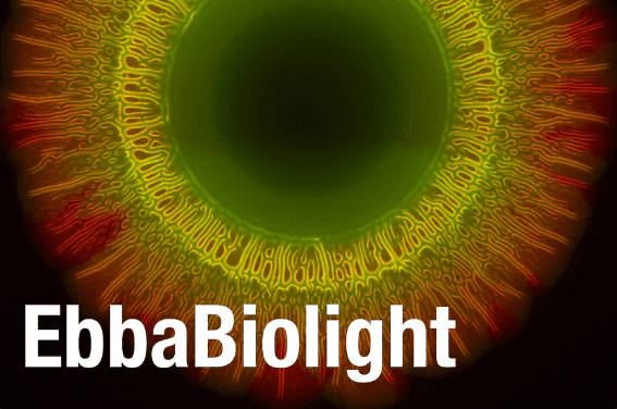
  • EbbaBiolight – to probe and detect bacteria & biofilm.
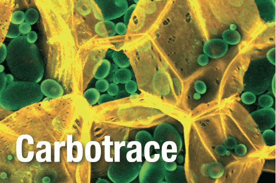
  • Carbotrace – for anatomical mapping of plants to understand their composition.
  • Amytracker – to search and detect amyloids and other protein aggregates.

Fluorescent tracers for amyloids and other protein aggregates
Amytracker are fluorescent tracer molecules providing high quality visualization of protein aggregates. Using Amytracker, pre-fibrillar states of amyloids from a variety of amyloidogenic proteins or peptides are detected in tissues and living cells.

Optotracers for carbohydrate analysis and anatomical mapping
Carbotrace are fluorescent tracer molecules targeting carbohydrate structures. Carbotrace can be used for non-destructive carbohydrate analysis, carbohydrate discrimination and anatomical mapping of carbohydrate structures in plants.

Optotracers for detection of bacteria and bacterial biofilm
EbbaBiolight are fluorescent tracer molecules allowing to identify components of bacteria and bacterial extracellular matrix. Using EbbaBiolight, it is now possible to track biofilm formation in living, growing cultures in liquid and on surfaces.

Amytracker

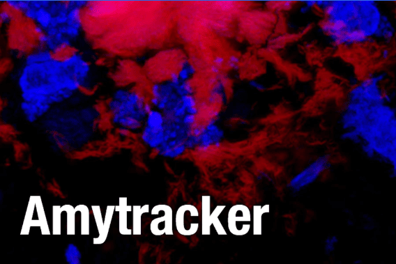
Amytracker are fluorescent tracer molecules for high-quality visualization of protein aggregates.

The molecules become vividly colorful – highly fluorescent – when bound to a target in a sample. Targets are amyloids or protein aggregates containing amyloid fibrils and proto-fibrils. Therefore, using Amytracker, you can achieve reliable fluorescent staining of early, pre-fibrillar states of amyloids arising from a variety of amyloidogenic proteins or peptides in tissues from a wide range of species and perform fibrillation assays and spectrophotometric detection of protein aggregates in liquid samples.

Amytracker molecules are non-toxic and are readily taken-up when applied to cells in culture. Further, they are photo- and thermostable and allow for easy handling in any application. Our ex vivo products will simplify your life when performing staining of tissue sections, live-cell imaging or fibrillation assays. The ex vivo products are available in aliquots of 50 µl, 100 µl, 150 µl or 200 µl (all delivered in 50 µl vials). Please, contact us to find out more about Amytracker for in vivo use.

Applications

  • Protocol I: Staining of protein aggregates in tissue sections
  • Protocol II: Staining of protein aggregates with Amytracker and antibody co-stain
  • Protocol III: Live-cell imaging
  • Protocol IV: Fibrillation assay

Quickstart Protocol

Amyloid staining in tissue sections (Protocol I)

  • use fixed- or fresh tissue
  • dilute Amytracker 1:1000 in PBS (pH 7.4)
  • apply generously to sample
  • incubate 5-30 min
  • wash with PBS (optional)
  • image at recommended excitation/emission

Live-cell imaging (Protocol III)

  • dilute Amytracker 1:500 in cell culture medium or PBS (pH 7.4)
  • apply generously to sample
  • incubate 30 min
  • wash with cell culture medium or PBS (optional)
  • image at recommended excitation/emission

Note

  • Amytracker is for research use only.
  • Amytracker is not for diagnostic use or use in humans.
  • Amytracker is not for resale.

Quick Facts

Amytracker fluorescent tracer molecules are:

  • provided as ex vivo variant with 1 mg/ml concentration in volumes of 50 µl, 100 µl, 150 µl and 200 µl and as a Mix&Try Kit with 5×10 µl
  • readily taken up into cells and tissues
  • ex vivo variants are diluted 1000-fold for staining of tissue sections and 500-fold for live-cell imaging
  • non-toxic
  • photo- and thermostable
  • fast
  • accurate

Storage

  • Store Amytracker at 4°C.
  • Use the opened vial within 12 month.

Products

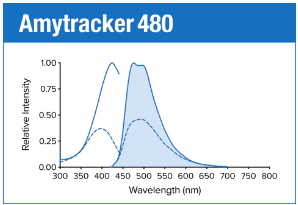
Amytracker 480 is our blue fluorescent optotracer for labeling protein aggregates with repetitive arrangement of β-sheets. It labels Aβ plaques and neurofibrillary tangles in tissue sections with AD pathology and α-synuclein aggregates in tissue sections with PD pathology. Specifically, Amytracker 480 has been used to study amyloid formation during abnormal coagulation. Contact us to learn more about Amytracker applications.

As all our optotracers, Amytracker 480 is exceptionally photostable and fluorogenic. When bound to a target, Amytracker 480 can be imaged using epifluorescence, confocal and superresolution microscopy. Spectral information can be acquired using a fluorescence spectrophotometer. Use recommended filter sets as well as excitation- and emission wavelengths according to the following table.

Table: Excitation- and emission wavelengths as well as recommended filter sets.

Exmax Emmax Recommended filter-sets
Amytracker 480 420 nm 480 nm DAPI

Amytracker 480 is available as a 1 mg/ml solution in volumes ranging from 10 – 200 µL (see volumes and prices in the list below). As our standard solvent, we use ultrapure water. Please contact us for custom options.

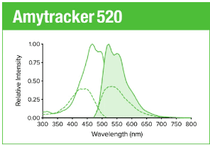
Amytracker 520 is our green fluorescent optotracer molecule for labeling protein aggregates with repetitive arrangement of β-sheets. It labels Aβ plaques and neurofibrillary tangles in tissue sections with AD pathology and α-synuclein aggregates in tissue sections with PD pathology. Contact us to learn more about Amytracker applications.

As all our optotracers, Amytracker 520 is exceptionally photostable and fluorogenic. When bound to a target, Amytracker 520 can be imaged using epifluorescence, confocal and superresolution microscopy. Spectral information can be acquired using a fluorescence spectrophotometer. Use recommended filter sets as well as excitation- and emission wavelengths according to the following table.

Table: Excitation- and emission wavelengths as well as recommended filter sets.

Exmax Emmax Recommended filter-sets
Amytracker 520 460 nm 520 nm FITC, GFP

Amytracker 520 is available as a 1 mg/ml solution in volumes ranging from 10 – 200 µL (see volumes and prices in the list below). As our standard solvent, we use ultrapure water. Please contact us for custom options.

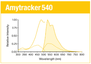
Amytracker 540 is our yellow fluorescent optotracer molecule for labeling protein aggregates with repetitive arrangement of β-sheets. It labels Aβ plaques and neurofibrillary tangles in tissue sections with AD pathology and α-synuclein aggregates in tissue sections with PD pathology. Contact us to learn more about Amytracker applications.

As all our optotracers, Amytracker 540 is exceptionally photostable and fluorogenic. When bound to a target, Amytracker 540 can be imaged using epifluorescence, confocal and superresolution microscopy. Spectral information can be acquired using a fluorescence spectrophotometer. Use recommended filter sets as well as excitation- and emission wavelengths according to the following table.
Table: Excitation- and emission wavelengths as well as recommended filter sets.

Exmax Emmax Recommended filter-sets
Amytracker 540 480 nm 540 nm FITC, GFP, YFP

Amytracker 540 is available as a 1 mg/ml solution in volumes ranging from 10 – 200 µL (see volumes and prices in the list below). As our standard solvent, we use ultrapure water. Please contact us for custom options.

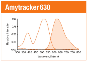
Amytracker 630 is our orange fluorescent optotracer for labeling protein aggregates with repetitive arrangement of β-sheets. It labels Aβ plaques and neurofibrillary tangles in tissue sections with AD pathology and α-synuclein aggregates in tissue sections with PD pathology. Contact us to learn more about Amytracker applications.

As all our optotracers, Amytracker 630 is exceptionally photostable and fluorogenic. When bound to a target, Amytracker 630 can be imaged using epifluorescence, confocal and superresolution microscopy. Spectral information can be acquired using a fluorescence spectrophotometer. Use recommended filter sets as well as excitation- and emission wavelengths according to the following table.
Table: Excitation- and emission wavelengths as well as recommended filter sets.

Exmax Emmax Recommended filter-sets
Amytracker 630 520 nm 630 nm PI, Cy3, TxRed, mCherry, Cy3.5

Amytracker 630 is available as a 1 mg/ml solution in volumes ranging from 10 – 200 µL (see volumes and prices in the list below). As our standard solvent, we use ultrapure water. Please contact us for custom options.

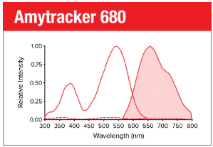
Amytracker 680 is our red fluorescent optotracer for labeling protein aggregates with repetitive arrangement of β-sheets. It labels Aβ plaques and neurofibrillary tangles in tissue sections with AD pathology and α-synuclein aggregates in tissue sections with PD pathology. Specifically, Amytracker 680 has been used to study amyloid formation during abnormal coagulation, Lewy body formation in a seeding based neuronal model, accumulation of misfolded proteins in the nucleolus and intracerebral formation of Aβ plaques using multiphoton microscopy. Contact us to learn more about Amytracker applications.

As all our optotracers, Amytracker 680 is exceptionally photostable and fluorogenic. When bound to a target, Amytracker 680 can be imaged using epifluorescence, confocal and superresolution microscopy. Spectral information can be acquired using a fluorescence spectrophotometer. Use recommended filter sets as well as excitation- and emission wavelengths according to the following table.
Table: Excitation- and emission wavelengths as well as recommended filter sets.

Exmax Emmax Recommended filter-sets
Amytracker 680 530 nm 680 nm PI, mCherry, Cy3.5

Amytracker 680 is available as a 1 mg/ml solution in volumes ranging from 10 – 200 µL (see volumes and prices in the list below). As our standard solvent, we use ultrapure water (MilliQ). We also provide Amytracker 680 dissolved in DMSO. Please contact us for custom options.

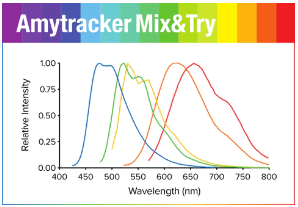
Amytracker Mix&Try Kit is our recommended option for starting out with using Amytracker. It contains a sample volume (10 µL) of each variant. Testing each variant will allow you to determine which Amytracker is best suited for your experiments and available instruments.

All Amytracker variants label Aβ plaques and neurofibrillary tangles in tissue sections with AD pathology and α-synuclein aggregates in tissue sections with PD pathology. The optotracers are exceptionally photostable and fluorogenic. The variants differ with regards to affinity, cellular uptake and excitation and emission wavelengths (see Table below). When bound to a target, Amytracker can be imaged using epifluorescence, confocal and superresolution microscopy. Spectral information can be acquired using a fluorescence spectrophotometer. Contact us to learn more about Amytracker applications.

Table: Excitation- and emission wavelengths as well as recommended filter sets.

Exmax Emmax Recommended filter-sets
Amytracker 480 420 nm 480 nm DAPI
Amytracker 520 460 nm 520 nm FITC, GFP
Amytracker 540 480 nm 540 nm FITC, GFP, YFP
Amytracker 630 520 nm 630 nm PI, Cy3, TxRed, mCherry, Cy3.5
Amytracker 680 530 nm 680 nm PI, mCherry, Cy3.5

Carbotrace

Carbotrace are optotracer molecules providing means for anatomical mapping of carbohydrate structures in plants and for non-destructive composition analysis of polysaccharides in bio-based materials and biofuels.

The optotracing technology has been developed by a team of researchers at Karolinska Institutet (KI), Royal Institute of Technology (KTH) and Linköping University in Sweden and is a result of many years of cutting-edge nanoscience and organic chemistry research. Optotracing has been defined by the researchers as the use of structure-responsive fluorescent molecules which become highly fluorescent when binding and exhibit unique spectral signatures dependent on the structure of their binding partner. The unique combination of fluorescence and the target-specific signal can be used to clearly identify biomolecules in various types of materials.

Carbotrace optotracers are designed for anatomical mapping of carbohydrate structures in plants and for non-destructive composition analysis of polysaccharides in bio-based materials and biofuels. The structure-responsive optotracer Carbotrace differentiates between different types of glycosidic linkages in carbohydrates. The products are available in aliquots of 50 µl, 100 µl, 150 µl or 200 µl (all delivered in 50 µl vials).

Spectral Imaging

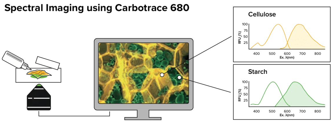
Identification of Cellulose and Starch Structures in Potato

  • prepare freshly sliced potato (1 mm)
  • dilute Carbotrace 680 1:1000 in PBS (pH 7.4)
  • immerse potatoes in diluted Carbotrace 680 for ca. 30 min
  • wash with PBS
  • mount on microscope slide
  • image with the following excitation/emission settings: Ex: 405 nm Em: 430–450 nm, Ex: 473 nm Em: 490–540 nm, Ex: 535 nm Em: 575–620 nm and Ex: 635 nm Em: 655–755 nm or acquire spectra with a plate reader or spectral microscope.

Applications

  • Protocol I: Anatomical mapping of cellulose in plant tissues using Carbotrace 680
  • Protocol II: Measurement of cellulose content in a sample using Carbotrace 680

Storage

  • Store Carbotrace at 4°C.
  • Use the opened vial within 12 month.

Quick Facts

Carbotrace optotracers are:

  • Provided as 1 mg/ml solution in volumes of 50 µl, 100 µl, 150 µl and 200 µl and as a Mix&Try Kit with 5×10 µl
  • Fast
  • Simple and easy to handle
  • Photo- and thermostable
  • Non-toxic
  • Accurate


Note

  • Carbotrace is for research use only.
  • Carbotrace is not for diagnostic use or use in humans.
  • Carbotrace is not for resale.

Products

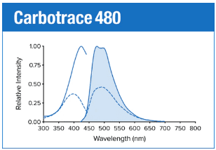
Carbotrace 480 is our blue fluorescent optotracer molecule for labeling glucans and some hemicelluloses. Generally, labeling is strongest with homopolymers compared to highly branched polysaccharides. Optotracers are structure-responsive molecules which display a unique spectral signature when binding to a target structure. Therefore, depending on the sample composition, it is possible to determine the purity of cellulose in the sample.

Specifically, Carbotrace 480 has been used to optimize a sustainable extraction method for cellulose hydrogels derived from microalgae. Contact us to learn more about applications for Carbotrace 480.

Table: Excitation- and emission wavelengths as well as recommended filter sets.

Exmax Emmax Recommended filter-sets
Carbotrace 480 420 nm 480 nm DAPI

Carbotrace 480 is available as a 1 mg/ml solution in volumes ranging from 10 – 200 µL (see volumes and prices in the list below). As our standard solvent, we use ultrapure water. Please contact us for custom options.

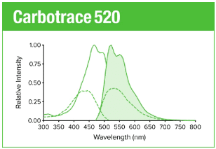
Carbotrace 520 is our green fluorescent optotracer molecule for labeling glucans and some hemicelluloses. Generally, labeling is strongest with homopolymers compared to highly branched polysaccharides. Optotracers are structure-responsive molecules which display a unique spectral signature when binding to a target structure. Therefore, depending on the sample composition, it is possible to determine the purity of cellulose in the sample. Contact us to learn more about applications for Carbotrace 520.

Table: Excitation- and emission wavelengths as well as recommended filter sets.

Exmax Emmax Recommended filter-sets
Carbotrace 520 460 nm 520 nm FITC, GFP

Carbotrace 520 is available as a 1 mg/ml solution in volumes ranging from 10 – 200 µL (see volumes and prices in the list below). As our standard solvent, we use ultrapure water. Please contact us for custom options.

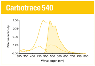
Carbotrace 540 is our yellow fluorescent optotracer molecule for labeling glucans and some hemicelluloses. Generally, labeling is strongest with homopolymers compared to highly branched polysaccharides. Optotracers are structure-responsive molecules which display a unique spectral signature when binding to a target structure. Therefore, depending on the sample composition, it is possible to determine the purity of cellulose in the sample.

Specifically, Carbotrace 540 has been used to determine the purity of cellulose extracted from macroalgae by allowing analysis of extracted carbohydrates in their polymeric state. In addition, Carbotrace 540 has shown to bind to the hemicellulose xyloglucan. Contact us to learn more about applications for Carbotrace 540.

Table: Excitation- and emission wavelengths as well as recommended filter sets.

Exmax Emmax Recommended filter-sets
Carbotrace 540 480 nm 540 nm FITC, GFP, YFP

Carbotrace 540 is available as a 1 mg/ml solution in volumes ranging from 10 – 200 µL (see volumes and prices in the list below). As our standard solvent, we use ultrapure water. Please contact us for custom options.

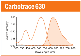
Carbotrace 630 is our orange fluorescent optotracer molecule for labeling glucans and some hemicelluloses. Generally, labeling is strongest with homopolymers compared to highly branched polysaccharides. Optotracers are structure-responsive molecules which display a unique spectral signature when binding to a target structure. Therefore, depending on the sample composition, it is possible to determine the purity of cellulose in the sample. Contact us to learn more about applications for Carbotrace 630.

Table: Excitation- and emission wavelengths as well as recommended filter sets.

Exmax Emmax Recommended filter-sets
Carbotrace 630 520 nm 630 nm PI, Cy3, TxRed, mCherry, Cy3.5

Carbotrace 630 is available as a 1 mg/ml solution in volumes ranging from 10 – 200 µL (see volumes and prices in the list below). As our standard solvent, we use ultrapure water. Please contact us for custom options.

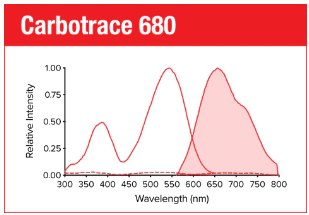
Carbotrace 680 is our red fluorescent optotracer molecule for labeling glucans and some hemicelluloses. Generally, labeling is strongest with homopolymers compared to highly branched polysaccharides. Optotracers are structure-responsive molecules which display a unique spectral signature when binding to a target structure. Therefore, depending on the sample composition, it is possible to determine the purity of cellulose in the sample.

Specifically, Carbotrace 680 has been used to image cellulose and starch components in plant cells and differentiate between those components using multi-laser/multi-detector analysis with confocal microscopy. Using a fluorescence spectrophotometer to acquire excitation and emission spectra, a unique spectral signature allows to differentiate between α(1-3), α(1-4), α(1-6), β(1-3), β(1-4) and β(1-6) linked glucans. As such, it labels cell wall components such as Cellulose, Chitin, Laminarin and the hemicellulose xyloglucan. Further, Carbotrace 680 has been used to determine relative carbohydrate content and elasticity of cell walls in plant material. Contact us to learn more about applications for Carbotrace 680.

Table: Excitation- and emission wavelengths as well as recommended filter sets.

Exmax Emmax Recommended filter-sets
Carbotrace 680 530 nm 680 nm PI, mCherry, Cy3.5

Carbotrace 680 is available as a 1 mg/ml solution in volumes ranging from 10 – 200 µL (see volumes and prices in the list below). As our standard solvent, we use ultrapure water (MilliQ). We also provide Carbotrace 680 dissolved in DMSO Please contact us for custom options.

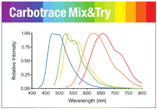
Carbotrace Mix&Try Kit is our recommended option for starting out with using our fluorescent optotracers. It contains a sample volume (10 µL) of each variant. Testing each variant will allow you to determine which Carbotrace is best suited for your experiments and available instruments.

All Carbotrace variants label glucans and some hemicelluloses. Generally, labeling is strongest with homopolymers compared to highly branched polysaccharides. The optotracers are structure-responsive molecules which display a unique spectral signature when binding to a target structure. Therefore, depending on the sample composition, it is possible to determine the purity of cellulose in the sample. Contact us to learn more about Carbotrace applications.

Table: Excitation- and emission wavelengths as well as recommended filter sets.

Exmax Emmax Recommended filter-sets
Carbotrace 480 420 nm 480 nm DAPI
Carbotrace 520 460 nm 520 nm FITC, GFP
Carbotrace 540 480 nm 540 nm FITC, GFP, YFP
Carbotrace 630 520 nm 630 nm PI, Cy3, TxRed, mCherry, Cy3.5
Carbotrace 680 530 nm 680 nm PI, mCherry, Cy3.5

EbbaBiolight

EbbaBiolight are optotracers for detection of bacteria & components of bacterial biofilm.

EbbaBiolight molecules allow direct visualisation of bacteria or bacterial biofilm, without using antibodies or any toxic chemicals. The molecules become highly fluorescent when bound to a target, are non-toxic, and do not interfere with bacterial growth or biofilm formation at recommended concentrations so EbbaBiolight can be added to live cultures to track bacterial growth or biofilm formation in real-time using spectrophotometric analysis. Further, EbbaBiolight are photo- and thermostable and allow for easy handling in any application.

We offer the EbbaBiolight optotracers in five different variants that might be screened for applications in various bacterial species and strains. All molecules are available in aliquots of 50 µl, 100 µl, 150 µl or 200 µl (all delivered in 50 µl vials). We also offer an EbbaBiolight Mix&Try kit, that contains 10 µl of each of the five optical tracer molecules from the EbbaBiolight series.

Optotracing

Optotracing is an innovative visualization method developed by scientists for scientists. With optotracing – using fluorescent tracer molecules – Ebba Biotech provides an innovative method for expanding areas of research. The optotracing technology has been developed by a scientific expert team of researchers at Karolinska Institutet, Linköping University and the Royal Institute of Technology (KTH) in Sweden and is a result of many years of cutting-edge nanoscience and organic chemistry research.

We supply and deliver the fluorescent tracer (optotracer) molecules for detection of bacteria and bacterial biofilm in the EbbaBiolight product series. The EbbaBiolight optotracers are a tool to search and detect bacteria & biofilm in hospitals and microbiology laboratories, as well as in industry with high demands to detect bacterial contamination and biofilm formation.

Quick Facts

EbbaBiolight optotracer molecules are:

  • Provided as 1 mg/ml solution in volumes of 50 µl, 100 µl, 150 µl and 200 µl and as a Mix&Try Kit with 5×10 µl
  • Should be diluted 1000-fold before use
  • Non-toxic
  • Photo- and thermostable

Applications for EbbaBiolight 680

EbbaBiolight 680 is our optotracer for biofilm visualization. EbbaBiolight 680 binds to cellulose and amyloid components in the biofilm of Salmonella enterica, serovars S. Enteritidis and Salmonella Typhimurium.

Quickstart Guide

Depending on the bacterial species or strain, EbbaBiolight optotracer molecules will bind to components in the bacterial biofilm. We recommend to follow this Quickstart Guide when using any of the EbbaBiolight molecules for the first time.

  • Dilute EbbaBiolight 1:1000 in growth medium (pH 7.4).
  • Dilute a bacterial over-night culture 1:100 with growth medium containing EbbaBiolight.
  • Fill (at least) two wells of a 96-well microtiter plate with 100 μl of the diluted over-night culture containing EbbaBiolight.
  • Prevent drying by filling unused wells with sterile water and seal the plate with a cover or adhesive.
  • Incubate the plate at optimal growth temperature and measure absorbance as well as fluorescence at regular intervals during a period of at least 48h.
  • Take samples (10 µl) during exponential- and stationary phase and pipet onto a microscope slide for imaging. Cover with a glass coverslip and seal the edges to prevent drying. Image at recommended excitation/emission to detect fluorescent bacteria or biofilm.

Storage

  • Store EbbaBiolight at 4°C.
  • Use the opened vial within 12 month.

Note

  • EbbaBiolight is for research use only.
  • EbbaBiolight is not for diagnostic use or use in humans.
  • EbbaBiolight is not for resale.

Products

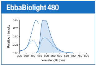
EbbaBiolight 480 is our blue fluorescent optotracer for labeling extracellular and intracellular bacterial amyloids as well as certain glucans produced as part of the extracellular matrix. Specifically, EbbaBiolight 480 has been used to label extracellular matrix produced by P. aeruginosa but not C. albicans in a mircofluidic device during mono- and co-culture. Contact us to learn more about EbbaBiolight applications.

As all our optotracers, EbbaBiolight 480 is exceptionally photostable and fluorogenic. When bound to a target, EbbaBiolight 480 can be imaged using fluorescence microscopy and spectral information can be acquired using a fluorescence spectrophotometer. As spectral information can give hints of the nature of the target, we recommend to acquire excitation and emission spectra whenever possible. Use recommended filter sets as well as excitation- and emission wavelengths according to the table below. EbbaBiolight is intended for use in live-cultures and are not compatible with fixation. If fixation needs to be performed, we recommend to apply fixation after labelling.

Table: Excitation- and emission wavelengths as well as recommended filter sets.

Exmax Emmax Recommended filter-sets
EbbaBiolight 480 420 nm 480 nm DAPI

EbbaBiolight 480 is available as a 1 mg/ml solution in volumes ranging from 10 – 200 µL (see volumes and prices in the list below). As our standard solvent, we use ultrapure water. Please contact us for custom options.

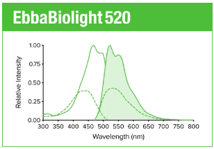
EbbaBiolight 520 is our green fluorescent optotracer for labeling extracellular and intracellular bacterial amyloids as well as certain glucans produced as part of the extracellular matrix. Contact us to learn more about EbbaBiolight applications.

As all our optotracers, EbbaBiolight 520 is exceptionally photostable and fluorogenic. When bound to a target, EbbaBiolight 520 can be imaged using fluorescence microscopy and spectral information can be acquired using a fluorescence spectrophotometer. As spectral information can give hints of the nature of the target, we recommend to acquire excitation and emission spectra whenever possible. Use recommended filter sets as well as excitation- and emission wavelengths according to the table below. EbbaBiolight are intended for use in live-cultures and are not compatible with fixation. If fixation needs to be performed, we recommend to apply fixation after labelling.

Table: Excitation- and emission wavelengths as well as recommended filter sets.

Exmax Emmax Recommended filter-sets
EbbaBiolight 520 460 nm 520 nm FITC, GFP

EbbaBiolight 520 is available as a 1 mg/ml solution in volumes ranging from 10 – 200 µL (see volumes and prices in the list below). As our standard solvent, we use ultrapure water. Please contact us for custom options.

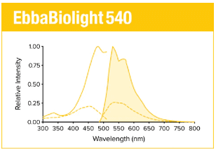
EbbaBiolight 540 is our yellow fluorescent optotracer for labeling extracellular and intracellular bacterial amyloids as well as certain glucans produced as part of the extracellular matrix. Contact us to learn more about EbbaBiolight applications.

As all our optotracers, EbbaBiolight 540 is exceptionally photostable and fluorogenic. When bound to a target, EbbaBiolight 540 can be imaged using fluorescence microscopy and spectral information can be acquired using a fluorescence spectrophotometer. As spectral information can give hints of the nature of the target, we recommend to acquire excitation and emission spectra whenever possible. Use recommended filter sets as well as excitation- and emission wavelengths according to the table below. EbbaBiolight are intended for use in live-cultures and are not compatible with fixation. If fixation needs to be performed, we recommend to apply fixation after labelling.

Table: Excitation- and emission wavelengths as well as recommended filter sets.

Exmax Emmax Recommended filter-sets
EbbaBiolight 540 480 nm 540 nm FITC, GFP, YFP

EbbaBiolight 540 is available as a 1 mg/ml solution in volumes ranging from 10 – 200 µL (see volumes and prices in the list below). As our standard solvent, we use ultrapure water. Please contact us for custom options.

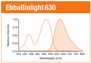
EbbaBiolight 630 is our orange fluorescent optotracer for labeling extracellular and intracellular bacterial amyloids as well as certain glucans produced as part of the extracellular matrix. Specifically, EbbaBiolight 630 labels peptidoglycan and lipoteichoic acids in the cell wall of certain gram positives like Staphylococci and E. faecalis. Contact us to learn more about EbbaBiolight applications.

As all our optotracers, EbbaBiolight 630 is exceptionally photostable and fluorogenic. When bound to a target, EbbaBiolight 630 can be imaged using fluorescence microscopy and spectral information can be acquired using a fluorescence spectrophotometer. As spectral information can give hints of the nature of the target, we recommend to acquire excitation and emission spectra whenever possible. Use recommended filter sets as well as excitation- and emission wavelengths according to the table below. EbbaBiolight are intended for use in live-cultures and are not compatible with fixation. If fixation needs to be performed, we recommend to apply fixation after labelling.

Table: Excitation- and emission wavelengths as well as recommended filter sets.

Exmax Emmax Recommended filter-sets
EbbaBiolight 630 520 nm 630 nm PI, Cy3, TxRed, mCherry, Cy3.5

EbbaBiolight 630 is available as a 1 mg/ml solution in volumes ranging from 10 – 200 µL (see volumes and prices in the list below). As our standard solvent, we use ultrapure water. Please contact us for custom options.

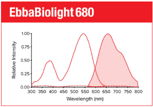
EbbaBiolight 680 is our red fluorescent optotracer for labeling extracellular and intracellular bacterial amyloids as well as certain glucans produced as part of the extracellular matrix. Specifically, EbbaBiolight 680 labels curli in the extracellular matrix produced by Salmonella as well as β-glucans and chitins in yeast- and hyphal forms of C. albicans. Furthermore, it has been used to track pellicle biofilm formation by B. cenocepacia. Contact us to learn more about EbbaBiolight applications.

As all our optotracers, EbbaBiolight 680 is exceptionally photostable and fluorogenic. When bound to a target, EbbaBiolight 680 can be imaged using fluorescence microscopy and spectral information can be acquired using a fluorescence spectrophotometer. As spectral information can give hints of the nature of the target, we recommend to acquire excitation and emission spectra whenever possible. Use recommended filter sets as well as excitation- and emission wavelengths according to the table below. EbbaBiolight are intended for use in live-cultures and are not compatible with fixation. If fixation needs to be performed, we recommend to apply fixation after labelling.

Table: Excitation- and emission wavelengths as well as recommended filter sets.

Exmax Emmax Recommended filter-sets
EbbaBiolight 680 530 nm 680 nm PI, mCherry, Cy3.5

EbbaBiolight 680 is available as a 1 mg/ml solution in volumes ranging from 10 – 200 µL (see volumes and prices in the list below). AAs our standard solvent, we use ultrapure water (MilliQ). We also provide EbbaBiolight 680 dissolved in DMSO Please contact us for custom options.

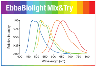
EbbaBiolight Mix&Try Kit is our recommended option for starting out with using EbbaBiolight. It contains a sample volume (10 µL) of each variant. Testing each variant in the EbbaBiolight Mix&Try Kit will allow you to determine which one of our optotracers is best suited for your experiment.

All EbbaBiolight variants label extracellular and intracellular bacterial amyloids as well as certain glucans produced as part of the extracellular matrix. Specifically, the optotracers have been used as follows: EbbaBiolight 480 labeling extracellular matrix produced by P. aeruginosa but not C. albicans in a mircofluidic device during mono- and co-culture. EbbaBiolight 630 labeling peptidoglycan and lipoteichoic acids in the cell wall of certain gram positives like Staphylococci and E. faecalisEbbaBiolight 680 labeling curli in the extracellular matrix produced by Salmonella as well as β-glucans, chitin and intracellular amyloid aggregates in yeast- and hypal forms of C. albicans. Furthermore, it has been used to track pellicle biofilm formation in B. cenocepacia. Contact us to learn more about EbbaBiolight applications.

All EbbaBiolight variants are exceptionally photostable and fluorogenic. When bound to a target, the optotracers can be imaged using fluorescence microscopy and spectral information can be acquired using a fluorescence spectrophotometer. As spectral information can give hints of the nature of the target, we recommend to acquire excitation and emission spectra whenever possible. Excitation and emission wavelengths for each molecule are shown in Table 1. EbbaBiolight are intended for use in live-cultures and are not compatible with fixation. If fixation needs to be performed, we recommend to apply fixation after labelling.

Table 1: Excitation- and emission wavelengths as well as recommended filter sets.

Exmax Emmax Recommended filter-sets
EbbaBiolight 480 420 nm 480 nm DAPI
EbbaBiolight 520 460 nm 520 nm FITC, GFP
EbbaBiolight 540 480 nm 540 nm FITC, GFP, YFP
EbbaBiolight 630 520 nm 630 nm PI, Cy3, TxRed, mCherry, Cy3.5
EbbaBiolight 680 530 nm 680 nm PI, mCherry, Cy3.5

Optotracer Science

Our Optotracers: EbbaBiolight, Carbotrace and Amytracker

Optotracing is an innovative visualization method developed by scientists for scientists ideal to use in fluorescence microscopy. With optotracing – using fluorescent tracer molecules – Ebba Biotech provides an innovative method for several expanding areas of research. The optotracing technology has been developed by a scientific expert team of researchers at Karolinska Institutet, Linköping University and the Royal Institute of Technology (KTH) in Sweden and is a result of many years of cutting-edge nanoscience and organic chemistry research.

Ebba Biotech has commercialized distribution and sales of fluorescent tracer molecules and today supply these in three different series:

Optotracers for labeling amyloids and other protein aggregates

Optotracers for carbohydrate analysis and anatomical mapping

Optotracers for detection of bacteria and bacterial biofilm

Video clips about Optotracing

On the first of June 2021, Ferdinand Choong, Ebba Biotech’s co-founder, and Assistant Professor at Karolinska Institutet and AIMES (Center for the Advancement of Integrated Medical and Engineering), presented his research using Ebba Biotech’s optotracers at the digital event Lab & Diagnostics of the Future 2021, held by Life Science Sweden. At this event, Ferdinand spoke about Ebba Biotech’s optotracers multifunctional tracer for disease research and diagnostics. He explains the technical concept in large and Ebba Biotech’s three product series.

The video clip about optotracing illustrates why our current model of Take, Make, Dispose is draining our planet of natural resources and why circular economy and renewable resources are the answer. Learn how Optotracers can be used to identify renewable resources in plant biomass and support circular economy in the food & beverage, pharmaceutical and chemical industry.

In the first episode of our webinar series about EbbaBiolight, Dr. Karen Butina present her research findings using optotracers for detection and quantification of Staphylococci. You can read more about Dr. Butina’s work here. DISCLAIMER: Dr. Butina describes her work using HS-167 molecules. These are predecessor molecules to what is now commercialised under the product family name EbbaBiolight, which are marketed and sold by Ebba Biotech.