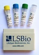
LifeSpan BioSciences provides high-quality antibodies, proteins, biochemicals, ELISA and Assay kits, immunohistochemistry data and services to researchers worldwide.


Product Catalogue

Antibodies
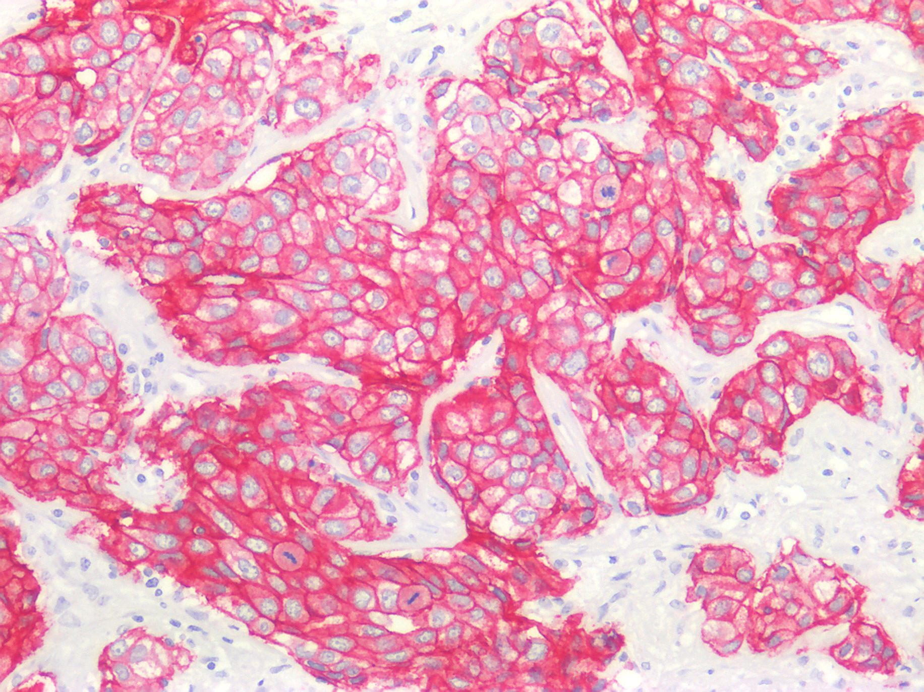
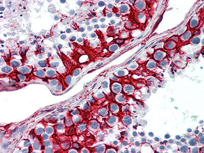
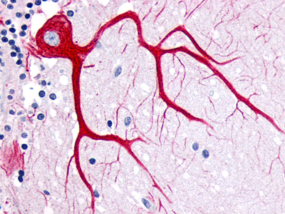
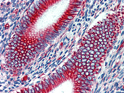
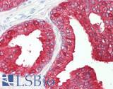
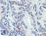
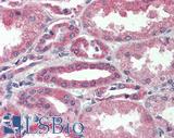
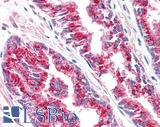
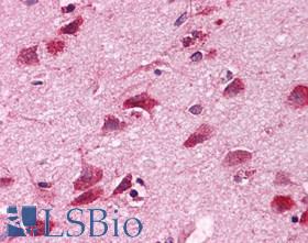
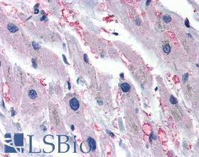
We have a collection of 565,545 monoclonal and polyclonal antibodies to most target proteins. They cover all major research species, applications, and are available in multiple conjugated forms. Our IHC-plus™ antibodies are highly characterized for use in FFPE human tissue Immunohistochemistry.

ELISA and Assay Kits
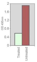
Our traditional Sandwich, Competitive EIA, and Direct ELISA kits provide a means to quantitatively measure thousands of targets of interest, while our Cell-Based and DNA-Binding ELISA kits are ideal for in-vitro studies and transcription factor analysis. Chemiluminescent CLIA kits provide high-sensitivity detection of low-copy targets and our Development kits enable researchers to cost-effectively run large numbers of assays. Our growing collection of Assay kits provide researchers with the means to monitor a variety of biological processes, such as apoptosis, cell proliferation, metabolism, and more.

Proteins
Proteins can be used in a wide variety of applications, such as for the development of functional assays, small-molecule screening, receptor activation in-situ, or simply as controls for a Western Blot. We offer extracted native proteins and recombinant proteins in the form of cell lysates, or purified from bacterial or mammalian expression systems. Many proteins are bio-active and certified low-endotoxin.

Immunohistochemistry Reagents
LSBio offers all of the components needed to design and execute custom Immunohistochemistry experiments, from antigen retrieval to coverslipping. High-quality, trusted brand blocking agents, detection plumers and Avidin-Biotin systems for AP or HRP substrate development. Make a challenging project easy with our mouse-on-mouse, and multi-plex detection systems.

Product Name Catalogue Number
VECTASTAIN® Elite® ABC-HRP Reagent, R.T.U. (Peroxidase, Ready-to-Use) LS-J1026
VECTASTAIN® Elite® ABC-HRP Kit (Peroxidase, Universal), R.T.U. (Ready-to-Use) LS-J1027
Antigen Unmasking Solution, Citric Acid Based LS-J1040
VECTASHIELD Antifade Mounting Medium LS-J1032
VECTASTAIN® ABC-AP Staining Kit (Alkaline Phosphatase, Rabbit IgG) LS-J1001
VECTASTAIN® Elite® ABC-HRP Kit (Peroxidase, Rabbit IgG) LS-J1019
ImmPRESS® HRP Anti-Mouse IgG (Peroxidase) Polymer Detection Kit, made in Goat LS-J1067
ImmPACT™ DAB Peroxidase (HRP) Substrate LS-J1075
VECTASHIELD HardSet Antifade Mounting Medium with DAPI LS-J1035
Vector® Hematoxylin QS LS-J1045
BLOXALL™ Endogenous Peroxidase and Alkaline Phosphatase Blocking Solution LS-J1031
VECTASTAIN® ABC-HRP Kit (Peroxidase, Rabbit IgG) LS-J1010
VECTASTAIN® Elite® ABC-HRP Kit (Peroxidase, Standard) LS-J1018
VECTASTAIN® ABC-HRP Kit (Peroxidase, Standard) LS-J1009
TMB SUBSTRATE KIT LS-J1079
Vector Red™ Alkaline Phosphatase (Red AP) Substrate Kit LS-J1086
Hematoxylin and Eosin Stain Kit LS-J1047
VECTASTAIN® Elite® ABC-HRP Kit (Peroxidase, Goat IgG) LS-J1023
ImmPRESS® HRP Universal Antibody (Anti-Mouse IgG/Anti-Rabbit IgG, Peroxidase) Polymer Detection Kit, made in Horse LS-J1068
ImmPRESS® Excel Amplified HRP Polymer Staining Kit (Anti-Rabbit IgG) LS-J1069
ImmPRESS*-VR HRP ANTI-RABBIT IgG KIT (15 ml) LS-J1058
ImmPRESS® HRP Anti-Rabbit IgG (Peroxidase) Polymer Detection Kit, made in Goat LS-J1066
Vector® Nuclear Fast Red LS-J1044
Antigen Unmasking Solution, Tris-Based LS-J1041
Vector® Methyl Green LS-J1043
VECTASHIELD Antifade Mounting Medium with DAPI LS-J1033
VECTASTAIN® ABC-AP Staining Kit (Alkaline Phosphatase, Standard) LS-J1000
VECTASTAIN® ABC-AP Staining Kit (Alkaline Phosphatase, Rat IgG) LS-J1004
VECTASTAIN® Elite® ABC-HRP Kit (Peroxidase, Mouse IgG) LS-J1020
VECTASTAIN® ABC-AP Staining Kit (Alkaline Phosphatase, Human IgG) LS-J1003
VECTASTAIN® ABC-AP Staining Kit (Alkaline Phosphatase, Goat IgG) LS-J1005
VECTASTAIN® ABC-HRP Kit (Peroxidase, Human IgG) LS-J1012
VECTASTAIN® ABC-HRP Kit (Peroxidase, Sheep IgG) LS-J1015
VECTASTAIN® ABC-AP Staining Kit (Alkaline Phosphatase, Mouse IgM) LS-J1007
ImmPRESS®-AP Anti-Rabbit IgG (Alkaline Phosphatase) Polymer Detection Kit LS-J1053
ImmPRESS®-AP Anti-Goat IgG (Alkaline Phosphatase) Polymer Detection Kit LS-J1056
ImmPRESS®-AP Anti-Mouse IgG (Alkaline Phosphatase) Polymer Detection Kit LS-J1054
ImmPRESS® HRP Anti-Rabbit IgG (Peroxidase) Polymer Detection Kit, made in Horse LS-J1060
ImmPRESS® HRP Anti-Mouse IgG (Peroxidase) Polymer Detection Kit, made in Horse LS-J1061
ImmPRESS® HRP Anti-Goat IgG (Peroxidase) Polymer Detection Kit, made in Horse LS-J1063
ImmPRESS® Excel Amplified HRP Polymer Staining Kit (Anti-Mouse IgG) LS-J1070
ImmPRESS® Duet Double Staining HRP/AP Polymer Kit (anti-rabbit IgG-brown, anti-mouse IgG-red) LS-J1071
AEC Peroxidase (HRP) Substrate Kit, 3-amino-9-ethylcarbazole LS-J1076
DAB Peroxidase (HRP) Substrate Kit (with Nickel), 3,3’-diaminobenzidine LS-J1073
ImmPACT™ AEC Peroxidase (HRP) Substrate LS-J1077
ImmPACT™ AMEC Red Peroxidase (HRP) Substrate LS-J1078
ImmPRESS®-AP Anti-Rat IgG (Alkaline Phosphatase) Polymer Detection Kit, Made in Goat LS-J1055
ImmPRESS®-AP Anti-Rat IgG, Mouse Adsorbed (Alkaline Phosphatase) Polymer Detection Kit, Made in Goat LS-J1057
ImmPRESS*-VR HRP ANTI-MOUSE IgG KIT (15 ml) LS-J1059
ImmPRESS® Duet Double Staining HRP/AP Polymer Kit (anti-mouse IgG-brown, anti-rabbit IgG-red) LS-J1072
ImmPACT™ DAB EqV Peroxidase (HRP) Substrate LS-J1074
ImmPRESS® HRP Anti-Rat IgG (Peroxidase) Polymer Detection Kit, made in Goat LS-J1062
ImmPRESS® HRP Anti-Mouse IgG, Rat adsorbed (Peroxidase) Polymer Detection Kit, made in Horse LS-J1064
ImmPRESS® HRP Anti-Rat IgG, Mouse adsorbed (Peroxidase) Polymer Detection Kit, made in Goat LS-J1065
ImmPrint Permanent Marking Pen LS-J1049
ImmPACT™ VIP Peroxidase (HRP) Substrate LS-J1081
Vector® VIP Peroxidase (HRP) Substrate Kit LS-J1080
Vector® SG Peroxidase (HRP) Substrate Kit LS-J1082
ImmPACT™ SG Peroxidase (HRP) Substrate LS-J1083
Vector® Black Alkaline Phosphatase (AP) Substrate Kit LS-J1088
Animal-Free Blocker™ (5x) LS-J1090
ImmPACT™ Vector Red™ Alkaline Phosphatase (AP) Substrate LS-J1087
Vector® Blue Alkaline Phosphatase (Blue AP) Substrate Kit LS-J1089
Vector® NovaRED™ Peroxidase (HRP) Substrate Kit LS-J1084
ImmPACT™ NovaRED™ Peroxidase (HRP) Substrate LS-J1085
VectaMount™ AQ Aqueous Mounting Medium LS-J1038
DAB Enhancing Solution LS-J1039
Alcian Blue (pH 2.5) Stain Kit LS-J1046
Vector® Hematoxylin LS-J1042
VECTASHIELD HardSet Antifade Mounting Medium LS-J1034
VECTASHIELD HardSet Antifade Mounting Medium with Phalloidin LS-J1036
VectaMount™ Permanent Mounting Medium LS-J1037
Levamisole Solution LS-J1030
Normal Chicken Serum Blocking Solution LS-M7
VECTASTAIN® ABC-HRP Kit (Peroxidase, Mouse IgG) LS-J1011
VECTASTAIN® ABC-HRP Kit (Peroxidase, Rat IgG) LS-J1013
VECTASTAIN® ABC-HRP Kit (Peroxidase, Goat IgG) LS-J1014
VECTASTAIN® ABC-HRP Kit (Peroxidase, Mouse IgM) LS-J1017
VECTASTAIN® ABC-HRP Kit (Peroxidase, Guinea Pig IgG) LS-J1016
Normal Goat Serum Blocking Solution LS-M1
2.5% Normal Goat Serum Blocking Solution LS-M2
Normal Horse Serum Blocking Solution LS-M3
VECTASTAIN® ABC-AP Staining Kit (Alkaline Phosphatase, Mouse IgG) LS-J1002
2.5% Normal Horse Serum Blocking Solution LS-M4
VECTASTAIN® Elite® ABC-HRP Kit (Peroxidase, Human IgG) LS-J1021
VECTASTAIN® Elite® ABC-HRP Kit (Peroxidase, Rat IgG) LS-J1022
VECTASTAIN® Elite® ABC-HRP Kit (Peroxidase, Sheep IgG) LS-J1024
VECTASTAIN® Elite® ABC-HRP Kit (Peroxidase, Universal) LS-J1025
Normal Swine Serum Blocking Solution LS-M5
VECTASTAIN® ABC-AP Staining Kit (Alkaline Phosphatase, Universal) LS-J1008
Normal Rabbit Serum Blocking Solution LS-M6

Molecular Biology
LSBio offers a broad selection of over 13,000 cDNAs, each available in either a cloning vector or one of fourteen different ready-to-use expression vectors. cDNA/ORF clones are fundamental to the study of protein function and gene expression. Verified Expression-Ready ORF clones give a researcher the option to directly initiate protein analysis and expression studies without spending valuable time on RNA isolation, cDNA synthesis, cloning in-frame, and sequencing.

LSBio Advantages

  • All clones are sequence-verified 100% match against GenBank, Ensembl, and Swiss-Prot
  • Each clone is available in 14 different READY-TO-USE Expression Vectors
  • Choice of 6 Epitope Tags, N- or C-terminal FLAG, His, Myc, GFP, HA, or OFP

Our goal is to offer reliable and comprehensive molecular biology products to accelerate your research and help prioritize your research requirements.

cDNA Clones

LSBio offers a selection of over 13,000 cDNA clones in cloning vectors covering a variety of research species. These cDNA clones can be used as gene probes or as stock material for subcloning or expression vector construction.

Expression-Ready ORF Clones

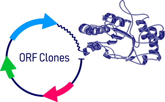
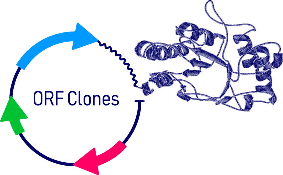
Cloning cDNAs into expression vectors requires expertise, takes time, and adds cost to your project. Accelerate your research by ordering your cDNA already cloned into one of fourteen Ready-to-Use pCMV3-Series mammalian expression vectors.

Virology Research Products

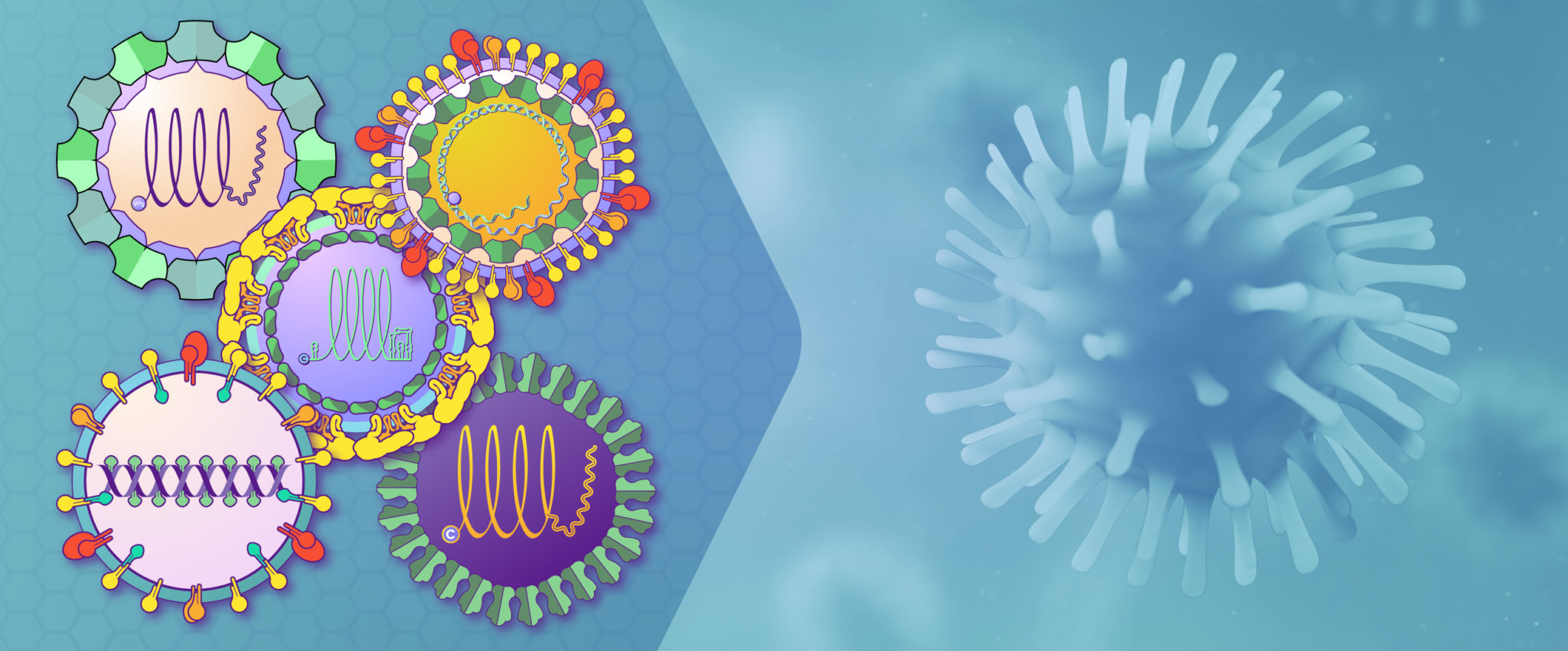
Viruses are obligate intracellular parasites that contain either an RNA or DNA genome. They are classified on the basis of morphology, chemical composition, and mode of replication. The viruses that infect humans represent only a narrow slice of the wide variety that infect hosts from protozoa to plants, fungi, bacteria, and vertebrates, but are responsible for causing many human and animal diseases. LSBio offers antibodies, proteins, ELISA kits, assay kits, and chemical compounds used by researchers to study and develop diagnostics and therapeutics for major viral diseases.

DNA versus RNA Viruses

Viruses are initially grouped on the type of genetic material, RNA or DNA. RNA virus genomes can be double stranded, positive single stranded, or negative single stranded, whereas DNA virus genomes can be single stranded, linear double stranded, or circular double stranded. Positive stranded ssRNA genomes can be directly translated into protein by the host, while negative stranded ssRNA genomes must be converted to a positive strand by RNA-dependent RNA polymerase before translation. Retroviruses have RNA genomes and use reverse transcriptase to generate a DNA intermediate during replication, which is integrated into the host genome and transcribed back into RNA.

DNA viral genomes can be large, encoding hundreds of viral proteins, whereas RNA viruses usually encode only a few proteins. DNA viruses have lower mutation rates due to the editing functions of the DNA polymerase and some are capable of integrating into the genomes of the host cell. RNA viruses undergo frequent mutation, enabling them to diversify to escape host immune mechanisms. Both DNA and RNA viruses manipulate a broad number of host cellular processes by interacting with host proteins involved in the cell cycle, transcription, nuclear transport, viral processing, packaging, and virion release.

Neurodegenerative Disease
Alzheimer’s Disease

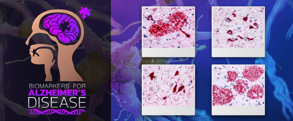
Alzheimer’s Disease (AD) is the most common progressive form of dementia. It is typically late onset (95% age >60 years), characterized by neuroimaging findings of cerebral cortical atrophy with cerebral amyloid angiopathy and CSF fluid measurements of amyloid-beta peptide, total or phosphorylated tau (Cohen Ad 2018). Neuropathological findings that are diagnostic include neuritic plaques containing beta-amyloid, neurofibrillary tangles consisting of hyper-phosphorylated microtubule associated protein tau, and amyloid angiopathy, described by accumulations of amyloid-beta proteins in the vascular smooth muscle of small cerebral arteries and capillaries.

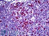
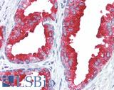
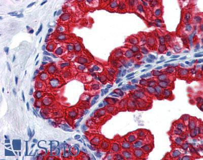
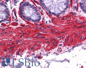
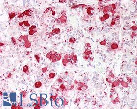
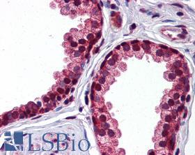
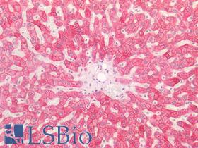
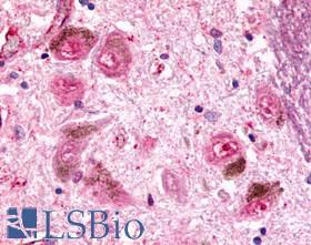
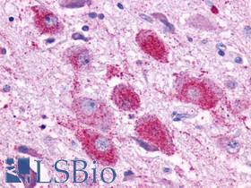
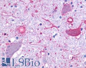
LSBio’s Immunohistochemistry (IHC) Validation

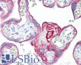
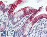
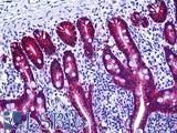
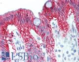
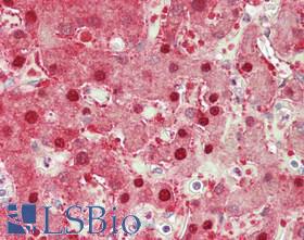
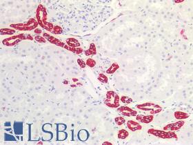
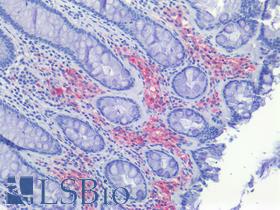
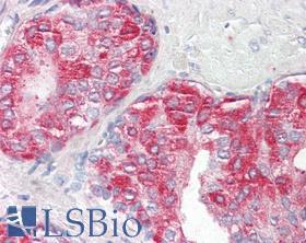
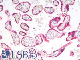
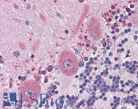
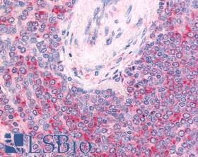
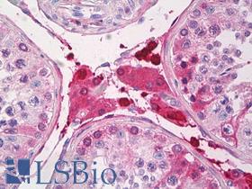
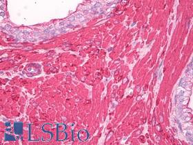
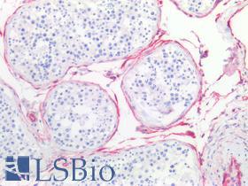
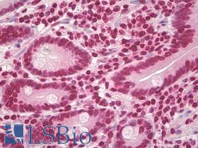
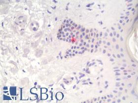
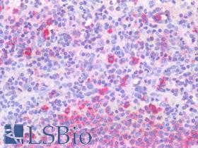
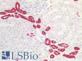
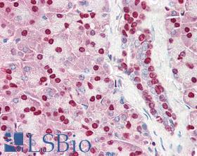
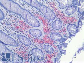
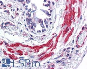
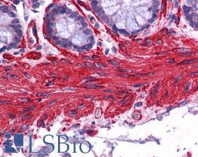
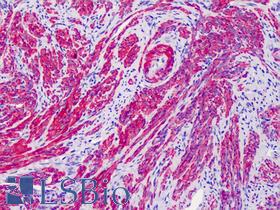
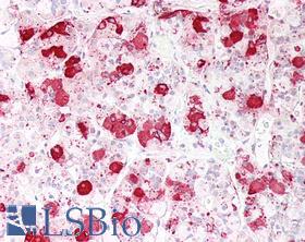
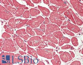
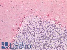
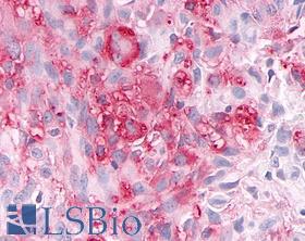
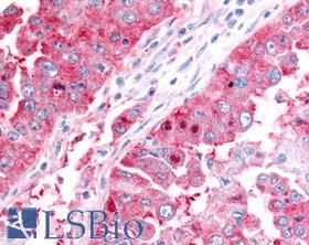
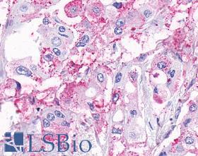
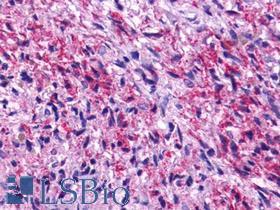
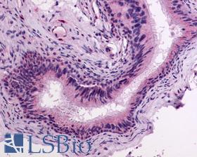
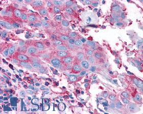
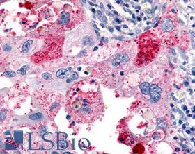
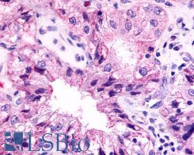
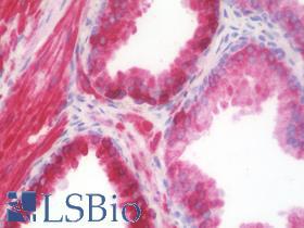
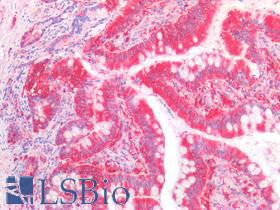
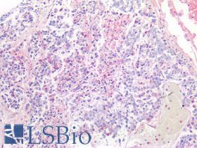
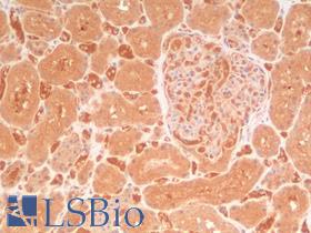
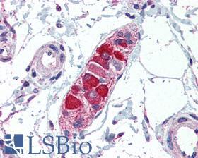
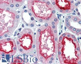
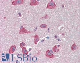
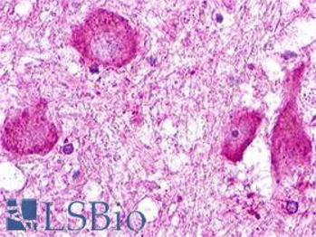
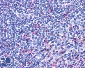
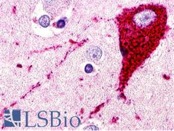
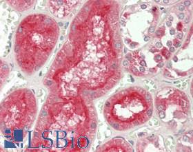
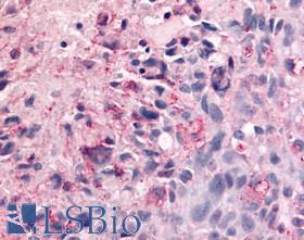
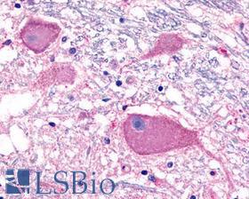
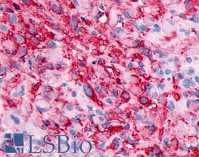
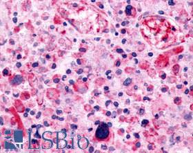
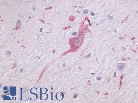
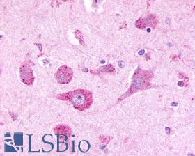
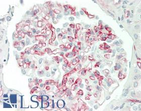
SBio (LifeSpan BioSciences) offers a catalog of 130,000 antibodies that have been tested for use in a variety of research applications, including immunohistochemistry, ELISA, Western blot, and flow cytometry. Antibodies can often be used in multiple assays, but they do not perform equally well in all assays. This is particularly true for immunohistochemistry (IHC). Many antibodies that perform well in other assays do not work well in IHC against formalin-fixed paraffin-embedded tissues (FFPE-IHC). In immunohistochemistry, antibodies may produce no signal, produce a weak signal, show nonspecific background staining that interferes with analysis, or show false positive signals. LSBio’s goal in immunohistochemistry validation is to identify for our customers those antibodies that perform well in FFPE-IHC. Out of the 130,000 antibodies in the LSBio catalog, 35,000 have been tested and received validation for use in IHC by LSBio or through collaborators or suppliers. Of these, 9,900 antibodies have been extensively tested in our Seattle laboratory and awarded IHC-plus™ brand validation. IHC-plus™ antibodies have been identified as the best reagents for use in FFPE-IHC.LSBio has tested and validated monoclonal antibodies (mouse, rabbit, rat), polyclonal antibodies (rabbit, goat, sheep, llama, chicken), and human single and double chain antibodies for immunohistochemistry. After fifteen years of experience performing contract IHC research, creating immunohistochemistry reports, and producing and testing over 12,500 antibodies in IHC, we have acquired a substantial body of experience regarding the best methods for validation of an IHC antibody. The purpose of this section is to share our knowledge of immunohistochemistry validation with you. The following summarizes our methods and approach toward immunohistochemistry (IHC) antibody validation.

Antibodies

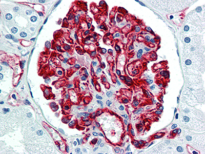
LSBio’s IHC-plus™ antibodies have been tested and identified as being optimal for use in immunohistochemistry (IHC) against formalin-fixed paraffin-embedded (FFPE) human tissues under LSBio’s standardized IHC-plus™ immunohistochemistry protocol.

Each antibody is tested at multiple concentrations on more than 20 normal human tissue types, and when appropriate, multiple normal brain regions and/or cancer types. LSBio’s IHC protocol has been developed over the past 15 years as the most optimal method of immunolabeling FFPE tissues, the most common fixation method used by pathology labs worldwide. A LifeSpan pathologist, with extensive experience evaluating IHC, analyzes the localization profile of each antibody, identifying positive and negative cell types, signal strength, subcellular and extracellular staining, and staining artifacts. This information is then compared with all published expression and localization data available for the protein. This enables LSBio to evaluate how each antibody behaves in IHC, including its specificity to the target protein, its sensitivity of detection, and any non-specific staining characteristics that it may display. In order to be selected as an IHC-plus™ brand antibody, antibodies must have a close correlation to the published literature, be high affinity, display minimal staining artifacts, and have a high signal-to-noise ratio, such that its specific staining is considerably higher than its level of nonspecific background staining.

LSBio’s catalog of primary antibodies covers more than 20,000 target molecules. Use the search box to list all of our antibodies to your target, and then use the filters to narrow your search by species, clonality, application, conjugate, etc. Alternatively, enter a search term such as “EGFR IHC monoclonal”. Every antibody is backed by our superior Technical Support staff, and every purchase is protected by our 100% satisfaction guarantee

LSBio offers a wide variety of secondary antibodies designed to complement the diverse needs of today’s researchers. Secondary antibodies are available to all primary antibody host species, such as anti-mouse, anti-rabbit, anti-goat, and anti-chicken; all major isotypes, such as IgA, IgE, IgG, IgG1, IgG2, IgM, and IgY; heavy chain and light chains, both kappa and lambda; as well as fragments Fc, Fab, Fab’2, and Fc5mu. We carry all of the common conjugates used for detection, for example AP, HRP, Biotin, and FITC, as well as a spectrum of specialty conjugate series, such as ATTO, Alexa Fluor, Cy, DyLight, and HiLyte. Search below for the secondary antibodies you need for use in ELISA, ICC, IHC, Western, Flow and IF.
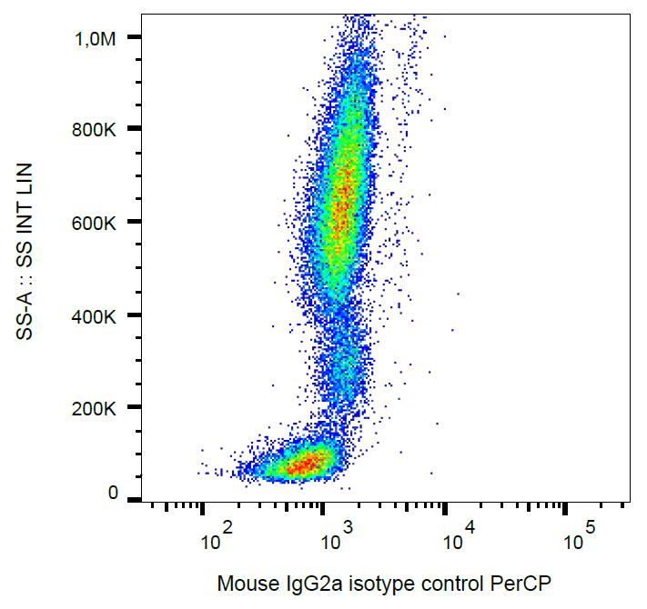
Including an Isotype Control in your IHC or Flow cytometry experiment is an important step in identifying potential non-specific binding of your primary antibody. Primary antibodies can bind to FC receptors expressed on non-target cell types, as well as non-specifically to other cellular proteins, sugars, and/or lipids. An Isotype Control should be selected that matches the host species and isotype of the primary antibody. Isotype Controls can also be used for a variety of other applications, such as ELISA, Western blot, and Immunoprecipitation. LSBio offers Isotype Controls from all major species, isotypes, and with a variety of conjugates.
Use the filters below to narrow your search by species, isotype, clonality, application, conjugate, etc.

ELISA and Assay Kits

LSBio’s growing collection of Assay kits provide researchers with the tools necessary to monitor various biological processes in the course of their life science research, drug discovery & development, or environmental monitoring studies. Assay kits are available for monitoring cell proliferation, damage, and oxidative stress, cytotoxicity, apoptosis, metabolism, signaling pathways and signal transduction. Each kit includes all of the necessary reagents to conduct the assay, and a clear, detailed protocol. LSBio’s experienced technical staff is also available to address any questions researchers may have about each kit and how they may be used to generate optimal results.
LSBio offers over 20,000 cost-effective, ready-to-use ELISA kits for the quantitative or qualitative detection of thousands of targets of interest. Each kit includes all of the necessary reagents to perform the assay as well as a clear protocol that walks you through the entire process, from sample preparation to analysis of the results. LSBio’s ELISA kits use traditional Sandwich, Competitive EIA, and Direct assay formats. Sandwich ELISA kits are a fast and simple way to quantify specific target antigens in a variety of sample types including urine, plasma, and cell culture medium. Competitive EIA kits use a single, antigen specific capture antibody rather than paired antibodies, allowing for detection of molecules where no antibody pair exists. Direct ELISA kits focus on the detection of antibodies developed as part of an auto-immune response, as well as anti-viral or anti-bacterial antibodies.

Features:

  • Ready-to-use kit includes all necessary reagents
  • Available for a broad selection of target antigens
  • Suitable for use with a wide variety of sample types
  • Multi-species reactivity (human, mouse and rat)
  • Excellent sensitivity, specificity, and reproducibility
  • Built on standard 96-well microtiter plate format
  • 450 nm or 405 nm colorimetric detection

Kit Components:

  • 96-well microtiter plate and sealers
  • Sample Diluent
  • Capture and/or detection antibodies
  • Avidin/HRP conjugate
  • Wash buffer
  • Substrate solution
  • Detailed protocol

Sandwich ELISAs

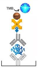
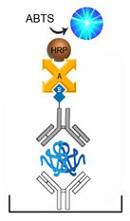
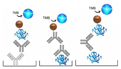
There are several types of Sandwich ELISA assays, all of which start with an antigen-specific capture antibody bound to the plate. When a sample is applied, the target antigen binds to this capture antibody. The plate is then washed and the captured antigen detected. The specific detection system varies between assays but generally consists of a detection antibody directly or indirectly linked to HRP. Often Avidin-biotin complexes are used to amplify the amount of HRP. The HRP then modifies a chromogen substrate, such as TMB, producing a color that is detectable at a specific light wavelength.

Competitive EIA

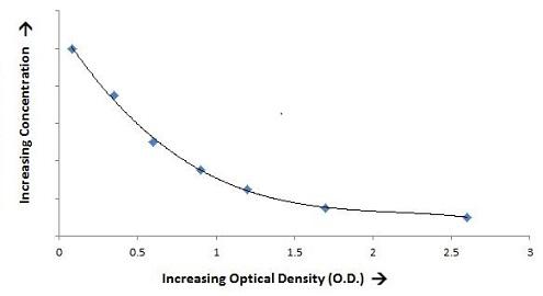
Competitive EIA kits are a diverse group of assays, all of which are based upon competitive binding of the antibody or antigen. Unlabeled target antigen from the sample must compete for binding with a fixed amount of biotin- or HRP-conjugated target antigen supplied with the kit. The greater the amount of target antigen within the sample, the more it is able to out-compete the conjugated antigen. This causes a reduction in the detectable conjugated antigen. Unlike Sandwich or Direct ELISAs, the lower the detectable signal in a Competitive EIA assay, the greater the amount to target antigen in the sample.

Direct ELISA Principle

Direct ELISA assays are ideal for the detection of antibodies in samples. There are two primary types of Direct assay, those that capture the target-specific antibody using the target antigen bound to the plate, or those that first capture all antibodies using a universal capture antibody bound to the plate, then detecting the captured target-specific antibody using the target antigen. Both assays employ a conjugated HRP which then modifies a chromogen substrate, such as TMB, producing a color that is detectable at a specific light wavelength.

Detection and Quantitation

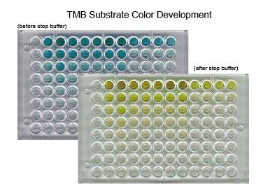
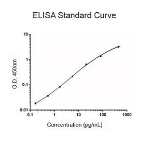
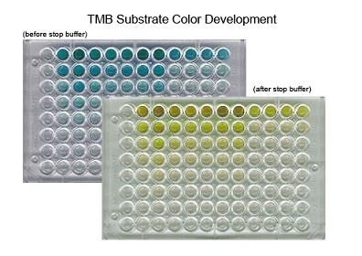
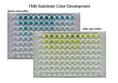
Most ELISAs use 3,3′,5,5′-tetramethylbenzidine (TMB) as their detection chromogen. When modified by Horseradish Peroxidase (HRP), TMB yields a blue color (370nm and 652nm) which then changes to yellow (450nm) upon the addition of a sulfuric acid stop solution. By running a dilution series of a known concentration standard, an OD450 standard curve can be generated. The OD450 for each unknown sample can then be compared to this standard curve in order to determine the concentration of target antigen.
Cell-Based ELISA kits are a fast and cost-effective way to quantify specific target antigens in vitro. Additionally, phospho-specific cell-based ELISA kits enable researchers to quickly evaluate changes in the phosphorylation state of specific proteins in whole cells under various treatment conditions. In a single experiment the activation state of 96 treated and untreated cells can be evaluated simultaneously. Each kit is ready-to-use with all the necessary reagents and a clear, concise protocol that will step you through the process, from sample preparation to analysis of the results. Cell-Based ELISA kits are available for both the detection of both phosphorylated and non-phosphorylated targets.

Features:

  • Ready-to-use kit includes all necessary reagents
  • Direct in vitro detection on un-lysed cultured cells
  • Phospho- and non-Phospho specific kits available
  • Excellent sensitivity, specificity, and reproducibility
  • Built on standard 96-well microtiter format
  • 450 nm colorimetric detection

Kit Components:

  • 96-well microtiter plate
  • Fixing Solution
  • Quenching buffer
  • Blocking Buffer
  • Wash Buffers
  • Primary Detection Antibody
  • HRP-Conjugated Secondary Antibody
  • TMB Substrate Solution
  • Stop Buffer
  • Plate Sealer Sheets

Cell-Based ELISA Kit Protocol

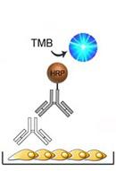
1) Cell monolayers are cultured in the 96-well microtiter plate. Each well can then be treated with stimulants, inhibitors, etc. The cells are then fixed and blocked.

2) Antigen specific primary antibody is added and binds to the target antigen on the cells surface.

3) The wells are washed and a HRP-conjugated secondary antibody is added which binds to the primary antibody.

4) The wells are washed and a solution containing TMB is added. The TMB reacts with the HRP changing from colorless to a blue color. An acid stop solution is then added, the reaction stops, and the blue color changes to yellow.

Phospho-Specific Cell-Based ELISA Kit Protocol

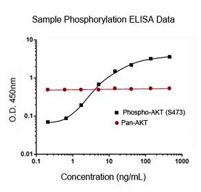
1) Cell monolayers are cultured in the 96-well microtiter plate. Each well can then be treated with stimulants, inhibitors, etc. to induce expression of the phosphorylated target. The cells are then fixed and blocked.

2) Phospho-specific or pan-specific primary antibody is added and binds to the target antigen on the cells surface.

3) The wells are washed and a HRP-conjugated secondary antibody is added which binds to the primary antibody.

4) The wells are washed and a solution containing TMB is added. The TMB reacts with the HRP changing from colorless to a blue color. An acid stop solution is then added, the reaction stops, and the blue color changes to yellow.


The plate can then be read in a spectrophotometer at a wavelength of 450 nm.

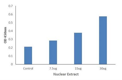
DNA-Binding ELISA Kits enable researchers to quickly and easily detect active transcription factors in eukaryotic nuclear extracts or cell lysates without the use of harmful radioactive reagents. Additionally our Phospho-DNA Binding kits can be used to study the effects of phosphorylation on transcription factor activation. Each kit is ready-to-use with all the necessary reagents and a clear, concise protocol that will step you through the process, from sample extraction to analysis of the results. DNA-Binding kits are available for both the detection of both phosphorylated and non-phosphorylated targets.

Features:

  • Ready-to-use kit includes all necessary reagents
  • Nuclear extraction protocol and reagents included
  • Phospho- and non-Phospho specific kits available
  • Detection of active transcription factors
  • Excellent sensitivity, specificity, and reproducibility
  • Built on standard 96-well microtiter plate format
  • 450 nm colorimetric detection

Kit Components:

  • 96-well microtiter plate
  • Anti-Phospho and Anti-Target Primary Antibodies
  • Nuclear Lysate Positive Control
  • Wild-Type Consensus dsDNA Oligonucleotide
  • Mutant Consensus dsDNA Oligonucleotide
  • Wash Buffers
  • Binding Buffer
  • Nuclear Wash Buffer
  • Nuclear and Cytoplasmic Extraction Buffers
  • HRP-Conjugated Secondary Antibody
  • TMB Substrate Solution
  • Stop Buffer
  • Plate Sealer Sheets

DNA-Binding ELISA Protocol

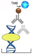
1) The 96-well microtiter plate comes pre-bound with specific double-stranded (dsDNA) oligonucleotides.
2) Following a blocking step samples are added to each well and transcription factor binds to the oligonucleotides.
3) The wells are washed and primary antibody is added which binds activated transcription factor.
4) The wells are washed and a HRP-conjugated secondary antibody is added which binds to the primary antibody.
5) The wells are washed and a solution containing TMB is added. The TMB reacts with the HRP changing from colorless to a blue color. An acid stop solution is then added, the reaction stops, and the blue color changes to yellow.

Phospho-Specific DNA-Binding ELISA Kit Protocol

1) The 96-well microtiter plate comes pre-bound with specific double-stranded (dsDNA) oligonucleotides.
2) Following a blocking step samples are added to each well and phosphorylated transcription factor binds to the oligonucleotides.
3) The wells are washed and a phospho-specific primary antibody is added which binds activated transcription factor.
4) The wells are washed and a HRP-conjugated secondary antibody is added which binds to the primary antibody.
5) The wells are washed and a solution containing TMB is added. The TMB reacts with the HRP changing from colorless to a blue color. An acid stop solution is then added, the reaction stops, and the blue color changes to yellow.

The plate can then be read in a spectrophotometer at a wavelength of 450 nm.
LifeSpan offers a number of different types of ELISA kit that are specific for the detection of phosphorylated target proteins. Phospho-specific kits are available for standard ELISA detection, in vitro detection in adherent cells, and detecting transcription factor activity in nuclear extracts and cell lysates. Each kit is ready-to-use with all the necessary reagents and a clear, concise protocol that will step you through the process, from sample preparation to analysis of the results.

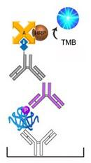
Phospho-Specific Sandwich ELISA Kit Protocol

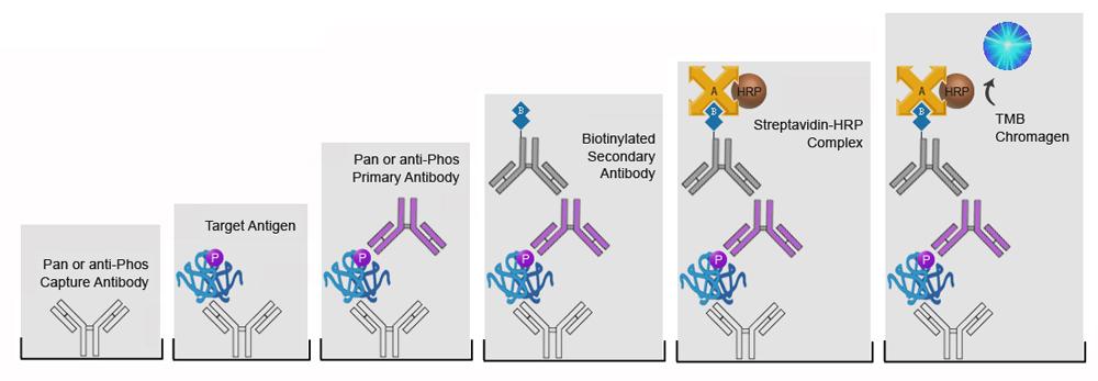
1) The supplied 96-well microtiter plate is pre-coated either pan- or phospho-specific capture antibody.
2) Control standards or test samples are added to the well and the target antigen is captured by the antibodies.
3) Phospho-specific or pan-specific primary antibody is added and binds to the target antigen.
4) The wells are washed and a biotinylated secondary antibody is added and binds to the primary antibody.
5) The wells are washed and a solution containing HRP-conjugated Avidin is added. The Avidin binds to the biotinylated secondary antibody.
6) The wells are washed and a solution containing TMB is added. The TMB reacts with the HRP changing from colorless to a blue color. An acid stop solution is then added, the reaction stops, and the blue color changes to yellow.

Phospho-Specific DNA-Binding ELISA Kit Protocol

1) The 96-well microtiter plate comes pre-bound with specific double-stranded (dsDNA) oligonucleotides.
2) Following a blocking step samples are added to each well and phosphorylated transcription factor binds to the oligonucleotides.
3) The wells are washed and a phospho-specific primary antibody is added which binds activated transcription factor.
4) The wells are washed and a HRP-conjugated secondary antibody is added which binds to the primary antibody.
5) The wells are washed and a solution containing TMB is added. The TMB reacts with the HRP changing from colorless to a blue color. An acid stop solution is then added, the reaction stops, and the blue color changes to yellow.

Phospho-Specific Cell-Based ELISA Kit Protocol

1) Cell monolayers are cultured in the 96-well microtiter plate. Each well can then be treated with stimulants, inhibitors, etc. to induce expression of the phosphorylated target. The cells are then fixed and blocked.
2) Phospho-specific or pan-specific primary antibody is added and binds to the target antigen on the cells surface.
3) The wells are washed and a HRP-conjugated secondary antibody is added which binds to the primary antibody.
4) The wells are washed and a solution containing TMB is added. The TMB reacts with the HRP changing from colorless to a blue color. An acid stop solution is then added, the reaction stops, and the blue color changes to yellow.


The plate can then be read in a spectrophotometer at a wavelength of 450 nm.
Chemiluminescent immunoassay (CLIA) offers significant advantages over traditional assay detection methods. Rather than using messy isotopic detection, or chromogens such and TMB or ABTS that require absorption spectroscopy, CLIA kits utilize chemical reactions that produce light. Measurement of light is highly useful because the concentration of an unknown can be inferred from the rate at which light is emitted. Extraordinary sensitivity, low background, a wide dynamic range, and inexpensive instrumentation distinguish luminometry from other analytical methods.

Features:

  • Ready-to-use kit includes all necessary reagents
  • Available for a broad selection of target antigens
  • Suitable for use with a wide variety of sample types
  • Multi-species reactivity (human, mouse and rat)
  • Excellent sensitivity, specificity, and reproducibility
  • Built on standard 96-well microtiter plate format
  • Sensitive luminescent detection

Kit Components:

  • 96-well microtiter plate and sealers
  • Sample Diluent
  • Capture and/or detection antibodies
  • Avidin/HRP Conjugate
  • Wash Buffer
  • Substrate Solution
  • Detailed Protocol

Sandwich ELISA Based CLIA Protocol

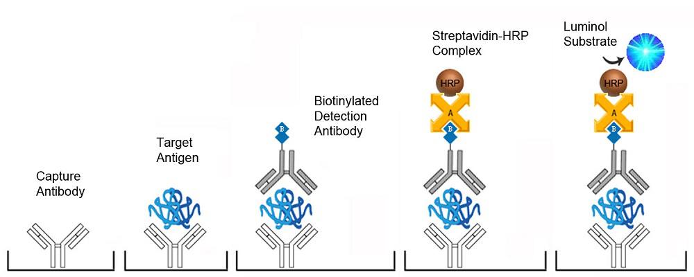
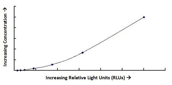
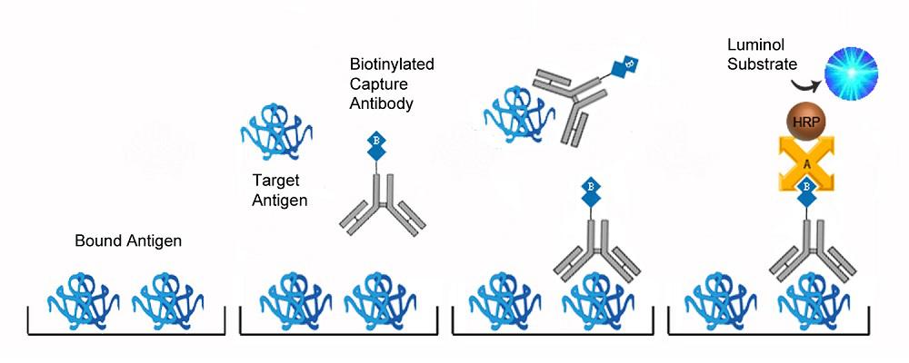
This assay is based on the sandwich CLIA principle. Each well of the supplied microtiter plate has been pre-coated with a target specific capture antibody. Standards or samples are added to the wells and the target antigen binds to the capture antibody. Unbound Standard or sample is washed away. A biotin-conjugated detection antibody is then added which binds to the captured antigen. Unbound detection antibody is washed away. An Avidin-Horseradish Peroxidase (HRP) conjugate is then added which binds to the biotin. Unbound Avidin-HRP conjugate is washed away. A chemiluminescent substrate is then added which reacts with the HRP enzyme resulting in light development. The relative light units (RLU) of each well are measured using a luminometer. The RLU of an unknown sample can then be compared to an RLU standard curve generated using known antigen concentrations in order to determine its antigen concentration.

Competition EIA Based CLIA Protocol

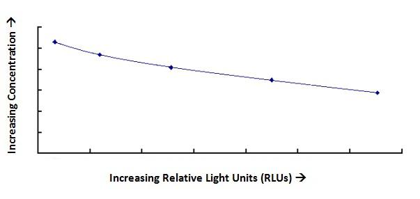
This assay is based on a competitive principle. Each well of the supplied microtiter plate has been pre-coated with target antigen. Standards or samples are added to the wells as well as a Biotin-conjugated detection antibody. The free antigens (in the standards or samples) and antigens bound to the plate compete to bind to the detection antibody. All but the bound Biotin-conjugated detection antibody is washed away. An Avidin-Horseradish Peroxidase (HRP) conjugate is then added which binds to the biotin then unbound HRP-conjugate is washed away. A chemiluminescent substrate is then added which reacts with the HRP enzyme resulting in light development. The relative light units (RLU) of each well are measured using a luminometer. The RLU of an unknown sample can then be compared to an RLU standard curve generated using known antigen concentrations in order to determine its antigen concentration. Unlike typical Sandwich assays, in competition assays the greater the amount of antigen in the sample, the lower the light development and relative light unit (RLU) measurement.
LSBio’s ELISA Development Kits allow researchers to prepare their own ELISA plates as needed, and at a lower cost per plate than off-the-shelf ready-to-use kits. Each ELISA DevKit includes principal components required to assemble and run a sensitive and specific Sandwich ELISA, such as the antibody pair, standard, and conjugate. LSBio also supplies all other ancillary products needed, such as plates, wash buffers and diluents, secondary detection antibodies, substrates and stop solutions. Before you begin, read through the LSBio ELISA Kit Guide to learn what things to consider when assembling, testing, and validating your own ELISA kit.

DevKit Duo

 (Antibody Pairs)

DevKit Duo Kits include the most critical components of the ELISA, the capture and detection antibody pair (biotinylated or non-biotinylated). Each antibody pair has been selected for specificity to their target, validated for use in ELISA, and provide researchers with the foundation upon which to build their own customized ELISA. Researchers can couple these antibodies with standards, conjugates, and substrates of their own choosing in order to optimize their assay for specific applications.

DevKit DuoPlus

 (Antibody Pairs plus Standard)

DevKit DuoPlus kits include the capture antibody, detection antibody (biotinylated or non-biotinylated), as well as the standard (typically a full-length recombinant protein) used to validate the antibodies. These development kits provide the foundational elements of an ELISA and save the hassle of having to source and validate a standard, but allow the research the freedom to select a signaling system particular to their specific needs.

DevKit Core

 (Antibody Pairs plus Standard and Conjugate)

DevKit Core kits contain all four key components required to construct reliable Sandwich ELISA’s for the quantitative measurement of target antigen. Each kit contains the capture antibody, a biotinylated detection antibody, an antigen standard, and Avidin-HRP or Streptavidin-HRP conjugate for use with ABTS or TMB substrates respectively. Each kit contains enough material to assay 2 or 5 standard 96-well ELISA plates.

ELISA Kit Reagents and Accessories

LSBio offers all the reagents and accessories you’ll need in order to build a better ELISA assay. Our catalog of coating buffers, blocking buffers, wash buffers, substrates, diluents, and much more allow the most flexibility for your research needs. Our reagents are specifically formulated to help you achieve the best signal while decreasing background noise. Explore the reagents and accessories we have that will help build your ELISA assay every step of the way.

Proteins

LSBio offers an extensive selection of high-quality recombinant proteins of human, murine, and rat origin, produced in a variety of expression systems that include bacteria, yeast, baculovirus infected insect cells, and mammalian cells. Protein categories include Growth Factors, Cytokines, Chemokines, Neurotrophins, Proteases, and other miscellaneous proteins, all of which are rigorously tested to meet the research and development demand for excellent quality and uncompromising biological activity. Our recombinant proteins can be used for a variety of applications, including functional assays, high throughput screens, antigen generation, structural analysis, cell growth and differentiation, cell signaling, and studies involving disease onset and progression.
LSBio offers a diverse collection of native proteins that are carefully isolated from a variety of animal and cellular sources. Unlike proteins generated using recombinant techniques, native proteins are produced in vivo and are unaltered and retain their natural assembled state. These proteins are useful in a wide range of applications such as functional assays, ELISA, Western Blot, and Immunoprecipitation. All LSBio native proteins are supplied in a stable, non-pathogenic formulation.
LSBio offers a broad collection of full- and partial-length proteins in the form of over-expression lysates. These proteins are generated in a variety of cell types, including mammalian HEK293/293T cells and E.coli, and are designed with a tag such as GST and HIS for easy detection and isolation. Over-expression lysates can be used as assay standards in Western blot, ELISA and other antibody assays.
LSBio offers whole protein lysates from cells and tissues commonly used in research in a Ready-to-Go Western Blot format. More than 30 cell lysates are available from A549, 293, 293T, U251, 3T3, and other cell types, as well as tissue extracts from various normal mouse organs like brain, liver, kidney, and more. Each sample is supplied in an SDS loading buffer containing 0.3% beta-mercaptoethanol making them ideal for use as Western blot positive controls.
LSBio’s biologically active proteins have been tested to confirm that they retain their functional attributes. Each protein is uniquely tested based upon its particular function, such as measuring the ability of an enzyme to hydrolyze a substrate, using a cell proliferation assay to test a growth factor, or a cytotoxicity assay to test proteins that can cause cell death. These proteins are a cost-effective resource for use in a wide variety of in vitro research applications.
Synthetic peptides are small polymers of amino-acids useful in a wide variety of experimental applications. LSBio’s high-quality synthetic peptides include activator and inhibitor functional groups, as well as ligands and substrates.
Animal-free proteins offer an alternative to researchers concerned about the experimental variables caused by the use of reagents containing trace animal components and mammalian pathogens. All of our animal-free proteins are manufactured in dedicated facilities using only animal-free reagents and components. Animal-free proteins have comparable biological activity and purity to animal-derived proteins produced using standard techniques.

Virology Research

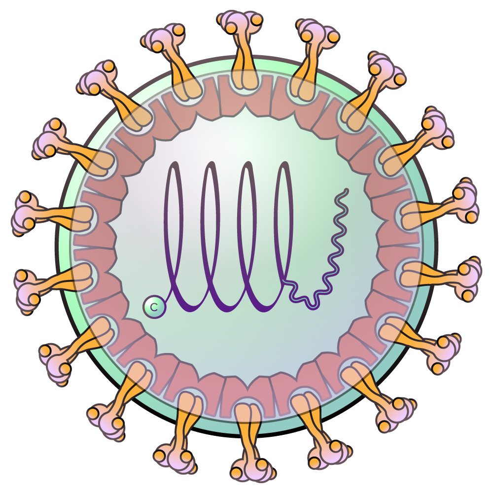
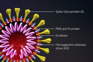
In support of the global research effort, we offer SARS-CoV-2 Antibodies, Detection Kits, Proteins, cDNA clones, Inhibitors, and Cytokine Release Syndrome research products.
All of our Coronavirus Research Products are for research use only.

Coronavirus Detection Kits

Real-Time qPCR Viral RNA Detection Kits
ELISA Kits

 Antibodies, Proteins, and cDNAs

Antibodies, Recombinant Proteins, Expression-Ready ORF cDNA Clones

Coronavirus Inhibitors

Small molecules and antibodies for the inhibition of coronavirus and SARS-CoV-2

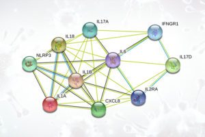
Cytokine Release Syndrome

Resources for studying the inflammatory response associated with COVID-19

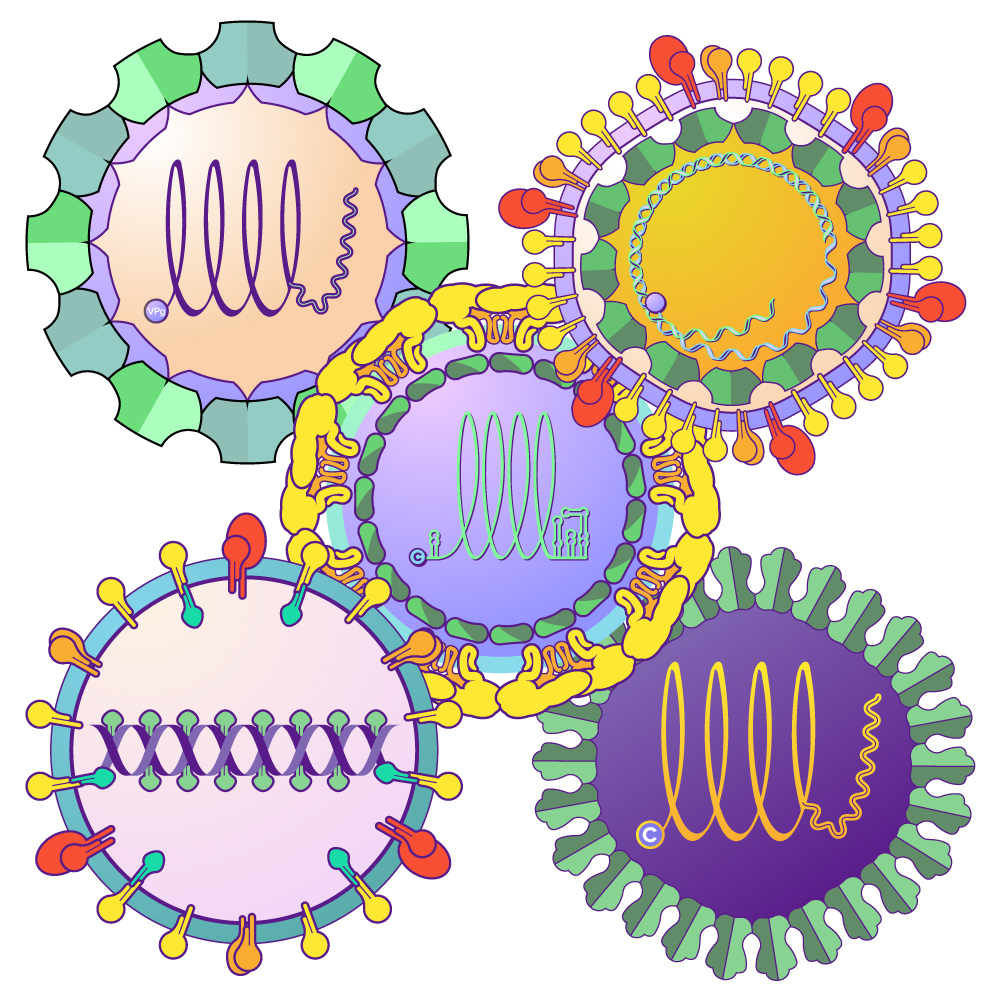
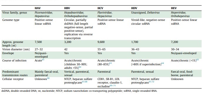
Hepatitis viruses cause 1.3 million deaths worldwide annually, and cause liver diseases including acute and chronic infections, liver failure, cirrhosis, and hepatocellular carcinoma. These viruses are a diverse group, and can be either dsDNA viruses such as Hepatitis B virus (family Hepadnaviridae), or ssRNA viruses such as Hepatitis A (family Picornaviridae), Hepatitis C (Flaviviridae), Hepatitis D (Deltavirus) and Hepatitis E (Hepeviridae).

About Hepatitis Viruses

They all have in common their ability to infect hepatocytes and cause jaundice and liver disease (Table 1 from Rasche 2019):

Table 1: Properties of Hepatitis Viruses

Hepatitis A

Hepatitis A virus belongs to the family Picornaviridae, which contains non-enveloped positive-sense ssRNA viruses that also include members that infect respiratory epithelium and cause the common cold (Rhinovirus), and those that infect the CNS cause meningitis and paralysis such as poliomyelitis (Poliovirus). Other diseases caused by Picornaviridae include encephalomyocarditis and foot-and-mouth disease (Gorbalenya 2020). This family includes important human and veterinary pathogens that can infect the nervous system, heart, liver, skin, gastrointestinal and respiratory tracts.

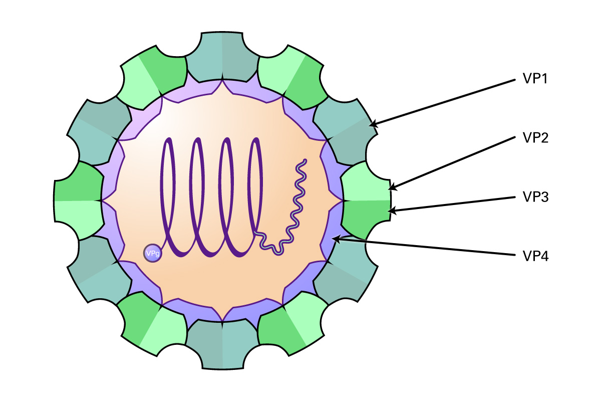
These viruses are approximately 30 nm in diameter, and contain an icosahedral capsid made of four proteins (VP1-4) that surround the 6-10 kb RNA genome. Proteins VP1-3 are located on the external side of the capsid, and VP4 is located internally. The genome consists of a single ORF encoding three groups of proteins: the P1 region which encodes structural polypeptides (L, VP1-3), and the P2 (2A, 2B and 2C) and P3 regions (3A, VPg, 3Cpro, RdRp) encoding proteins required for viral replication. In addition, the virus contains a long UTR at the 5’ end containing an internal ribosome entry site that allows direct translation of the polyprotein, and a shorter 3’ UTR that is important in negative-strand RNA synthesis. Some genera also contain an N-terminal leader protein (L) that can be a papain-like cysteine proteinase or perform some other function. After the virus attaches to host receptors and enters via endocytosis, the VP4 protein opens a pore in the host endosomal membrane, allowing the genomic RNA to enter the host cytoplasm. VPg is removed from the viral RNA, which is translated into the polyprotein. Replication occurs in membrane vesicles derived from the ER where dsRNA is initially synthesized, then transcribed and replicated into ssRNA(+) genomes and packaged into preassembled procapsids. Virus is then released via cell lysis.

Hepatitis A virus differs from other picornaviruses in that it is non-cytopathogenic and particles are released within membranous structures containing 1-4 virions. The virus infects the epithelial cells of the small intestine and hepatocytes of primates. Viral replication predominantly occurs in the liver, is excreted via the bile into the colon, and transmitted fecal-orally. Clinical manifestations include jaundice, fever, abdominal pain, light stools, and diarrhea. The infection is generally of a limited nature, and does not persist chronically. The virus is very stable, resistant to acid pH, and stable at elevated temperatures (60 degrees C) for short durations.

Hepatitis B

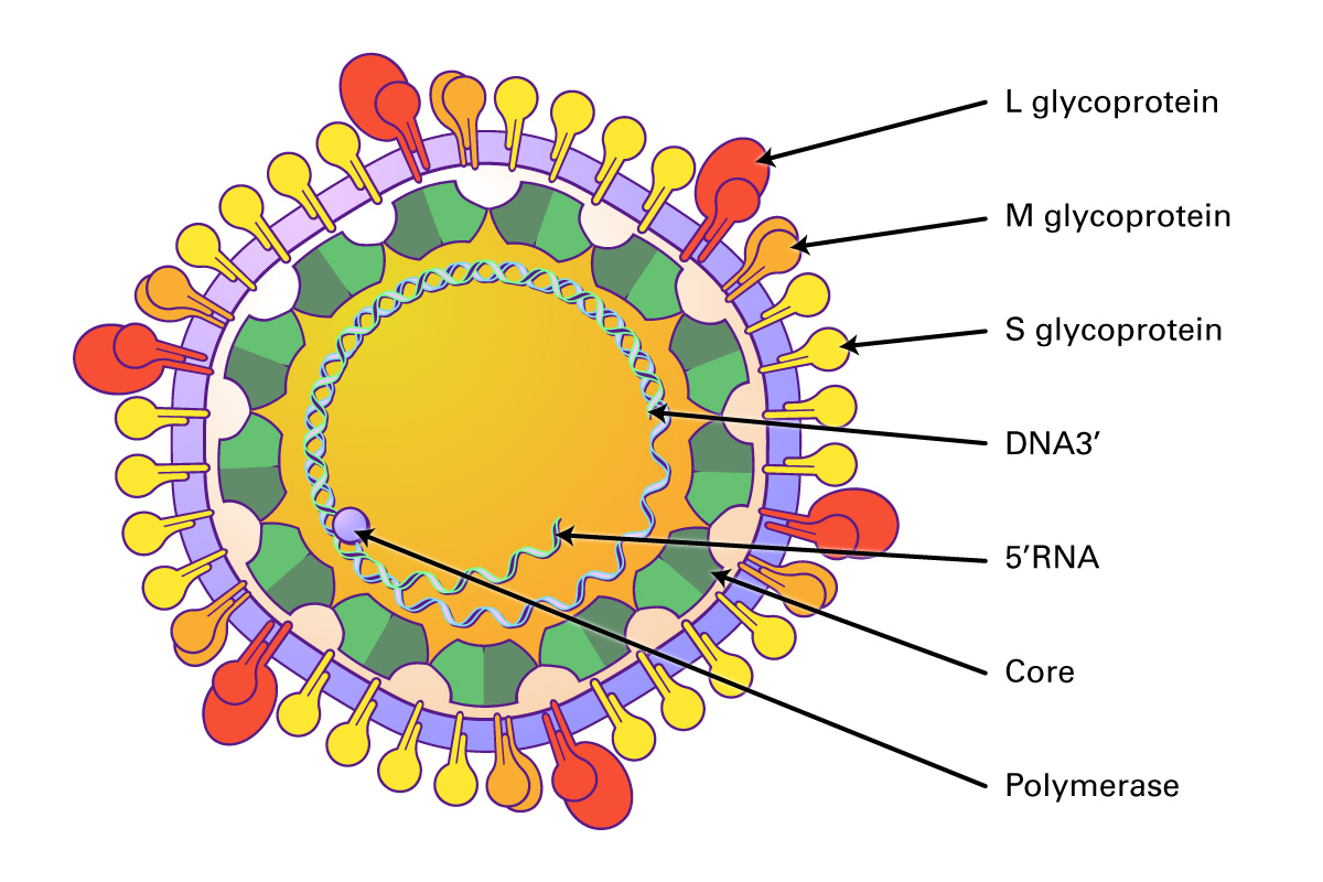
Hepatitis B virus (HBV) is a member of the Hepadnaviridae family of partially double stranded, enveloped DNA viruses with a circular genome and an icosahedral capsid. The family includes include Orthohepadnaviruses and Avihepadnaviruses. These viruses infect hepatocytes and cause liver injury and hepatocellular carcinomas in mammals and birds.

These viruses are approximately 30 nm in diameter, and contain an icosahedral capsid made of four proteins (VP1-4) that surround the 6-10 kb RNA genome. Proteins VP1-3 are located on the external side of the capsid, and VP4 is located internally. The genome consists of a single ORF encoding three groups of proteins: the P1 region which encodes structural polypeptides (L, VP1-3), and the P2 (2A, 2B and 2C) and P3 regions (3A, VPg, 3Cpro, RdRp) encoding proteins required for viral replication. In addition, the virus contains a long UTR at the 5’ end containing an internal ribosome entry site that allows direct translation of the polyprotein, and a shorter 3’ UTR that is important in negative-strand RNA synthesis. Some genera also contain an N-terminal leader protein (L) that can be a papain-like cysteine proteinase or perform some other function. After the virus attaches to host receptors and enters via endocytosis, the VP4 protein opens a pore in the host endosomal membrane, allowing the genomic RNA to enter the host cytoplasm. VPg is removed from the viral RNA, which is translated into the polyprotein. Replication occurs in membrane vesicles derived from the ER where dsRNA is initially synthesized, then transcribed and replicated into ssRNA(+) genomes and packaged into preassembled procapsids. Virus is then released via cell lysis.

In humans, HBV infection is typically transmitted via blood transfusion. The HBV vaccine can prevent disease, and nucleoside analogs (lamivudine, entecavir, adefovir, telbivudine, tenofovir) have been used for long term antiviral therapy to suppress viral genome replication (Yuen 2018).

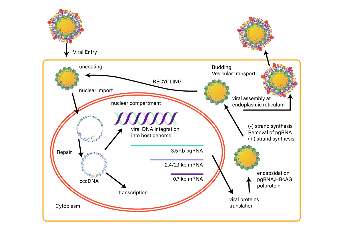
Figure 1: Hepatitis B Life Cycle

Hepatitis C

Hepatitis C virus is a small, enveloped, icosahedral, positive ssRNA virus that belongs to the Flaviviridae family. The Flaviviridae are vector-borne RNA viruses that cause a wide spectrum of diseases including hepatitis, encephalitis, vascular shock, acute flaccid paralysis, congenital abnormalities, and fetal death (Pierson, 2020). The family is subdivided into three genera: Flavivirus, Pestivirus, and Hepacivirus. The Flavivirus genus are zoonotic and insect-borne viruses, and responsible for Yellow fever, Dengue fever, Japanese encephalitis, West Nile encephalitis, and tick-borne encephalitis. The Pestivirus genus includes animal viruses causing bovine viral diarrhea and classical swine fever. The Hepacivirus genus (Hepatitis C) causes viral hepatitis and hepatocellular carcinoma in humans.

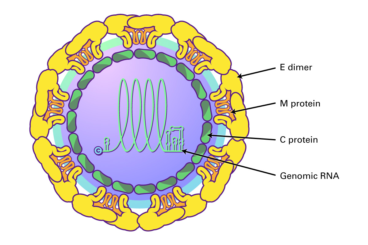
Flaviviruses share a common organization; they encode a single open reading frame that is translated into the ER as a polyprotein, which is cleaved by viral and host proteases into ten functional proteins, including three structural proteins (C, prM and E) and seven non-structural proteins (NS1, NS2A, NS2B, NS3, NS4A and B, and NS5 polymerase). The virus is assembled using the three structural proteins, a host envelope, and viral genomic RNA. Non-structural (NS) proteins include those involved in viral replication, such as the RNA-dependent RNA polymerase.

Hepatitis C virus (genus Hepacivirus) binds to two cell surface receptors, the low density lipoprotein receptor (LDLr) and heparin sulfate proteoglycans (HSPGs). This triggers the HCV outer E1/E2 heterodimer to bind to the scavenger receptor B1 and tetraspanin protein CD81. CD81 then interacts with claudin-1, triggering clathrin-mediated endocytosis and endosomal fusion. The released viral RNA is then translated and replicated via the NS5B RNA-dependent RNA polymerase to produce a negative-sense RNA template from which multiple positive-sense progeny are replicated. The newly synthesized HCV RNAS are then incorporated into nucleocapsid particles, fuse with a luminal lipid droplet containing ApoE proteins to create a high density HCV precursor, and then transit along with the VLDLs formed in the Golgi as multi-vesicular bodies to the cell surface where they are released as a lipoviral particle after fusing with the hepatocyte cell membrane (Dubuisson, J 2014; Grassi 2016).

Hepatitis C infection is the leading cause of liver-related mortality, and chronic infection can lead to cirrhosis, liver failure, hepatocellular carcinoma, liver transplants, and death. Direct-acting antivirals (DAAs), drugs that target multiple mechanisms of the HCV life cycle, have been successful in eradicating HCV from most patients, including those co-infected with HIV (Marks 2019, Morozov 2018). These include sofosbuvir/velpatasvir, which combines and NS5A and NS5B inhibitors, or glecaprevir/pibrentasvir, both of which are pangenotypic treatments that can be dosed once daily for twelve weeks for viral eradication.

Hepatitis D

Hepatitis delta virus (HDV) is a defective RNA virus that depends upon the expression of the hepatitis B virus surface antigen (HBsAg) in the same cell in order to complete its life cycle. HDV can infect hepatocytes that do not contain HBsAg, and can replicate its genome and express the hepatitis delta antigen, however, is unable to secreted infectious viral particles. When co-infection occurs with HBV, chronic hepatitis D is the most aggressive form of viral hepatitis, and is associated with cirrhosis, liver failure, and hepatocellular carcinoma. Co-infection can occur simultaneously with HBV, or can occur as a superinfection of a chronic carrier of HBV. Co-infection causes acute hepatitis, but most cases resolve both infections, although the chance of liver failure is higher than with HBV infection alone. In the case of superinfection, most progress to chronic dual infection with both viruses. There are no significant therapies that are successful in treating HDV infection, and nucleoside analogues that are useful for treating HBV have no effect on HDV replication.

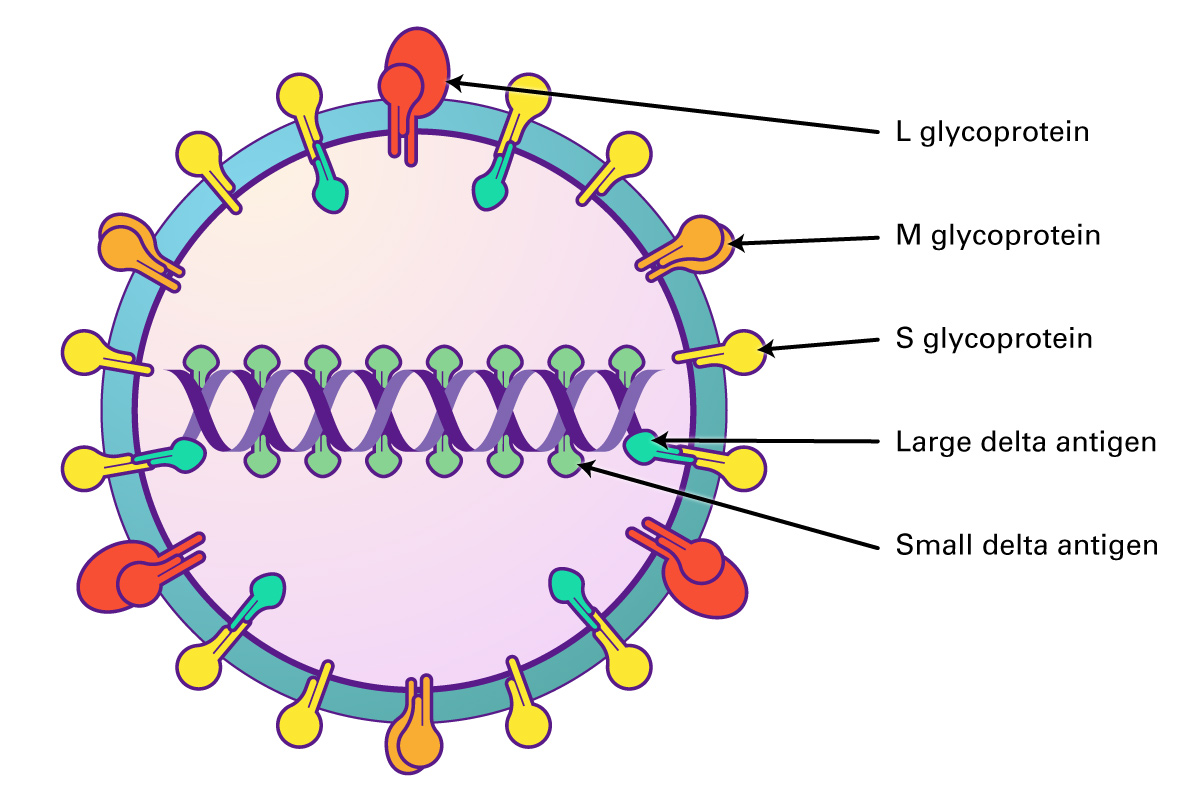
The virions are 35-37 nm, and comprised of an envelope and ribonucleoprotein. Similar in structure to HBV, both viruses use HBV surface proteins S, M, and HBsAg for assembly. The capsid is formed from multimerisation of core protein HBcAG, and contains one copy of the viral circular, ssRNA genome, enclosed by large and small delta antigen which form the viral ribonucleoprotein. The HDV genome is a circular, closed ssRNA which folds into a partially double stranded rod.

The viral life cycle begins when the virus enters hepatocytes through attachment to cell surface heparin sulfate proteoglycans (HSPGs) and SLC10A1, a receptor located at the basolateral membrane and involved in the uptake of bile acids. After entry, the HDV is released into to cytoplasm and transported into the nucleus, where transcription and replication occur. HDV replication is dependent upon host RNA-dependent RNA polymerase and perhaps RNA polymerase II. Replication is via a rolling-circle mechanism, resulting in the production of linear HDV antigenomic RNA, which circularizes and serves a template for genomic-strand RNA. An open reading frame in the antigenome is used to synthesize HDags (S and L-HDAg) that are required for replication and virion assembly. Both proteins then undergo post-translational modifications required for their function. After assembly into RNPs and encapsidation, infectious virions are then secreted via the Golgi or multivesicular body pathway (Mentha, 2019).

Hepatitis E

Hepatitis E virus (HEV) is the most common cause of hepatitis globally. A member of the Orthohepevirus family with four species (A to D), human disease is caused by strains of species A. HEV is a non-enveloped virus with an icosahedral capsid, a size of 27 to 34 nm, and contain a positive-sense, single-stranded, 7.2-kb RNA genome which is 5’ capped and 3’ polyadenylated.

The HEV genome contains three open reading frames (ORFs 1-3). ORF1 encodes a protein of 1,693 amino acids containing functional domains including a methyltransferase, cysteine protease, RNA helicase, and RNA-dependent RNA polymerase. ORF2 encodes the viral capsid protein of 660 amino acids that is responsible for virion assembly, interaction with target cells, and immunogenicity. The ORF2 protein consists of three linear domains: the shell domain (S), the middle domain (M), and the protruding domain (P, harboring the neutralizing epitope(s). ORF3, which overlaps ORF2, encodes a small protein of 113 or 114 amino acids that is involved in virion morphogenesis and release. HEV replicates in the cytoplasm, with a subgenomic RNA producing the ORF2 and ORF3 proteins and the full genomic RNA encoding nonstructural proteins and serving as a template for replication. HEV replicates in hepatocytes but also in the small intestine, colon, and lymph nodes. It can transmit disease either via the fecal-oral route, or through animal reservoirs.

HEV infection causes an acute, self-limited hepatitis in 5% of patients who contract the virus, but can have high mortality in pregnant women, the elderly, or those with pre-existing liver disease. Chronic infection can occur in the immunosuppressed or organ transplant patients. The virus can also cause extrahepatic manifestations including glomerulonephritis, pancreatitis, thrombocytopenia, autoimmune condition, and neurological conditions such as neuritis, neuropathy, Guillain-Barre syndrome, and encephalitis/myelitis.

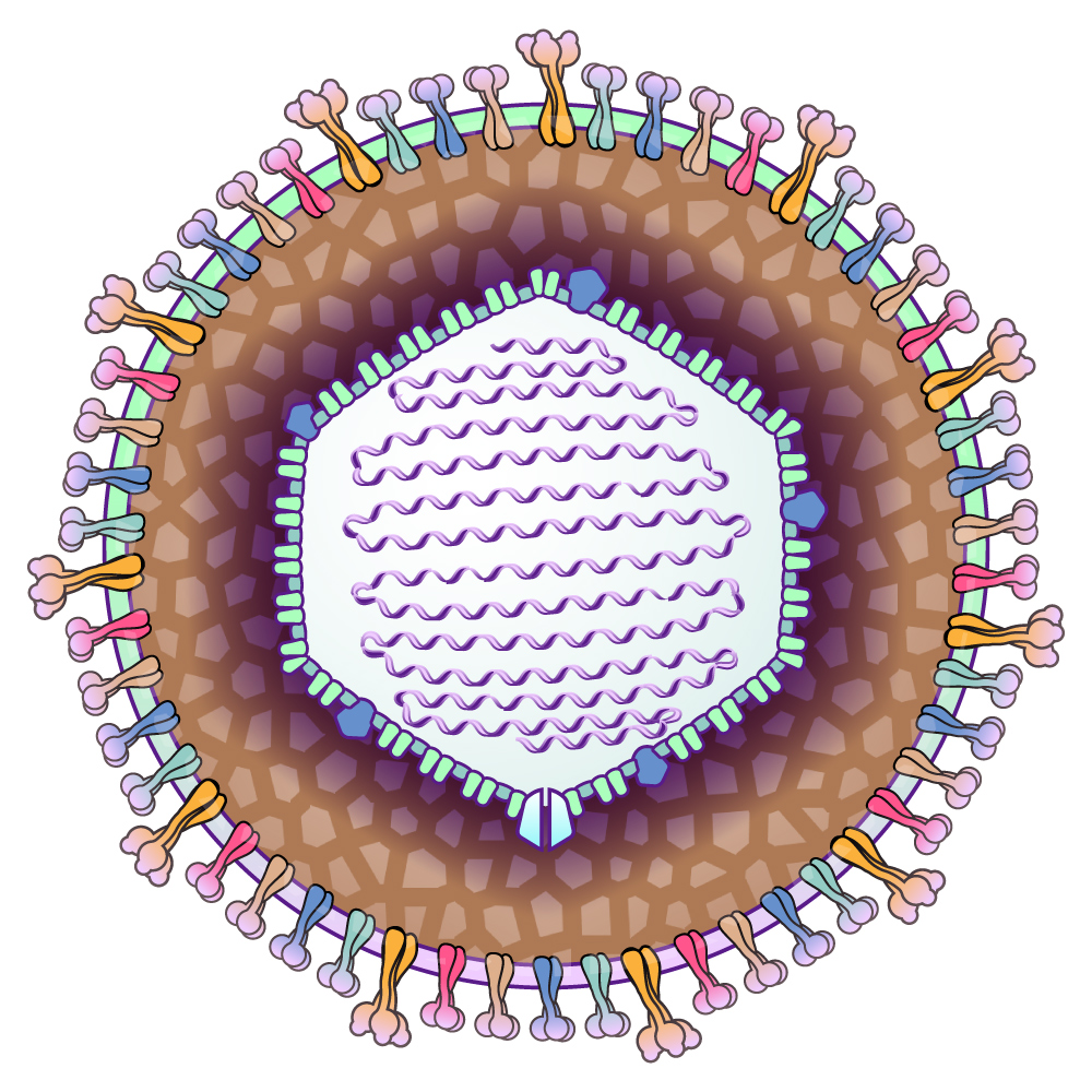
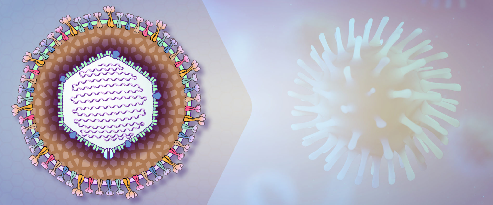
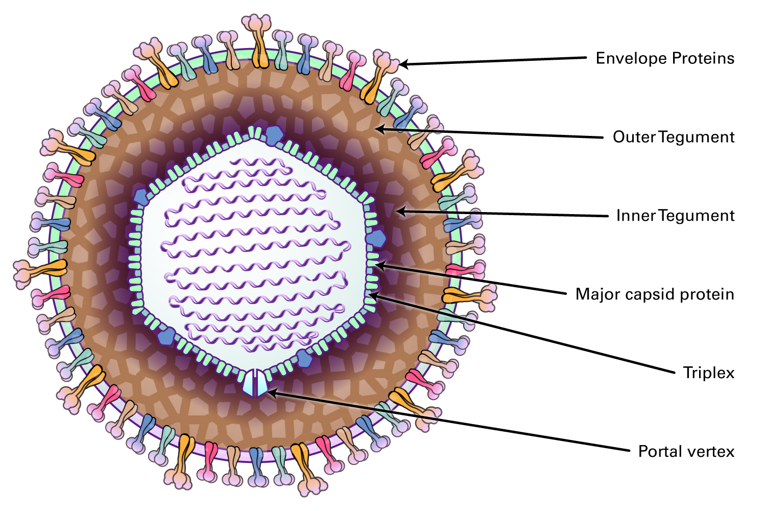
Herpesviruses are double stranded DNA viruses that are enclosed by an icosapentahedral capsid, surrounded by a protein coat (tegument), and encased in a glycoprotein-bearing bilayer envelope. They are divided into three groups: alpha-herpesviruses (herpes simplex virus types 1 and 2, and varicella-zoster virus), beta-herpesviruses (cytomegalovirus, HHV-6, and HHV-7) and gamma-herpesviruses (Epstein-Barr and HHV-8). Alpha-herpesviruses have a short reproductive cycle, cause lysis of the host cell, and have a wide host range, as well as the ability to establish latent infections in sensory nerve ganglia. Beta-herpesviruses have a long reproductive life cycle, a limited host range, establish latent infections, and can form accumulations within cells such as within secretory glands, the reticuloendothelial system, and the kidneys. The gamma-herpesviruses have a very limited host range, are replicated in lymphoblastoid cells, and can be both latent and lytic within host cells.

About Herpesviridae

After the virus enters the cell, the DNA is uncoated and transported to the nucleus, whereupon the virus undergoes transcription of immediate-early genes that code for regulatory proteins. This is followed by expression of early and late genes. The viral core and capsid are assembled in the nucleus, then enveloped at the nuclear membrane, transported through the endoplasmic reticulum and the Golgi apparatus, glycosylated, and mature virions are transported to the cell membrane inside vesicles. Release of virus is accompanied by cell death, with the release of non-infectious as well as infectious viral particles.

Figure 1: Herpesviridae Virus Genome
Figure 2: Structure of Herpesviridae Virus

The genomes of herpesviruses are linear double-stranded DNA molecules that range in size from 125 to 240 kbp, and contain direct or inverted repeats. The genome replicates by circularization, followed by production of concatemers and cleavage of unit-length genomes during packaging into capsids.

A characteristic of herpesviruses is their ability to establish latent infection in specific host cells, either extra-chromosomally or integrated into the host cell DNA. Latent virus may be reactivated to enter a replicative cycle at any point in time. Also, herpesviruses require intimate contact between the individual shedding the virus and the susceptible host.

Infection with an alpha-herpesvirus follows a characteristic pattern. Four to six days after inoculation onto the skin or the mucous membrane, the virus replicates in epithelial cells, causing cell lysis and inflammation with the formation of vesicles. Viremia follows, and the virus ascends the sensory nerves to reach the dorsal root ganglia. This is where latency is established.

During reactivation, replication in the ganglia is followed by retrograde axonal spread back to other skin surfaces via peripheral sensory nerves.

Diseases caused by herpesvirus include oral and genital herpes (HSV-1, HSV-2), herpetic keratitis and conjunctivitis, neonatal herpes simplex, herpes simplex encephalitis, chickenpox, shingles, cytomegalic inclusion disease, CMV mononucleosis syndrome, Epstein-Barr mononucleosis, HHV-6 and HHV-7 roseola, Kaposi’s sarcoma (HHV-8), and B virus encephalitis.

The only vaccine that is currently available is to the varicella-zoster virus; otherwise, therapy consists of treatment with acyclovir, valaciclovir, famciclovir, or ganciclovir for HSV, zoster, and CMV infections. There are no current antiviral agents for the treatment of EBV, HHV-6, HHV-7, or HHV-8.

Influenza viruses are members of the family Orthomyxoviridae, which are enveloped viruses with a segmented negative-sense, single-strand RNA genome. Of the four genera in this family (A, B, C, and D), types A and B are the most clinically relevant because they cause influenza pneumonia with potentially severe complications such as encephalitis, myocarditis, and death. The virus is transmitted by aerosol infection as well as by contaminated surfaces. It enters the body through the nasal and laryngeal mucosa, as well as the lower airways, before spreading systemically to infect a variety of cell types.

About Orthomyxoviridae

Although Influenza A viruses cause most cases of influenza and are responsible for pandemics, influenza B can cause morbidity during inter-pandemic periods, and up to 23% of influenza cases in a season. It is split into two antigenically distinct phylogenetic lineages (B/Victoria and B/Yamagata). In contrast to influenza A which can be found in multiple species including humans, birds, and pigs, influenza B is typically only found in humans. Treatment for influenza includes antiviral medications such as oseltamivir, marboxil, perimavir, and zanamivir. Vaccination for influenza often protects against strains H1N1 and H3N2 of influenza A and one or two strains of influenza B viruses.

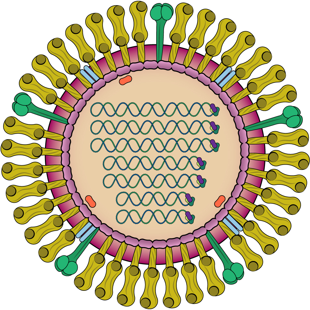
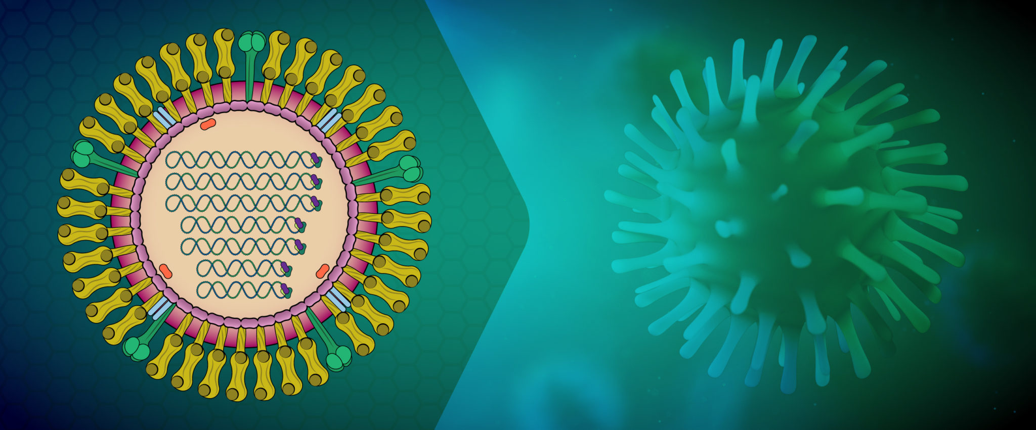
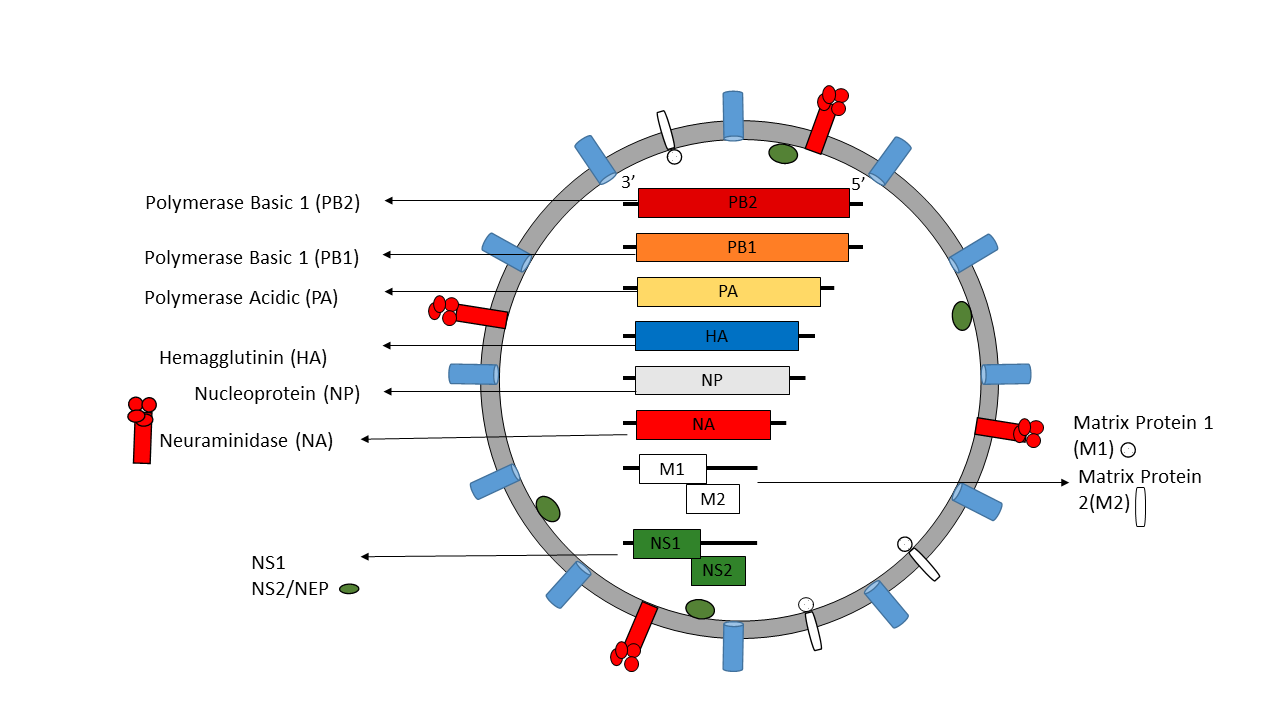
Influenza A virus (IAV) and B virus (IBV) both contain eight viral RNA gene segments, which code for ten essential viral proteins and accessory proteins. IAV and IBV contain the viral lipid membrane glycoproteins HA (hemagglutinin) and NA (neuraminidase) that determine the subtype of influenza virus. HA and NA are both targets for immune responses against the virus, and the NA protein is the target of antiviral drugs such as Relenza and Tamiflu. Also embedded in the lipid membrane is the M2 protein, the target of the antiviral drugs amantadine and rimantadine. Beneath the lipid membrane is M1, the matrix protein that forms the viral capsid, and inside are the viral RNA segments in tight association with polymerase B1 and B2 proteins, PA (polymerase acidic protein), and NP (nucleoprotein). The virion also includes 2 non-structural proteins, NS1 and NEP (nuclear export protein NS2).

Influenza Life Cycle

IAVs initiate infection by using hemagglutinin protein (HA) to bind to cell surface glycoconjugates that contain terminal sialic acid residues, which triggers endocytosis. The resulting endosome fuses with the viral membrane and releases viral ribonucleoproteins into the cytosol, which then recruit importins that facilitate transport through the nuclear pore complex into the cell nucleus. Inside the nucleus, the viral RNA-dependent RNA polymerase then transcribes and replicates the viral RNA. The newly replicated RNA associates with nucleoprotein molecules and a single copy of the viral polymerase to assemble into a viral ribonucleoprotein, which is then trafficked to the cell surface and budded from the plasma membrane. The complex, but fast (6 hour) replication cycle involves the participation of many host proteins that are involved in multiple aspects of the viral life cycle, including viral entry, uncoating, viral RNP nuclear import, transcription and translation, post-translational modification, and transport out of the cell. The viral neuraminidase (NA) cleaves terminal sialic acids to release virions and complete the infectious cycle.

Influenza A virions possess two highly antigenic surface glycoproteins (HA and NA) that play different critical roles in the viral life cycle. Both of these proteins can be neutralized by host antibodies, and inactivated IAV vaccines induce antibody responses to HA and NA. However, because of the high mutational tolerance of these surface glycoproteins and the segmented nature of the genome that facilitates genomic reassortment during coinfection with different strains, there is opportunity for both antigenic drift and antigenic shift that allows for escape from host immune responses.

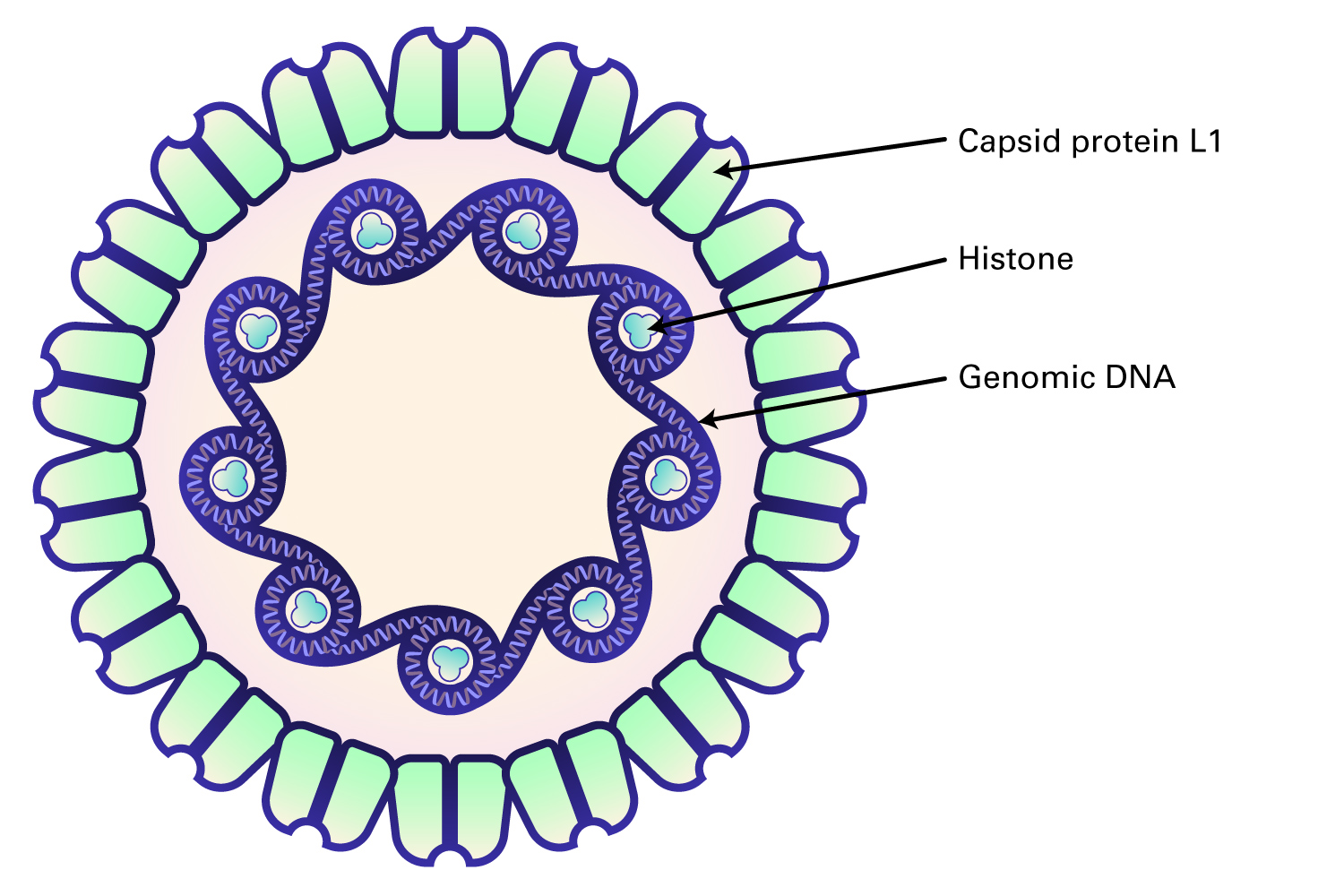
Papillomaviruses are non-enveloped, icosahedral double-stranded DNA viruses that range from 45 to 55 nm in diameter, and contain a limited number of genes (6-7).

About Papillomaviridae

Papillomaviruses are highly species-specific and cause tumors (papillomas and carcinomas) in their hosts. The papillomavirus genome contains at least six open reading frames; the early frames code for proteins that are involved in viral replication and cell transformation, whereas the late frames encode capsid proteins. Human papillomaviruses are a diverse group, and more than 200 types exist, associated with different pathologic conditions. They are particularly tropic for squamous epithelial cells, with some types (HPV 6, 11) producing papillomas (warts), and others (HPV 16, 18, 31) producing persistent subclinical infections or transforming the epithelium into oropharyngeal, anogenital, or cervical squamous cell carcinomas. Transmission occurs via direct mucosal contact with an infected person, by sexual transmission, or with viral particles left on a surface.

Papillomaviruses can either infect cells, replicate, and induce cell death upon lysis (permissive cells) or infect cells and transform them rather than undergoing viral replication (non-permissive cells). In permissive cells, the virion attaches to specific surface receptors, enters the cell and is transported to the nucleus, where viral DNA is released. Early phase proteins such as E6 and E7 drive the cell into S phase, then use cellular DNA synthesis proteins such as DNA polymerase and thymidine kinase to replicate. After transcription of late viral RNAs for structural proteins, progeny virions are assembled in the nucleus. Host cell lysis then releases the viral particles.

In non-permissive cells or permissive cells infected with defective viral genomes, the tumor antigens are synthesized, and cellular DNA synthesis is stimulated, but late viral capsid genes are not expressed. Instead, the viral genome integrates randomly into the host chromosome. In human papillomaviruses, the E6 and E7 proteins bind p53 and RB, respectively, leading to immortalization. These transforming proteins produce altered host cell morphology, increased growth rate, loss of contact inhibition, and anchorage independence, all of which promote tumorigenicity.

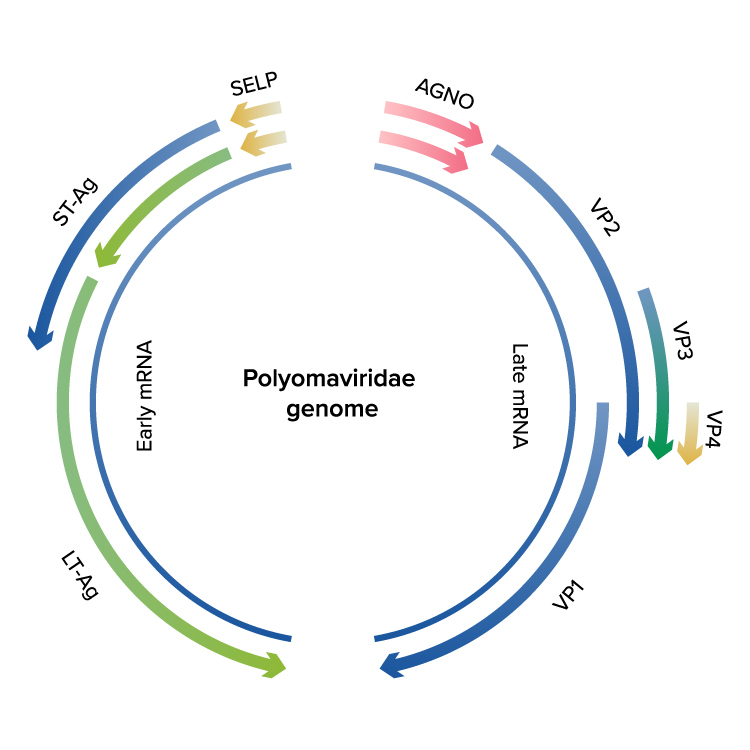
Polyomaviruses are non-enveloped, icosahedral double-stranded DNA viruses that range from 45 to 55 nm in diameter, and contain a limited number of genes (6-7).

About Polyomaviridae

Polyomaviruses include members such as SV40, JC, and BK viruses, and encode both early functions (nonstructural proteins) that can induce tumors in animals, and late functions that code for structural proteins such as the viral capsid. Most polyomaviruses produce harmless, persistent infections in their hosts, except for the JC virus, which has been implicated in progressive multifocal leukoencephalopathy, a demyelinating disease that occurs rarely in individuals with impaired immune systems. The BK virus can cause a mild respiratory infection in children, then persists in the kidney. In adults with impaired immunity or who have had renal transplants, the virus can produce nephropathy, ureteric stenosis, or cystitis.

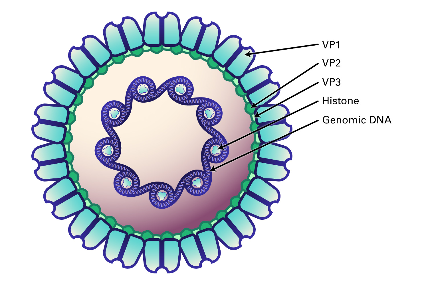
Polyomaviruses can either infect cells, replicate, and induce cell death upon lysis (permissive cells) or infect cells and transform them rather than undergoing viral replication (non-permissive cells). In permissive cells, the virion attaches to specific surface receptors, enters the cell and is transported to the nucleus, where viral DNA is released. Early phase proteins such as large T antigen drive the cell into S phase, then use cellular DNA synthesis proteins such as DNA polymerase and thymidine kinase to replicate. T antigen binding then initiates transcription of late viral RNAs for proteins such as VP1, VP2, and VP3, and progeny virions are assembled in the nucleus. Host cell lysis then releases the viral particles.

In non-permissive cells or permissive cells infected with defective viral genomes, the tumor antigens are synthesized, and cellular DNA synthesis is stimulated, but late viral capsid genes are not expressed. Instead, the viral genome integrates randomly into the host chromosome. In SV40, the large T antigen binds p53 and RB proteins to trigger immortalization. In other polyomaviruses, the middle T antigen binds cellular proteins such as c-src, and promotes cellular replication.

Figure 1: Polyomaviridae Virus Genome
Figure 2: Structure of Polyomaviridae Virus

Paramyxoviruses are negative-sense single stranded RNA viruses that cause a wide variety of diseases in human and animals. The majority of paramyxoviruses are respiratory pathogens and transmission occurs via aerosols or direct contact with infectious secretions, or in some cases, by fecal-oral contact.

About Paramyxoviridae

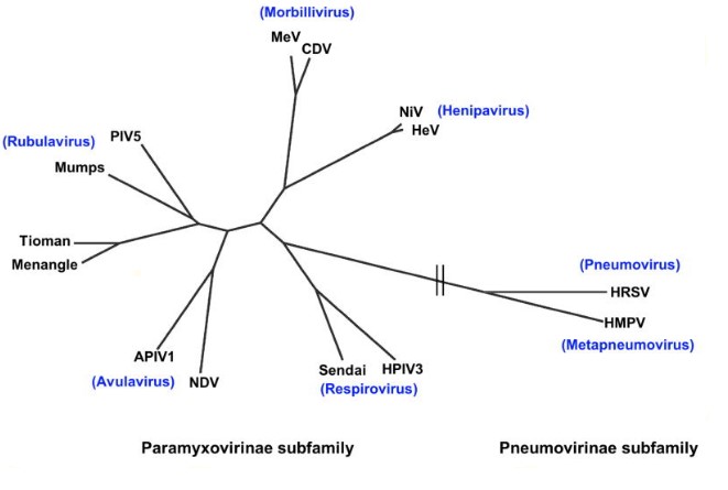
Paramyxoviridae contain two subfamilies: Paramyxovirinae and Pneumovirinae. The Paramyxovirinae subfamily contains five genera: Respirovirus (human parainfluenza virus 3, Sendai), Rubulavirus (Mumps, human parainfluenza virus 5), Avulavirus (Avian parainfluenza, Newcastle disease), Morbillivirus (Measles, canine distemper), and Henipavirus. The second subfamily, Pneumovirinae contains Pneumovirus and metapneumovirus as well as animal pathogens such as human and bovine respiratory syncytial viruses, and human and avian metapneumovirus.

HPIV can infect many different animals, including birds, primates and rodents. Some parainfluenzaviruses can infect poultry and penguins (Newcastle disease virus).

Clinical features of HPIV infection include both upper and lower respiratory tract illneses. Children present with fever, croup, laryngeal obstruction, and inspiratory stridor. The virus can also cause bonchiolitis in infants, pneumonia in children, tracheobronchitis, and giant-cell pneumonia in immunocompromised individuals. It has been associated with neurologic disease including seizures, encephalitis, and demyelinating syndromes, suggesting that the virus can be neurotropic.

Other important disease causing members of the Rubivirus family is the Mumps virus (MuV), which causes parotitis and orchitis and is also highly neurotropic and may cause encephalitis and meningitis. Although the disease is self-limiting and rarely fatal, long germ sequelae can occur, including seizures, deafness, and paralysis. The virus is transmitted by inhalation or oral contact with droplets, and the hallmark is salivary gland swelling, with viral replication occurring in the parotid glands bilaterally. Unilateral orchitis is the most common extra-salivary organ infected by mumps virus. Atrophy of the testicle can occur in half of cases, but sterility is a rare complication. The virus can also infect the kidneys and cause nephritis, as well as pancreatitis in a small percentage of cases. Half of cases of infection show virus within the cerebrospinal fluid and infect neurons in the brain, but mengingitis and encephalitis are rare complications. Deafness can also occur as a consequence of infection within the inner ear.

Henipavirus and Nipahvirus have broad tropism and extreme virulence and infect bats which can transmit to humans either directly or via an intermediate host. Due to their high virulence, they are classified as BSL4 pathogens.

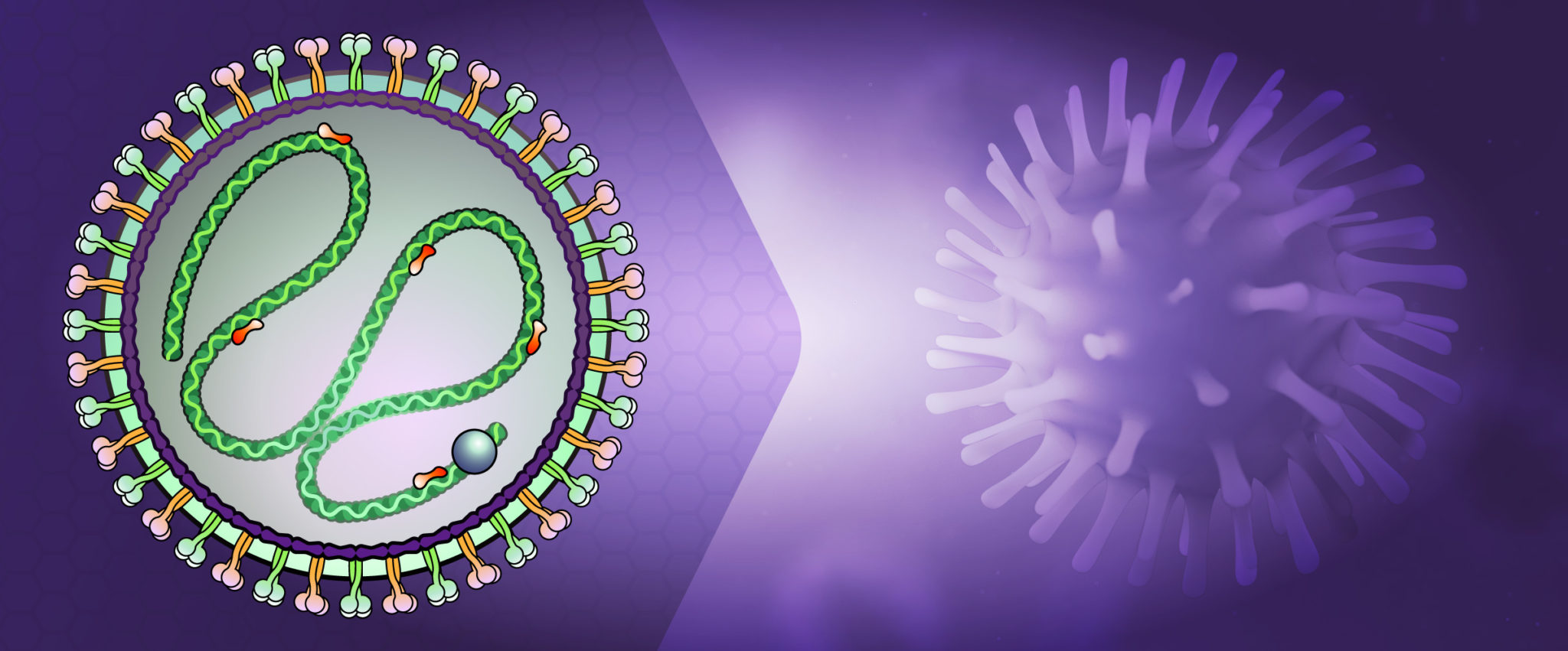
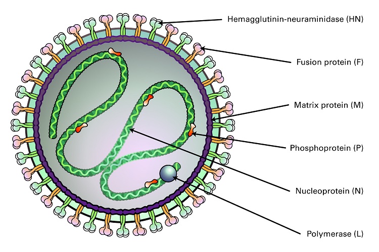
Figure 2: Structure of Paramyxoviridae Virus

Figure 1: Phylogeny of Paramyxoviridae (Aguilar and Lee, 2011). Legend: APIV1, avian parainfluenza virus 1; CDV, canine distemper virus; HeV, Hendra virus; HMPV, human metapneumovirus; HPIV3, human parainfluenzavirus3; HRSV, human respiratory syncytial virus; MeV, measles virus; NDV, Newcastle disease virus; NiV, Nipah virus; PIV-5, parainfluenzavirus 5.

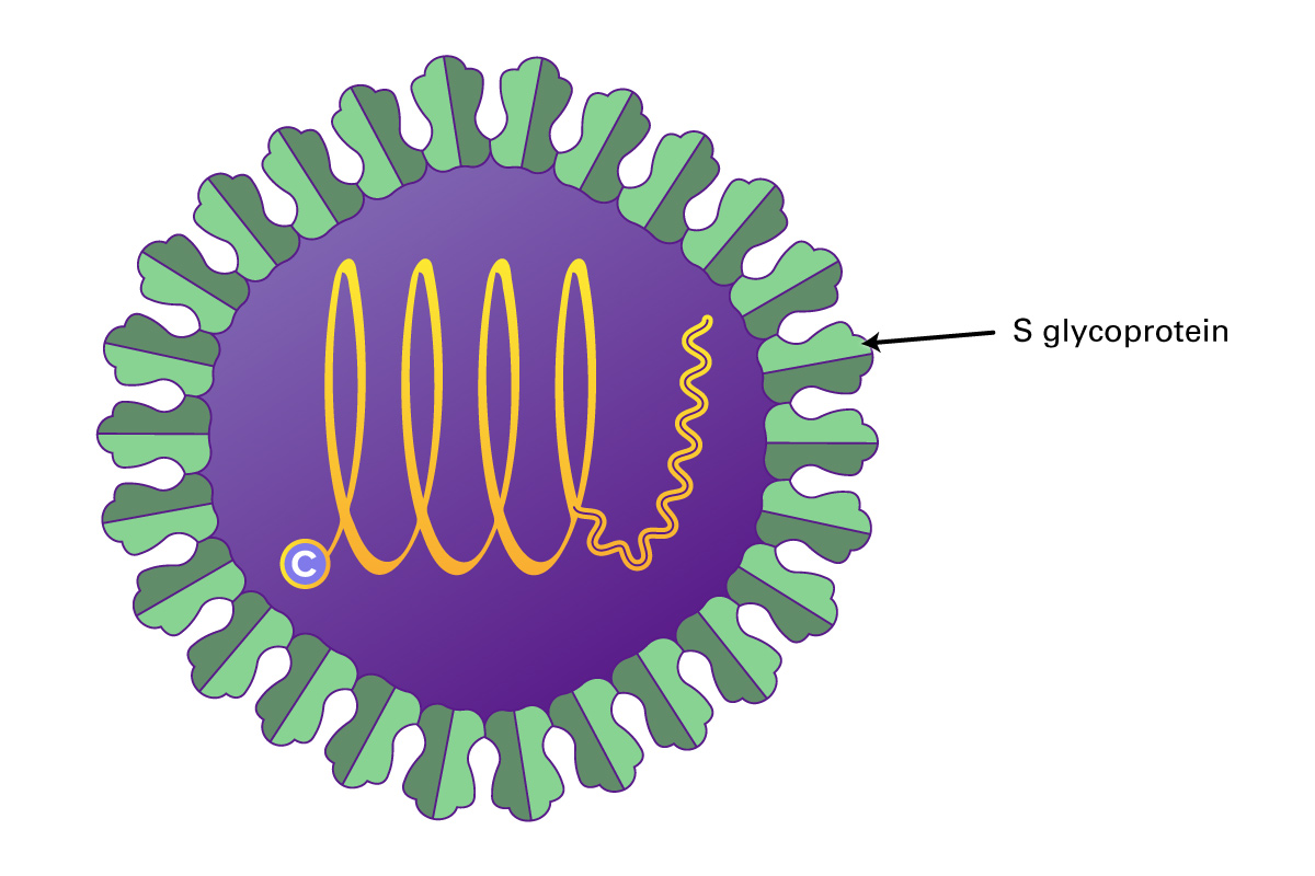
Parainfluenza viruses (HPIV) are divided into four groups (Types 1-4), and are all enveloped, negative-sense RNA genomes that cause lower respiratory infections in children, the immunocompromised, and the elderly. These viruses also cause significant veterinary disease. There are two genera of HPIV, the Respirovirus (HPIV-1 and HPIV-3) and Rubulavirus (HPIV-2 and HPIV-4). Both differ from influenza viruses by their non-segmented genomes. They also differ from other genera of Paramyxoviridae by the absence of a neuraminidase or nucleocapsid morphology. These viruses are between 150 and 250 nm, and contain a 15,000 nucleotide (-)ssRNA genome that encodes six common structural proteins (N, P, C, M, F, HN, L). Two surface glycoproteins are found in all HPIV: the hemagglutinin-neuraminidase (HN), and the fusion protein (Fo). Beneath the viral membrane is the M protein (membrane). The P gene produces several nonstructural proteins from overlapping reading frames that aid in viral replication or slowing the cell cycle. The V and C proteins may also be involved in inhibition of the interferon response by inducing degradation of STAT1 or STAT2. The nucleocapsid is comprised of viral RNA together with the N, P, and L proteins. The surface glycoproteins HN and F interact with the M protein to direct insertion and aggregation at the cell membrane, with participation of the M protein in cell budding.

The virus first enters the cell by fusing the viral and host cell lipid membranes, followed by release of the HPIV nucleocapsid into the cytoplasm. Replication than takes place by using viral RNA-dependent RNA polymerase (L protein). The cellular ribosomal machinery translates viral mRNA into poroteins, which then direct the full-length replication of the viral genome, first into (+)-sense RNA, then into the (-)-sense RNA strand. These (-)ssRNA starnds area then encapsdated with NP and packaged for export as a new virion.

The family of Pneumoviridae are large enveloped, non-segmented, negative-sense, single-stranded RNA viruses. The genus Orthopneumovirus includes human respiratory syncytial virus (HRSV) and bovine respiratory syncytial virus (BRSV), and the Metapneumovirus genus contains avian and human metapneumoviruses.

About Pneumoviridae

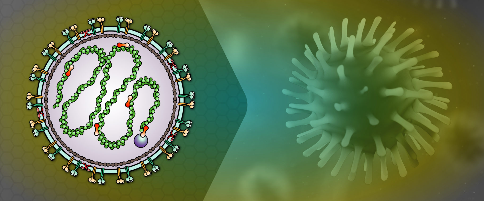
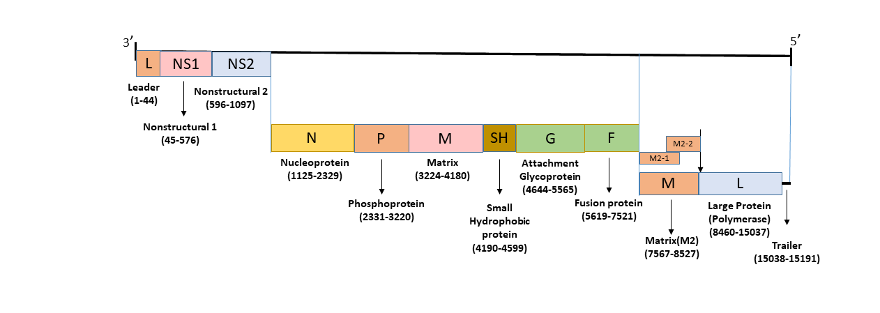
Pneumoviruses produce spherical and filamentous virions that range in size from 150-200 nm in diameter and contain a 115-kb genome that encodes 11 proteins. Included are three viral envelope proteins (G and F glycoproteins and the small hydrophobic protein) and eight other proteins, including the large (L) polymerase protein, nucleoprotein (N), phosphoprotein (P), three matrix proteins (M, M2-1, and M2-2), and two nonstructural proteins (NS1 and NS2).

The virus enters the cell by binding to the HN glycoprotein, fusing to the host plasma membrane, and then releasing the nucleocapsid. Viral mRNA is then transcribed by the virally encoded RNA-dependent RNA polymerase, and viral proteins are produced by host ribosomal machinery. After P, N, L, and M2 proteins are produced, a capsid is created around the newly replicated RNA genome to produce a ribonucleocapsid. The virion then buds from the cell membrane.

Respiratory syncytial viruses (RSV) are among the most important pathogenic infectious agents in children, and a leading cause of death globally in infants under 1 year of age. These viruses can cause upper respiratory infections similar to the common cold, but also can cause bronchiolitis, otitis media, pneumonia, and croup, and mortality in children with preexisting cardiac or lung disease. Reinfections, as well as co-infections with rhinovirus and adenoviruses, can also occur. In other cows, bovine respiratory syncytial virus (BRSV) is the major cause of respiratory disease in calves during the first year of life. There is currently no efficacious RSV vaccine and very few therapeutics to treat RSV.

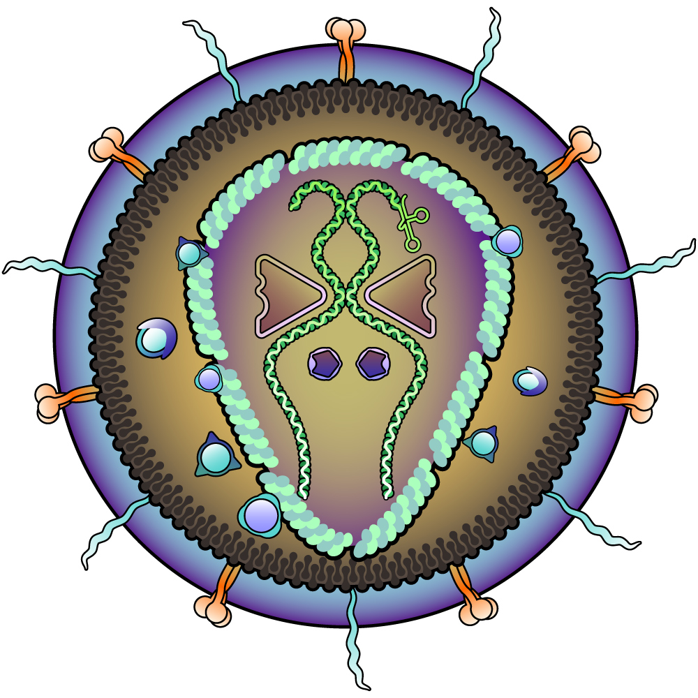
Retroviruses belong to a family of enveloped RNA viruses (Retroviridae) that infect vertebrates. Retroviruses are divided into five sub genera, with two members (Delta retroviruses HTLV-1 and HTLV-2 and Lentiviruses, HIV-1 and HIV-2) causing human disease. They can be exogenous, transmitted horizontally among hosts, or endogenous, inherited vertically through the genomes of their hosts. The virus requires a reverse transcriptase to convert the viral RNA genome into DNA, which integrates into host chromosomes and utilizes host proteins for gene expression and replication. Retroviruses can also be oncogenic through insertional activation of host genes. In humans, HIV is responsible for causing the acquired immunodeficiency syndrome (AIDS) and HTLV-1 and HTLV-2 are the retroviruses that cause human T-cell leukemia/lymphomas.

About Retroviridae

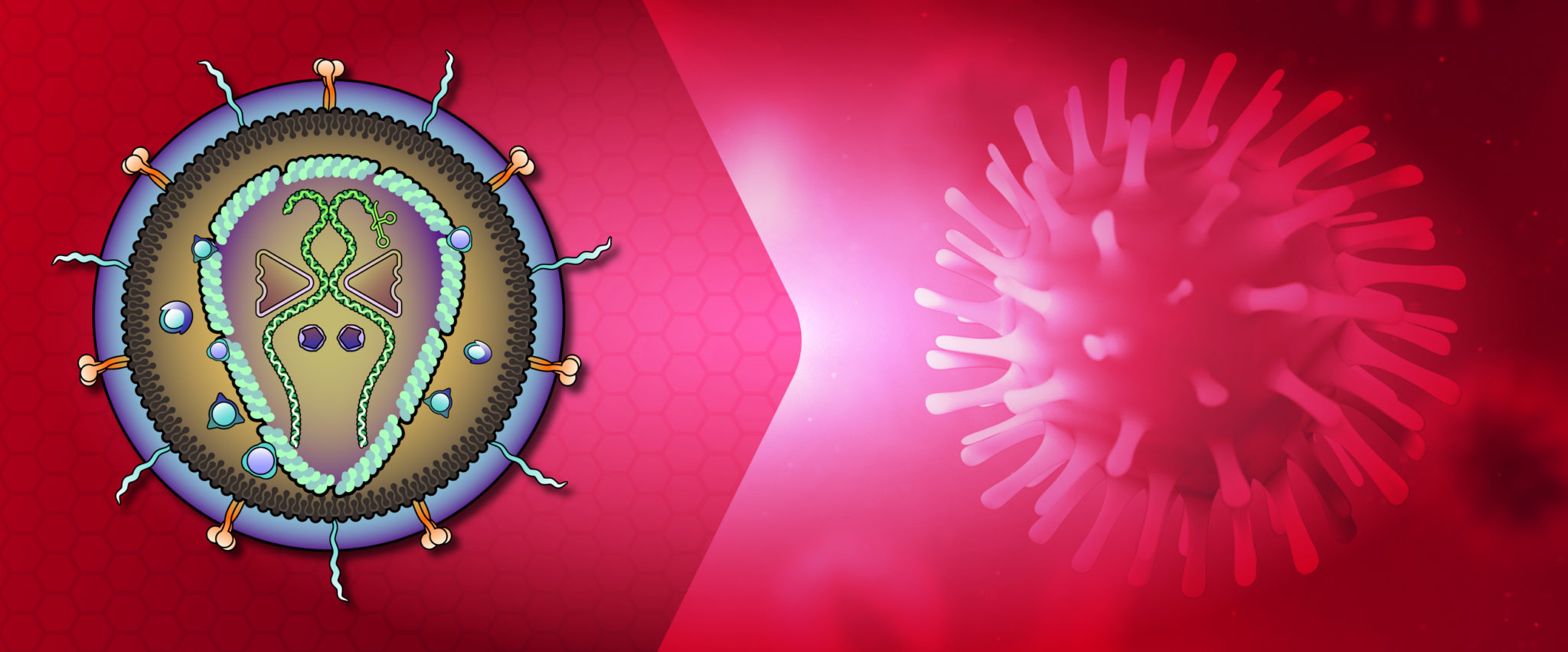
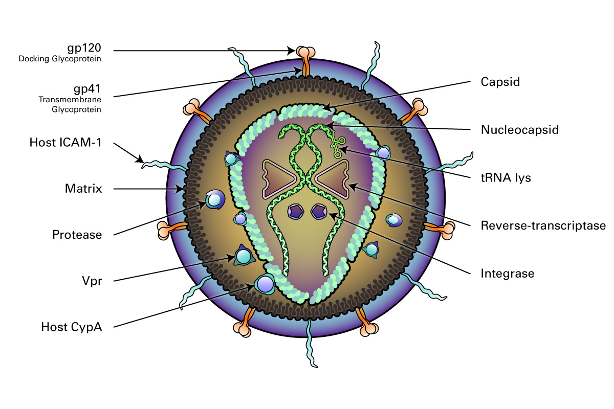
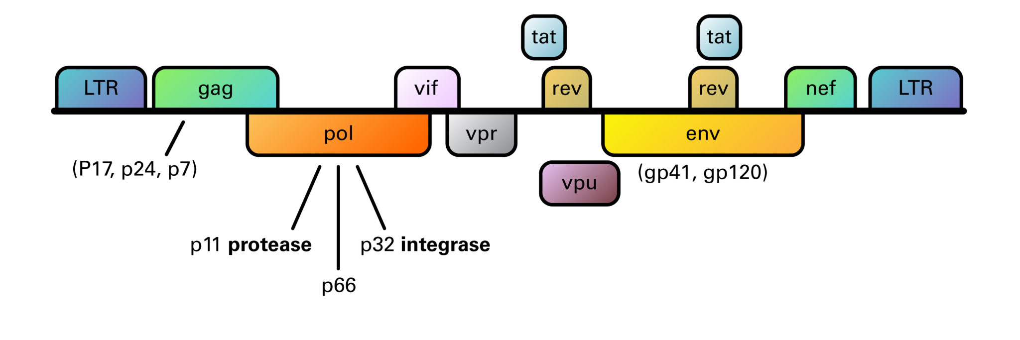
Structurally, retroviruses are enveloped viruses with an RNA genome. The virion contains a highly error-prone RNA-dependent DNA Polymerase (reverse transcriptase) that converts the viral RNA into DNA, which integrates into the host chromosome (Hu, 2012). They have a diploid genome, with 2 copies of RNA per viral particle. A lentiviral (HIV) genome is depicted here.

Organization of the HIV-1 genome showing the reading frames of the genes coding for structural and regulatory proteins: LTR = long terminal repeat; gag = group-specific antigen; pol = polymerase; env = envelope. The gag, pol, and env genes are expressed as precursor polyproteins, which are then cleaved to yield mature viral proteins. In the case of the regulator genes, the proteins of tat and rev are composed of two gene regions. The genome consists of 9,200-9,600 nucleotides in HIV-1.

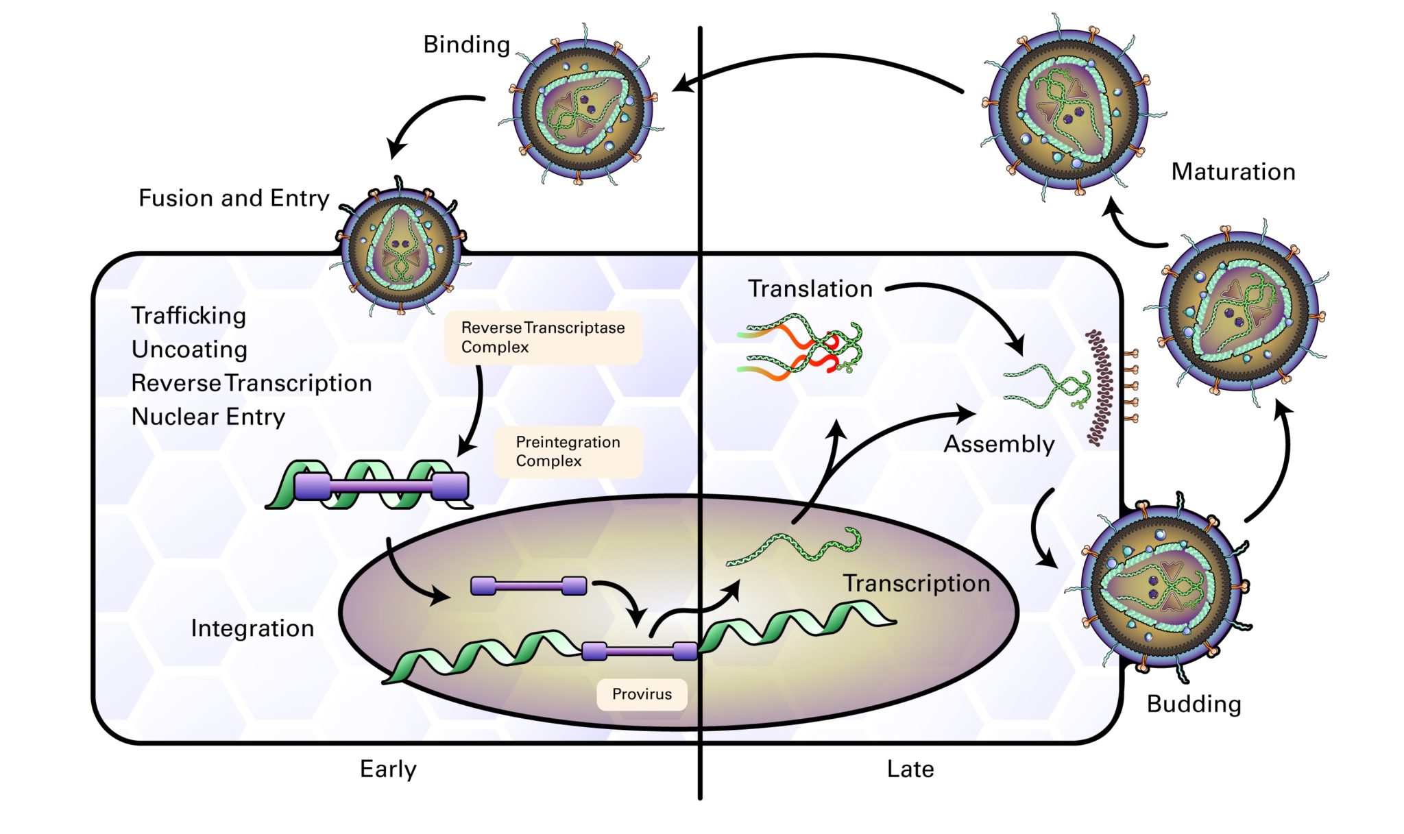
The life cycle of retroviruses can be divided into two phases: the early phase, which includes cell binding through integration of viral DNA into the cell genome, and the late phase, which includes expression of viral genes through the release of virions. Retroviral infection begins when the mature virion envelope glycoprotein interacts with co-receptors on the surface of a cell and fuses membranes of the host cell and virion (Wilen 2011). The viral reverse transcriptase (RT) then uses a host tRNA whose 3’ end is complementary to the sequence near the 5’ end of the viral RNA and acts as a binding site for DNA synthesis. The RT thereby creates an RNA-DNA duplex, which is a substrate for RNase H to remove the 5’ end of the viral RNA, resulting in a newly synthesized minus-strand DNA which can then act, through a complex series of steps, as a template for synthesizing linear double stranded DNA (reviewed by Hu, 2012). The double stranded DNA template is circularized and transported to the nucleus, where it integrates into the host genome, is then transcribed back into viral RNA which is translated to produce viral proteins, and the capsids are then assembled into viral particles and budded from the cell (Nisole, S 2004).

The Delta retroviruses (HTLV-1 and HTLV-2) have a world-wide distribution, and are commonly found in southwest Japan, the Caribbean, and West Africa, where the incidence rate can be 30% of the population. The virus is spread through sexual intercourse, blood transfusions, and by breast feeding. Infection is mostly asymptomatic, however, a subset of infected individuals can develop one of the following diseases: T-cell leukemia/lymphoma, an aggressive tumor of CD4 cells that occurs in 5% of infected individuals, HTLV-1 associated myelopathy (tropical spastic paraparesis), a non-demyelinating disorder with weakness in the lower limbs, infective dermatitis, or uveitis.

The Lentiviruses (HIV-1 and HIV-2) are the causative agent of acquired immune deficiency syndrome (AIDS). AIDS is a new disease that arose in 1980, and HIV-1 is highly homologous to the simian immunodeficiency viruses that infect African monkeys and have crossed the species barrier. HIV-2 is most homologous to an SIV strain that entered the human population in the 1940s. Between humans, both viruses are most commonly transmitted through sexual intercourse, but can also be transmitted vertically in utero, at childbirth, or through breast feeding.

The clinical course of HIV begins with a self-limiting febrile infection 2-4 weeks after exposure with symptoms of fever, lymphadenopathy, and diarrhea, during which time the host develops antibodies. This phase is followed by an asymptomatic phase that can last years, when the patient enters clinical latency. They are infectious but largely asymptomatic, and also test positive for HIV antibodies.

The prodromal phase begins with a drop in the CD4+ count, when patients begin to experience weight loss, fever, lymphadenopathy, oral candidiasis, and diarrhea. The disease then progresses to AIDS, which is characterized by fever, diarrhea, weight loss, skin rashes, neuropathy, myelopathy, dementia, and increased susceptibility to opportunistic infections such as TB, HSV, oral candidiasis, cryptosporidiosis, pneumocystis infections, other viral and fungal infections, and virally induced malignancies such as Kaposi’s sarcoma, hairy leukoplakia, and lymphomas.

The disease pathogenesis begins when HIV infects dendritic antigen processing cells (DC) in the oral and genital mucosa, attaching to these cells via a DC SIGN receptor on the cell surface. HIV particles are then transported to CD4+ helper T-cells where many cycles of infection are established in lymphoid tissues. Loss of function and death of CD4+ helper T-cells results in a drop in the helper T-cell count. Clinical disease progression is correlated with the loss of CD4+ cells and may take five to ten years to progress from primary infection to complete failure of the T-cell mediated immune response.

Early diagnosis is dependent upon the detection of viral DNA or RNA by PCR, or by the detection of p24 antigen in serum. The p24 antigen is usually cleared once patient antibodies appear, so this test is most useful during the early window period, which occurs six days before the development of antibodies and 3-5 weeks after initial exposure.

Treatment of HIV infection includes antiviral agents that disrupt the viral replication cycle. A regimen of three drugs (HAART) given simultaneously suppresses HIV replication, but these drugs are not cures; they must be taken for life to maintain viral suppression. These drugs include nucleoside and nucleotide reverse transcriptase inhibitors, non-nucleoside transcriptase inhibitors, and protease inhibitors. Short courses of anti-retroviral drugs have also been effective in preventing HIV infection following exposure.

There is no currently effective vaccine for the prevention of HIV infection, largely due to:

  1.    Variability of the envelope antigens.
  2.    Heavily glycosylated or hidden nature of epitopes of the envelope glycoprotein gp120.
  3.    Presence of specific antibodies have been shown to actually enhance infection because antibody coated viruses can bind to Fc receptors on the surface of cells.

Togaviridae are a family of single-stranded, positive-strand RNA viruses that include the genera Alphavirus and Rubivirus. The Alphaviruses within this family include arboviruses, which cause two types of diseases: the Eastern, Western, or Venezuelan equine encephalitis viruses cause fever, malaise, headache and encephalitis; and the viruses Chikungunya, Ross River, Mayaro, and Sindbis viruses cause fever, rash, and arthralgias. Rubella Virus is the only member of the genus Rubivirus, and humans are the only known host. It is the causative agent of rubella (German measles) in children, and can cause birth defects known as congenital rubella syndrome in early pregnancy.

About Togaviridae

The Togaviruses are enveloped, with an icosahedral capsid made of 240 copies of the nucleocapsid protein. The envelope contains 80 trimer spikes comprised of the E1 and E2 glycoproteins. The RNA genome is 9.7-11.8 kb, and is capped and polyadenylated. The virus infects cells by attachment of the viral E glycoprotein to host receptors, triggering clathrin-mediated endocytosis of the virus into the host cell. The virus membrane fuses with the host endosome, releasing the RNA genome into the cytoplasm. The positive-sense ssRNA is then translated into a polyprotein, which is cleaved by the viral nsP2 protease into non-structural proteins responsible for RNA replication and transcription. The positive sense ssRNA is transcribed into a complementary antisense RNA, which serves as a template for synthesizing genomic ssRNA. Transcription of subgenomic RNAs within the latter third of the genome give rise to a structural polyprotein, which is cleaved to produce the viral structural proteins. After capsid assembly, the virus is enveloped and budded at the plasma membrane.

The Alphaviruses include mosquito transmitted diseases such as Eastern, Western, or Venezuelan equine encephalitis, which are characterized by fever, malaise, headache and encephalitis, and diseases caused by the Chikungunya, Ross River, Mayaro, and Sindbis viruses, which produce arthralgias. The syndrome produced by viruses such as Chikungunya result in intense joint pain, high fevers, and a rash, and although infection is self-limited, symptoms may persist for years. In addition to mosquito transmission, the virus can be transmitted vertically as well as with infected blood transfusions.

Rubella Virus is the only member of the genus Rubivirus, and it is the causative agent of rubella (German measles) in children. It is transmitted by respiratory droplets. Rubella virus can act as a teratogen, and produce a spectrum of severe birth defects known as congenital rubella syndrome if contracted during the first trimester of pregnancy.

Figure 1: Structure of Togaviridae Virus


LSBio ELISA Development Kit Guide

Introduction to ELISA ELISA (enzyme-linked immunosorbent assay) is a type of immunoassay used for the detection and quantification of analytes Read More...

Alzheimer’s Disease

Alzheimer’s Disease (AD) is the most common progressive form of dementia. It is typically late onset (95% age >60 years), Read More...

Cancer Research

Breast Cancer Immunohistochemistry MarkersColorectal Cancer Immunohistochemistry MarkersEstrogen ReceptorERBB2 / HER2TP53EGFRBRCA1APOBEC3BKi-67BCL2Smooth Muscle Actin (SMA / ACTA2)Progesterone ReceptorSMMHCCK5CK7CK14CK20E-CadherinCTNND1FOXA1CNN1 (Calponin)GATA3VEGFACD31CD34SIAH2GCDFP-15Mammaglobin APIM1GPA33CK20CK7CalretininCDH17COX-2MUC2CDX2VillinMLH1mCEASATB2CA19-9TP53BRAFPIK3CARECQL4APCMOC-31 (EPCAM)MSH2MSH6PMS2AXIN1AXIN2TCF7L2Lung Cancer Read More...

Monoclonal Antibodies

At Stratech, we specialise in providing a comprehensive range of monoclonal antibodies (mAbs) for scientific research. We partner with state-of-the-art Read More...

Apoptosis

What is Apoptosis Apoptosis is a form of programmed cell death that occurs in multicellular organisms and has multiple functions Read More...

lifespan Biosciences COVID-19

Research Solutions for Coronavirus – Lifespan Biosciences

Contact usPlace an Order Coronavirus Research ProductsView AllSpike GlycoproteinsSARSIHC-Plus ACE2Coronavirus Detection LSBio’s library of coronavirus products includes numerous kits for Read More...

Coronavirus

COVID-19 Diagnostics Detection Kits (IgG/IgM Rapid test kits and RT-PCR)Viral Nucleic Acid Extraction Kits & Sample stabilisationCOVID-19 Research Products (Proteins, Read More...