The major advantages of plastic over aqueous mounting media are brightness, contrast, and longevity of the fluorescence. Cy2, Cy3, and Cy5 are available conjugated to a selection of secondary antibodies for multiple labelling, streptavidin, and purified IgG controls.
Figure 2 illustrates the discrete spectra that provide effective separation for multiple labelling. For single labelling protocols that do not require highly cross-adsorbed antibodies, choose Cy3-conjugated antibodies from the lists of whole IgG affinity-purified secondary antibodies. Note that Cy3 conjugates are bright and photostable in the aqueous environment as well as in non-polar media. This is in contrast to Cy2 and Cy5, which are outperformed in aqueous media by Alexa Fluor® conjugates.
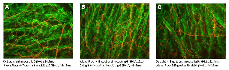
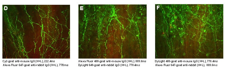
Figure 1. Sections of gastric mucosa were stained and mounted in DPX . Sections A, B, and C were stained with primary antibodies for abundant antigens (mouse anti-Type IV collagen [green] and rabbit anti-PGP 9.5 [red]). Sections D, E, and F were stained with primary antibodies for less abundant antigens (mouse anti-PGP 9.5 [green] and rabbit anti-substance P [red]). All sections were stained with different fluorophore-conjugated secondary antibodies from Jackson ImmunoResearch. All sections were exposed to light long enough to achieve approximately the same relative brightness. Exposure times are listed in millaseconds (ms). Differences in exposure time for each fluorophore are an indication of it’s relative degree of brightness. Note that Cy2 conjugates required significantly less exposure time in DPX than DyLight 488 and Alexa Fluor® 488, both with abundant and less abundant antigens. Cy2 conjugates also were brighter in DPX than in glycerol mounting media (not shown). Other antigens that are less visible in Cy2-stained sections are not visible with DyLight 488 and Alexa Fluor® 488. Similar differences were observed when the other cyanines, Cy3 and Cy5, were compared with corresponding Alexa Fluor® and DyLight dyes. Images and results are courtesy of Dr. Gwen Wendelschafer-Crabb, Kennedy Lab, University of Minnesota. Similar results were reported to us by Dr. Barbara Jones, Department of Neurology and Neurosurgery, McGill University.
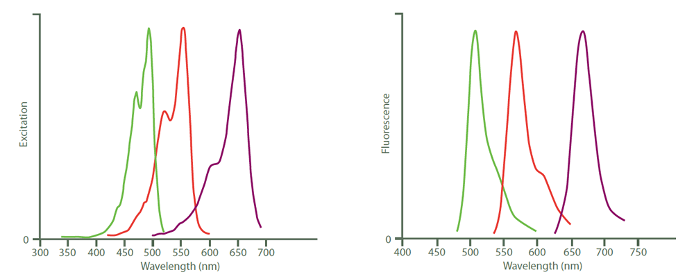
Figure 2. Excitation (left) and emission (right) spectra of Cy2 (green), Cy3 (red) and Cy5 (purple). Peak heights were normalized after the spectra were obtained with an M‑series spectrofluorometer system from Photon Technology International, Inc.
| Fluorophore | Excitation Peak (nm) | Emission Peak (nm) |
|---|---|---|
| Cyanine, Cy2 | 492 | 510 |
| Indocarbocyanine, Cy3 | 550 | 570 |
| Indodicarbocyanine, Cy5 | 650 | 670 |