TiterMax® adjuvants are used to produce cell mediated and humoral responses in research animals.
TiterMax® adjuvants were developed to meet the specific needs of investigators for immunoadjuvants that are at least as effective as Freund's Complete Adjuvant, safer and easier to use.
They are an attractive alternative to Freund's Complete Adjuvant for use by researchers in inducing antibody to diverse antigens.

The key to the potency of TiterMax® adjuvants lies both in the immunostimulatory activity of their components and in the fact that they form a stable water-in-oil emulsion.
TiterMax® adjuvants contain three essential ingredients: Proprietary block copolymers, squalene, a metabolizable oil, and a unique microparticulate stabilizer.

Like Freund's Complete Adjuvant, TiterMax® adjuvants can be used with a wide variety of antigens because they can entrap any antigen in a water-in-oil emulsion.
TiterMax® adjuvants aid in the antigens effective presentation to the immune system without the toxic effects of Freund's Complete Adjuvant.


Products

TiterMax® adjuvants are used to produce cell mediated and humoral responses in research animals.  TiterMax® adjuvants were developed to meet the specific needs of investigators for immunoadjuvants that are at least as effective as Freund’s Complete Adjuvant, safer and easier to use.  They are an attractive alternative to Freund’s Complete Adjuvant for use by researchers in inducing antibody to diverse antigens.  The key to the potency of TiterMax® adjuvants lies both in the immunostimulatory activity of their components and in the fact that they form a stable water-in-oil emulsion.  TiterMax® adjuvants contain three essential ingredients: Proprietary block copolymers, squalene, a metabolizable oil, and a unique microparticulate stabilizer.  Like Freund’s Complete Adjuvant, TiterMax® adjuvants can be used with a wide variety of antigens because they can entrap any antigen in a water-in-oil emulsion.  TiterMax® adjuvants aid in the antigens effective presentation to the immune system without the toxic effects of Freund’s Complete Adjuvant.

Products

TiterMax® is an adjuvant used to produce cell mediated and humoral responses in research animals. TiterMax® was developed to meet the specific needs of investigators for an immunoadjuvant that is at least as effective as Freund’s Complete Adjuvant, safer and easier to use. It is an attractive alternative to Freund’s Complete Adjuvant for use by researchers in inducing antibody to diverse antigens. The key to the potency of TiterMax® lies both in the immunostimulatory activity of its components and in the fact that it forms a stable water-in-oil emulsion. TiterMax® contains three essential ingredients: A proprietary block copolymer CRL-8941, squalene, a metabolizable oil, and a unique microparticulate stabilizer. Like Freund’s Complete Adjuvant, TiterMax® can be used with a wide variety of antigens because it can entrap any antigen in a water-in-oil emulsion. TiterMax® aids in the antigens effective presentation to the immune system without the toxic effects of Freunds Complete Adjuvant.

History

Freund’s Complete Adjuvant (FCA) has been used for over 50 years for producing antisera in animals. The severity of its toxicity was recognized immediately, but attempts to find a less toxic and equally effective alternative have not been successful. With advances in many areas of the biological sciences and increasing concern for the welfare of experimental animals, there was increased pressure to ban or restrict the use of FCA. In 1990, we introduced TiterMax®, which has the reliability and effectiveness of FCA without the toxic side effects.

After years of research to identify and characterize the scientific basis for adjuvant activity, Dr. Robert L. Hunter led the team which developed TiterMax®. It was designed to meet the specific needs of investigators for an immunoadjuvant that is at least as effective as FCA, but safer and easier to use. The key to the potency of TiterMax® lies both in the immunostimulatory activity of CRL-8941 and in its ability to form a stable water-in-oil emulsion. TiterMax® contains three essential ingredients:

Block copolymer CRL-8941
Squalene, a metabolizable oil
A unique microparticulate stabilizer

Like FCA, TiterMax® can be used with a wide variety of antigens because it can entrap any antigen in a water-in-oil emulsion.

History and Structure of Copolymers

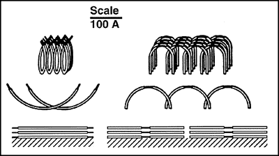
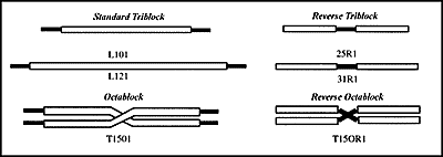
Robert L. Hunter, M.D., Ph.D., who led the team which developed TiterMax® #R-1 discovered the adjuvant action of nonionic block copolymers in the early 1980’s. The copolymers are composed of blocks or chains of hydrophilic polyoxyethylene (POE) and blocks of hydrophobic polyoxypropylene (POP) attached in one of four configurations (Figure 1). By varying the size of the blocks of POE and POP, dozens of preparations have been produced which span nearly the entire range of functional activities of available nonionic surfactants. The new copolymer CRL-8941 was developed specifically by CytRx Corporation to optimize the structural features essential to adjuvant activity while minimizing the inflammatory properties. The rationale for the synthesis of CRL-8941 evolved from the studies of Hunter and associates on the physicochemical parameters which contribute to adjuvant activity. These properties of such adjuvants are summarized here.

In initial studies with triblock copolymers, one of the preparations (L121) was found to serve as an adjuvant for stimulating antibody formation to BSA following injection in an oil-in-water emulsion. A second copolymer (L101) stimulated little antibody, but was more effective in inducing DTH and granulomas. The other triblock copolymers available at that time had lower adjuvant activity for either type of response. This demonstrated that small changes in the lengths of chains of POP and POE produced major changes in biologic activity and provided the foundation for further studies.

Figure 1. Structure of Block Copolymers.

The copolymers are composed of blocks of hydrophilic polyoxyethylene (POE, black) attached to blocks of hydrophobic polyoxypropylene (POP, white). The diagrams are drawn to scale for both length and thickness. Only one or two copolymers from each group is shown, but each copolymer family contains a series of members which differ in the length and relative proportion of POE vs. POP.

Adhesion and Adjuvant Activity

Hydrophile-lipophile balance (HLB) is a measure of the relative strength of the hydrophilic and hydrophobic activities of nonionic surface active agents which has been widely used to predict functional activities. The copolymers and other surfactants with adjuvant activity were found to have HLB values of less than 2 and were classified as spreading agents. Spreading agents are insoluble in water and do not emulsify or solubilize lipids or membranes. They are very different from common detergents which have HLB values greater than ten. They adhere to lipids and influence the interaction of soluble macromolecules with them. Hunter and associates demonstrated a correlation between the ability of copolymers to promote retention of soluble macromolecules on the surface of oil drops and their activity as adjuvants. The local concentration of antigen fixed to a surface is far higher than can be achieved by comparable amounts of material free in solution. Thus, the copolymers which were adjuvants produced a concentrated surface matrix which facilitated antigen presentation to cells of the immune system.

Copolymers which differ only in the lengths of chains of POP and POE demonstrate a great variety and intensity of immune and inflammatory responses. When a series of copolymers with similar low HLB values, but differing in the size of the component blocks and their mode of linkage, was tested, several patterns of immune response and inflammation were noted. The most effective adjuvants were the largest copolymers with hydrophilic POE on the ends (L121, L101, and T1501) Increasing the length of the POE chains even modestly caused the preparations to lose their activity as adjuvants. Shortening the length of the POP chains produced agents with lower adjuvant activity and greater toxicity as measured by inflammation at the site of injection. Placement of POE in the reverse configuration (at the center of the molecule flanked by POP) produced weak adjuvants which stimulated granulomatous inflammation. Interestingly, the intensity of the inflammatory responses did not correlate with antibody titers.

Analysis of the surfaces formed by selected copolymers and their interaction with plasma proteins provided interesting information (Figure 2). The copolymers all spontaneously form highly organized structures at an aqueous surface, and the heterogeneity of these structures contributes to the heterogeneity of their biologic activity. Thus, the copolymers which are adjuvants form a particular type of hydrophilic adhesive surface. If the blocks of POE are too long, the surface is not sufficiently adsorptive. If the blocks of POP are too short, they are unable to fold to produce a hydrophilic surface.

Figure 2. Model of surfaces of copolymers spread at varying concentrations.

Triblock copolymer L121 and reverse triblock copolymer 31R1 are drawn to scale with the POE portions black and the POP white. At low surface coverages, both copolymers lie flat producing largely hydrophobic surfaces (POP exposed). As the amount of copolymer on the surface increases, the hydrophobicity decreases demonstrating that the molecules become arranged with the POE oriented towards the surface. The surface of thick layers of L121 consists entirely of POE. It is hydrophilic and permits the underlying organizational tendencies of the copolymer to be expressed as fibers. The reverse copolymers cannot form hydrophilic surfaces because their POE portions are sterically inhibited. This accounts for their formation of spherical oil-like drops in saline. These orientations account, at least in part, for the biologic activity of the various copolymers.

Biologic Activities of Copolymers

Subsequently, it was found that copolymers with adjuvant activity not only bind antigen, but they activate macrophages as well. The ability to activate macrophages, however, was not sufficient to produce adjuvant activity. The antigen binding capacity of a given copolymer shows synergy with its potential to activate macrophages, suggesting that enhancement of antigen presenting capacity also plays a role in copolymer adjuvant activity. Finally, the adjuvant copolymers were shown to activate complement. This probably influences the localization and retention of antigen in lymphoid tissue and the activation of immunoreactive cells.

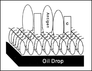
Figure 3. Model of an Immunogenic Particle.

The copolymer folds to form a hydrophilic adhesive surface. Antigen (BSA), complement (C) and mediators bound to the surface are presented to cells of the immune system in a particularly concentrated and effective form.

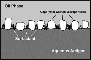
Figure 4. Model of Microparticle Stabilized Water-in-Oil Emulsion.

Microparticles potentiate emulsion stability and adjuvant activity while reducing the requirement for surfactants. Their localization at the interface between oil and aqueous compartments provides increased physical separation of adjacent droplets, retards the surface area which must be stabilized by surfactant, and reduces interfacial free energy. The orientation of the copolymer CRL-8941 on the surface of the microparticles is critical. It provides surface properties ideal for stabilizing water-in-oil emulsions. It also contributes to the expression of adjuvant properties by displaying the copolymer in a condensed fashion on bonded surfaces.

The TiterMax® formulation was developed to produce very stable emulsions, but the initial preparation of the emulsion is still highly dependent upon technique. Although TiterMax® emulsions can be prepared using any technique which works with Freund’s adjuvant, we recommend one of the procedures described in detail in the instruction sheet for TiterMax®. Procedures are available for the well-known two-syringe method, a micromethod for small volumes of antigen, as well as sonication and homogenization. The choice of method will depend on the equipment available and the amount of emulsion to be prepared. Transformation of your antigen/TiterMax® mixture into a water-in-oil emulsion can be tested by placing a drop on water.

Immunogens which contain high concentrations of surfactants or other materials may interfere with emulsification. We have found that SDS, which may be present in acrylamide gels, in concentrations greater than 1% or urea concentrations greater than 1.0 M significantly reduce the emulsifying capacity of TiterMax®. Other similar materials are likely to have the same effect. Some surface active agents serve as demulsifying agents which break emulsions. The modern emulsifiers and microparticulate stabilizer of TiterMax® are able to overcome the effects of most such agents present in moderate quantities. Glutaraldehyde does not affect the emulsion. TiterMax® is compatible with organic compounds and solvents such as DMF in moderate amounts.

Many of our customers ask about storing TiterMax® emulsions. A 50:50 water-in-oil emulsion can usually be stored at room temperature, 4° C, -20° C, or -70° C for as long as the antigen is stable. Upon storage, approximately 20% of the oil will dissociate from the emulsion. You may leave the emulsion in a syringe and simply reemulsify when ready to inject (provided the antigen is stable). TiterMax® emulsions have been repeatedly frozen and thawed or left at room temperature for several weeks with no loss of efficacy. Sodium azide or thimerosal which are present as preservatives in some antigen preparations will not affect the shelf life of TiterMax®, although at high concentrations of either, the emulsion could become toxic.

Examples of Successful Dosing Regimens
for TiterMax
® Classic


SPECIES INJECTION ROUTE TOTAL INJECTIONS VOLUME PER INJECTION SITE(S)
MICE IM 2  20 ul Each hind quadricep
SC 1  40 ul Base of tail
RATS IM 2  50 ul Each hind quadricep
GUINEA PIGS IM 2  50 ul Each hind quadricep
SC 4  50 ul Over both shoulders and both hind quadriceps
RABBITS IM 2  40 ul Each hind quadricep
SC 4 100 ul Over both shoulders and both hind quadriceps
ID 10  40 ul Along back
CHICKENS/ TURKEYS IM 2  50 ul Each breast
SC 1 100 ul Neck
CATS/DOGS IM 2 125 ul Each hind quadricep
SC 1 125 ul Along neck
RHESUS MONKEYS IM 2 100 ul Each hind quadricep
SC 4  50 ul Along inner thigh
GOATS/ SHEEP IM 4 250 ul 2 injections in each hind quadricep
COWS /HORSES SC 10 100 ul Along neck

Good immune responses have been achieved with Water to TiterMax® Classic ratios of 50:50 to 90:10, however the 50:50 water in oil emulsion is usually optimum.

*These are suggested routes and dosages that have proven successful and then reported to TiterMax USA, Inc. by numerous investigators. Dosing regimens that have been used with other adjuvants may certainly use TiterMax® Classic.

Introduction

After years of research to identify and characterize the scientific basis for adjuvant activity, CytRx Corporation released a research adjuvant, TiterMax®. It was originally designed to meet the specific needs of investigators for an immunoadjuvant that was at least as effective as Freunds Complete Adjuvant, but safer and easier to use. In 1996, CytRx Corporation released a new and improved water-in-oil adjuvant, TiterMax® Gold, which is now available for purchase.

The key to the potency of TiterMax® Gold lies in both the immunostimulatory activity of its new block copolymer, CRL-8300, and its ability to form a stable water-in-oil emulsion. TiterMax® Gold can be used with a wide variety of antigens which can be entrapped in a water-in-oil emulsion. Like TiterMax® Classic, TiterMax® Gold does not contain mineral oil, proteins, polysaccharides or other microbial products, thereby minimizing or eliminating the undesirable side effects caused by Freunds Complete Adjuvant.

Comparison Data

Procedures

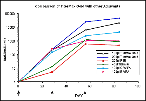
Studies were conducted using ovalbumin with various research adjuvants. Six to eight-week-old female C57BL/6 mice (10 per group) were immunized with a constant dose of 25 µg ovalbumin with each commercial adjuvant. Optimal dosing regimens for TiterMax® Gold were determined. Secondary immunizations were with antigen and adjuvant. All commercial adjuvants were prepared, injected, and boosted according to the manufacturers’ protocols. Serum samples were obtained at various times after immunization (days 28, 56, and 84) and IgG antibody titers against ovalbumin were determined using a microtiter plate ELISA. Data is expressed as units/ml against an in-house standard.

Results

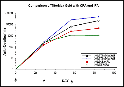
TiterMax® Gold injected intramuscularly, outperformed the other commercial adjuvants at all time points (days 28, 56 and 84). TiterMax® Gold was found to be effective following single and multiple immunizations. For polyclonal antibody production, we recommend immunizing mice with a 100 or 200 µl dose (TiterMax® Gold and antigen), injected intramuscularly into each hind quadricep.

For years researchers have been immunizing with Freunds Complete Adjuvant. Recently, many investigators have been searching for new adjuvants with less toxicities. We injected C57BL/6 mice with TiterMax® Gold, Freunds Complete Adjuvant, and Freunds Incomplete Adjuvant at Day 0. Mice initially injected with TiterMax® Gold were boosted with the same dose of TiterMax® Gold , while mice initially injected with FCA and IFA were boosted with IFA.

Results at day 28 were comparable. However, mice injected with TiterMax® Gold continued to have increasing titers at day 56 and 84, while the titers of mice injected with FCA and IFA began to level off.

Examples of Successful Dosing Regimens
for TiterMax
® Gold*


SPECIES INJECTION ROUTE TOTAL INJECTIONS VOLUME PER INJECTION SITE(S)
MICE IM 1 100 ul Hind quadricep
SC 1 100 ul Base of tail or neck
RATS IM 2 100 ul Each hind quadricep
GUINEA PIGS IM 2 100 ul Each hind quadricep
SC 4 100 ul Over both shoulders and both hind quadriceps
RABBITS IM 2 100 ul Each hind quadricep
SC 4 200 ul Over both shoulders and both hind quadriceps
CHICKENS/ TURKEYS IM 2 100 ul Each breast
SC 1 200 ul Neck
CATS/DOGS IM 2 200 ul Each hind quadricep
SC 1 200 ul Along neck
RHESUS MONKEYS IM 2 200 ul Each hind quadricep
SC 4 100 ul Along inner thigh
GOATS/ SHEEP IM 4 400 ul 2 injections in each hind quadricep
COWS /HORSES SC 10 200 ul Along neck

Good immune responses have been achieved with Water to TiterMax® Gold ratios of 50:50.

*These are estimated routes and dosages that have proven successful with the original formulation of TiterMax®. Dosing regimens that have been used with other adjuvants may certainly use TiterMax® Gold.

Technical Information

Freund’s Complete Adjuvant (FCA) has been used for over 50 years for producing antisera in animals. The severity of its toxicity was recognized immediately, but attempts to find a less toxic and equally effective alternative have not been successful. With advances in many areas of the biological sciences and increasing concern for the welfare of experimental animals, there is increased pressure to ban or restrict the use of FCA. In 1990, we introduced TiterMax®, which has the reliability and effectiveness of FCA without the toxic side effects.

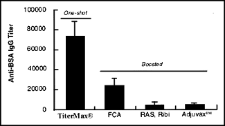
Now, data from an independent testing laboratory confirm that a single injection of TiterMax ®can produce significantly higher titers than two or more injections of antigen in other commercially available adjuvants. Based on our experience, TiterMax®can be expected to perform at least as well as FCA with almost any antigen. With many antigens, TiterMax®performs significantly better. Thus, it is an attractive alternative to FCA.

After years of research to identify and characterize the scientific basis for adjuvant activity, Dr. Robert L. Hunter led the team which developed TiterMax®. It was designed to meet the specific needs of investigators for an immunoadjuvant that is at least as effective as FCA, but safer and easier to use. The key to the potency of TiterMax®lies both in the immunostimulatory activity of CRL-8941 and in its ability to form a stable water-in-oil emulsion. TiterMax® contains three essential ingredients:

Block copolymer CRL-8941
Squalene, a metabolizable oil
A unique microparticulate stabilizer

Like FCA, TiterMax® can be used with a wide variety of antigens because it can entrap any antigen in a water-in-oil emulsion.

Figure 1. Effectiveness of various adjuvants in rabbits.

Rabbits were immunized with 50 µg of bovine serum albumin (peptide conjugated) in various adjuvants according to the manufacturers’ instructions. The TiterMax®group was not boosted, while the others were boosted according to the recommended procedures. Serum IgG antibody titers (mean±sem) were measured by ELISA after 8 weeks.

How Non-Toxic is TiterMax®?

Since TiterMax®#R-1 contains no mycobacteria or mineral oil, one would not expect it to induce adjuvant arthritis or the severe systemic granulomatous reactions characteristic of FCA. In our studies, we have seen no evidence of such reactions.

This included many years’ experience with block copolymer adjuvants and microparticulate stabilized emulsions in addition to work with the TiterMax®#R-1 formulation, including work done at an independent testing laboratory (Hazleton). TiterMax®has been optimized for induction of IgG antibody and has little ability to stimulate delayed type hypersensitivity reactions which are responsible for the most serious complications of FCA. Most of the studies conducted in development of the TiterMax® #R-1 formulation utilized subcutaneous foot pad injections in mice specifically to facilitate quantitative assessment of local reactions.

On several occasions we have observed inflammatory reactions which begin at two to three weeks after injection, intensify for a week and then gradually subside. They have the characteristics of local antigen-antibody-complement (Arthus) reactions rather than the delayed type hypersensitivity granulomas characteristic of FCA. With superficial subcutaneous injection, the reactions may resemble an abscess and drain. The most severe reactions were seen following injection of a total volume of 1 ml of adjuvant distributed equally among 4 sites of a 400 gram animal. Only mild reactions were observed when the same antigen was injected in doses of 20 to 50 µl per site. Other severe local reactions were observed in animals following injection of TiterMax®plus E. coli lipopolysaccharide (endotoxin). Consequently, TiterMax® does not counteract and may exacerbate the toxicity of some antigens.

While TiterMax®has been remarkably non-toxic in our hands, its production of high antibody titers may make the animals susceptible to localized Arthus reactions at the site of immunization, particularly when boosting. This can be minimized without compromising titers by injecting the antigen emulsion in multiple sites using small volumes. Many investigators find that TiterMax®produces highly acceptable titers without boosting. If boosting is desired, however, it is advisable to measure the antibody titer first. If it is high, then boosting should be done with soluble antigen without adjuvant and/or distributed among several sites.

TiterMax® contains no mineral oil and no protein, polysaccharide or other microbial products. In order to achieve successful immunization, the antigen must be delivered to the lymph node together with the adjuvant. The most effective way to accomplish this is to incorporate both into a water-in-oil emulsion. TiterMax® has been especially formulated to maximize the desirable effects of water-in-oil emulsions while minimizing or eliminating the undesirable side effects of FCA. Table 1 summarizes the composition of several commercially available adjuvants.

Adjuvant Emulsion Type Incorporation Immunomodulator
TiterMax®* water-in-oil encapsulation in oil with long retention copolymer CRL-8941
Freund’s Complete Adjuvant water-in-oil encapsulation in oil with long retention Killed whole Mytobacteria
Ribi Adjuvant Systems oil-in-water adsorption to oil with brief retention Mycobacterial product (TDM) and/or endotoxin product (MPL)
Lipovant(TM) oil-in-water adsorption to oil with brief retention lipid
Adjuvax(TM) none incorporation into polysaccharide matrix with brief retention glucan polysaccharide
Alhydrogel(TM) none adsorption to aluminum hydroxide none


*TiterMax® contains no mineral oil and no protein, polysaccharide, or other microbial products.

Adjuvants influence the titer, duration, isotype and avidity of antibody in addition to their well-known effects on cell mediated immunity. Recent investigations have shown that their influence extends even to inducing class I-restricted CD-8 positive cytotoxic T lymphocytes and modulating the specificity of antibody among available epitopes on protein antigens. It has been known for many years that FCA preferentially induces antibody against epitopes on denatured proteins while other adjuvants preferentially stimulate antibody against epitopes on native proteins. B epitope selection by adjuvants is seldom considered in experimental protocols even though it has been correlated with biologic activity such as protection against simian immunodeficiency virus. TiterMax®was designed to maximize production of high avidity antibody titer representing the spectrum of IgG subclasses while minimizing the undesirable induction of granulomatous delayed-type hypersensitivity.

Comparison Data

Procedures

Studies were conducted with various research adjuvants. Six to eight-week-old female C57BL/6 mice (10 per group) were immunized with a constant dose of 25 µg ovalbumin with each commercial adjuvant. Optimal dosing regimens for TiterMax® Goldwere determined. Secondary immunizations were with antigen and adjuvant. All commercial adjuvants were prepared, injected, and boosted according to the manufacturers’ protocols. Serum samples were obtained at various times after immunization (days 28, 56 and 84) and IgG antibody titers against ovalbumin were determined using a microtier plate ELISA. Data is expressed as units/ml against an in-house standard.

Results

Optimizing Immunization with TiterMax®

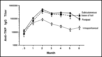
Effects of injection route on efficacy ofTiterMax®

Figure 1 shows the time course of anti-TNP antibody titers achieved using TiterMax®in mice immunized with a single dose of antigen using one of three routes: subcutaneous at the base of the tail, intraperitoneal, or intraplantar in one hind footpad. The data show that TiterMax®works well without boosting by several injection routes, with the intraplantar route superior. Four weeks after a single injection, titers of >100,000 were readily achieved. The subcutaneous route yielded a titer of >60,000, while the intraperitoneal route achieved titers >40,000. After eight weeks, the intraplantar route had produced titers >500,000, with the subcutaneous injection yielding titers >300,000 and the intraperitoneal route producing antibody titers >80,000. In general, during the first month after immunization, both the titers and the slope of their rise were comparable, regardless of route. However, the intraperitoneal route did not produce the persistent titers reproducibly seen with the other routes, probably because of shortened survival of the antigen depot.

Figure 1. Effects of Injection Route on Efficacy of TiterMax®#R-1.

Mice were immunized with a single dose of TNP-HEA (50 µl) in TiterMax® (total volume 40 µl) using one of three routes. Sera were collected at intervals and antibody titers were measured via ELISA. Antibody titers are expressed as mean ± sem. Immunization with multiple small volumes ofTiterMax®/antigen emulsion.

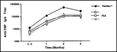
FCA is frequently injected in multiple subcutaneous sites on the back and flank of animals in order to increase titers and reduce local inflammation. Although we saw essentially no signs of inflammation with TiterMax® in any of the experiments described here, we evaluated protocols using antigen with TiterMax®in multiple injection sites. These might be useful for investigators using more toxic antigens. In the experiment shown in Figure 2, animals injected with identical volumes of TNP-HEA distributed over four subcutaneous sites gave higher antibody titers than those in which the antigen was distributed between only two sites. Thus, distributing antigen among several sites may facilitate immunization with TiterMax®.

Figure 2. Effect of Number of Injection Sites on Antibody Titer.

Mice were immunized with a single dose of TNP-HEA (50 µg) in TiterMax®(total volume 40 µl). The antigen emulsion was injected subcutaneously into either two or four flanks. Sera were collected at intervals and antibody titers (mean ± sem) were measured via ELISA.

Characterization of Immune Response Induced with TiterMax®

IgG antibody subclass distribution

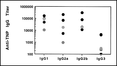
Figure 3 illustrates the IgG subclass distribution of anti-TNP antibody using TiterMax® in mice immunized with a single dose of antigen injected in one hind footpad. Sera were collected after 28 days and antibody titers were measured. Unlike the IgG1 predominance seen with FCA, TiterMax® produced considerable proportions of the IgG2a and IgG2b subclass as well. Interestingly, in these studies with outbred mice, the range of IgG2a responses was broader than that of the other subclasses, suggesting a stronger genetic component to the regulation of IgG2a production. Protection against a variety of infectious agents appears to be subclass-dependent, with the IgG2 isotypes frequently implicated in effective immunity.

Figure 3: IgG Subclass Distribution Following TiterMax®/TNP-HEA Immunization.

Mice were immunized with a single dose of antigen in TiterMax® injected in one hind footpad. Plasma samples were collected 28 days later and antibody titers were measured via ELISA. Each point represents data from an individual animal.

Studies on delayed-type hypersensitivity

TiterMax® has not been extensively evaluated for induction of delayed-type hypersensitivity (DTH), although the nature of the adjuvant and preliminary data strongly suggest that it preferentially stimulates humoral immunity. In one experiment to evaluate DTH, three Hartley guinea pigs were immunized with 100 µg TNP-HEA in TiterMax® (80 µl intramuscularly, divided between the hind flanks). Animals were boosted on day 28 with 100 µl soluble TNP-HEA (intramuscularly, divided between the hind flanks). Skin tests were performed by injection TNp-HEA (50 µg in 50 µl intradermally) six days and, into different sites, 14 days later. After 24 hrs, no site injected on day six showed erythema or induration, while those injected on day 14 showed only about 1 cm. of erythema. After 48 hrs, all sites injected on day six remained negative, no induration. Antibody titers on day 14 were 45,299±3,261 (mean ± sem). In other experiments, we have failed to induce adjuvant arthritis in Lewis rats under conditions where all of the control FCA-treated rats succumb.

Role of Boosting in TiterMax® Immunizations

TiterMax® can produce high titers without boosting

The duration of an antibody response has been related to the persistence of antigen. One would predict that an adjuvant such as TiterMax® which forms stable depots of water-in-oil emulsion in tissue might produce long-lasting responses. In addition, it should facilitate the generation of an antibody response with only a single injection of antigen. This was found to be the case with TiterMax®(Figure 4). Whether mice were injected with only a single dose of TNP-HEA in TiterMax®, or boosted after 28 days, the antibody titers reached high levels by six weeks. In unboosted animals the titers began to decline after a few months, but they rose again after seven months and, at one year, were comparable to levels in boosted animals.

Figure 4. High, Long-Lasting Titers Can Be Achieved with TiterMax®without boosting.

Mice were immunized with 100 μg of TNP-HEA in TiterMax®(80 μl, split between two hind footpads). One group was boosted with 100 μl of TNP-HEA in TiterMax®(80 μl, split between two hind footpads), while the other group was not. Antibody titers (mean ± sem) were measured via ELISA.

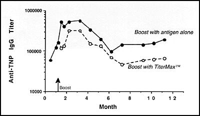
Figure 5. High, Long-Lasting Titers Can Be Achieved with TiterMax®When Boosted with Soluble Antigen.

Mice were immunized with 50 μg of TNP-HEA in TiterMax®(40 μl, split among the four footpads). On day 35, one group was boosted with 25 μg of TNP-HEA in TiterMax®(20 μl, split between two hind footpads), while the other group was boosted with 25 μg of TNP-HEA in saline (20 μl, split between two hind footpads). Antibody titers (mean ± sem) were measured via ELISA.

Boosting may be accomplished with soluble antigen

We evaluated the relative efficacy of boosting with soluble antigen vs. boosting with antigen in TiterMax®in the following experiment. Mice were injected with TNP-HEA and after 35 days they were boosted with either antigen in saline or antigen in TiterMax®emulsion. Interestingly, antibody titers showed a transient depression after boosing with antigen in TiterMax®. However, both groups showed strong titers which persisted throughout the six months of the experiment (Figure 5).

Efficacy of TiterMax® in Various Species

Antibodies to TNP

The efficacy of TiterMax® in various species was evalutated by measuring antibody titers 14 and 28 days following a single immunization. Groups of mice received 50 μg of TNP-HEA emulsified with 20 μl of TiterMax® in a total volume of 40 μl injected subcutaneously at the base of the tail. Rats, guinea pigs, and rabbits each received 100 μg of antigen emulsified with 40 μl of TiterMax®in a total volume of 80 μl equally distributed between two sites (except for the intraperitoneal route). Rabbits and guinea pigs received intramuscular flank injections while rats received intraplantar, subcutaneous, or intraeritoneal injections. The results are summarized in Table 3. All four species produced significant antibody titers, although the magnitude and duration of the response varies with species and route of immunization. The antibody titers of both rabbits and mice increased 5 to 6-fold between days 14 and 28, but were essentially unchanged in rats and decreased in guinea pigs. In rats, the intraplantar route was superior to either the subcutaneous or intraperitoneal routes. Thus TiterMax®was effectuve in four different species of experimental animals. Immunization protocols should be tailored to each experimental system.

Table 3. Efficacy of TiterMax® in Various Species

Injection
Anti-TNP Titerc
Local
Speciesa Routeb Day 14 Day 28 Toxicity
mice subcutaneous base of tail 23,857±3,944 130,374±22,825 None
rats intraplantar 2 hind foot pads 43,998±7,168 48,997±18,227 1 mm swelling & erythema
rats intraperitoneal 11,866±3,433 10,201±4,368 None
rats subcutaneous 2 hind flanks 4,335±873 4,584±984 None
guinea pigs intramuscular 2 flanks 45,228±3261 6,388±1,385 None
rabbits intramuscular 2 flanks 26,262±9,554 158,892±33,016 None

a Number of animals per group: 5 mice, 4 rats per route, 2 guinea pigs, 3 rabbits
b Mice received a total volume of 40 µl; all other groups received 80 µl
c Antibody titers (mean ± sem) were measured using an ELISA

Antibodies to BSA

Studies performed by Hazleton Laboratories evaluated TiterMax®against several other commercially available adjuvants in rabbits, mice, and goats immunized with LHRH-BSA. IgG antibody titers against BSA are shown in Table 4. Eight weeks after immunization, mice who recieved TiterMax®had 1.5-fold higher antibody titers than those who received FCA, while rabbits had 3.5-fold higher titers. Goats showed the highest antibody titers with FCA, but in all three species, TiterMax®induced at least 4.5-fold higher titers than either Adjuvax(TM) or RAS.

Table 4. Anti-BSA Titers in Different Species

Species TiterMax® FCA Adjuvax(tm) RAS
Rabbits 71,000 ± 16,921 18,950 ± 8,900 3,400 ± 1,482 3,283 ± 2,588
Mice 50,000 ± 16,202 34125 ± 11,091 163 ± 41 11,528 ± 6,748
Goats 1,895 ± 899 15,250 ± 6,263 100 ± 0 355 ± 120

Groups of experimental animals were immunized with a total of 50 µl LHRH-BSA in several adjuvants according to the manufacturers’ protocols. All groups were injected on day 1 and boosted on day 28 (TiterMax®and FCA groups), day 21 (RAS group) or day 21 and 35 (Adjuvax(tm) group). The second injection in the FCA group was with incomplete Freund’s adjuvant. Results from week 8 are shown. IgG antibody titers (mean ± sem) against the carrier BSA were measured using an ELISA.

Antibodies to peptide

Antibody activity against the LHRH peptide was measured using a radioimmunoassay with labeled peptide. Table 5 shows that immunization with TiterMax® induced anti-peptide titers comparable to FCA in all three species.

Table 5. Antibody to LHRH Peptide in Different Species

Species TiterMax® FCA Adjuvax(tm) RAS
Rabbits 61 ± 4.0 63 ± 3.7 5 ±1.7 18 ± 12.6
Mice 48 ± 11.0 57 ± 10.2 31 ± 7.5 34 ± 3.8
Goats 46 ± 6.4 60 ± 5.1 5 ± 0.6 12 ± 1.0

Groups of experimental animals were immunized as described in Table 4. Results using sera from week 8 are shown. Antibody activity against the LHRH peptide was measured using a radioimmunoassay with labeled peptide. Data (mean ± sem) for the percent peptide bound using a serum dilution (final concentration 1:200 for rabbits; 1:100 for mice; 1:5 for goats) in the linear portion of the dose-response curves are shown. TiterMax®induced anti-peptide titers were comparable to FCA in all three species.

Can TiterMax® be used to make Hybridomas?

Principles

Many investigators have asked us for a TiterMax® immunization protocol suitable for priming spleen cells for infusion with myeloma cells to produce monoclonal antibodies. There is no standard protocol for monoclonal antibody production; many different methods have been effective. Furthermore, monoclonal antibody protocols do not depend on the adjuvant except insofar as the adjuvant is used to increase the number of antibody-producing cells. TiterMax®can be expected to work with any antigen which has been successfully used in immunizations with FCA. For weak antigens or antigens available in minute amounts, the immunization protocols must be adapted so that the animals are producing detectable antibodies prior to fusion, a prerequisite for success. While no protocol can be written to anticipate every immunogen of interest, certain principles related to immunization can be considered in fine-tuning a general protocol for a specific immunogen.

Factors to consider in choosing the immunogen include the degree of purity, whether denaturation will impact the desired application, and the need to chemically aggregate small molecules or attach them to carriers to enhance their immunogenicity. These are beyond the scope of this document, but detailed information can be obtained from review articles, monoclonal antibody directories, or specific publications about monoclonal antibodies against immunogens similar to the one of interest.

The choice of animal species and strain as immune spleen cell donor for fusion depends on the myeloma cell line available and the origin of the immunogen. Mice are most commonly used, with the BALB/c strain preferred since most murine myelomas competent for fusion were derived from that strain. If low response to a particular antigen in BALB/c mice requires immunization of other inbred strains, the hybridomas can be propagated in the appropriate F1 hybrid. Rats (LOU/C or LOU/M strain) are used when monoclonal antibodies against murine antigens are desired, or to produce large amounts of ascites. Hamsters have been used to produce monoclonal antibodies against mouse antigens to which rats do not respond. In specialized cases, protocols have been designed to eliminate unwanted monoclonal antibodies against particularly immunogenic epitopes by treating the animals with cytotoxic agents immediately after the initial immunization.

Immunization procedures used by various investigators to produce monoclonal antibodies differ widely. Immunization is started by injecting 1-125 µg antigen per mouse or rat (in groups of 5 to 10) either intraperitoneally or subcutaneously. Soluble antigens have usually been administered with an adjuvant such as FCA. The animals are boosted after 10 days to three weeks, generally with the same dose as used in the original immunization. Multiple booster injections (as many as seven times weekly or every other week) have been used with weak antigens. Most protocols boost with antigen in incomplete Freund’s adjuvant, although some investigators have used multiple injections with FCA. This will result in severe inflammatory reactions and is to be discouraged.

Five to seven days following a booster immunization, serum may be tested for antibodies. This guides the investigator in deciding how long to pursue the immunization. It also ensures that the screening assay which will be used to screen the hybridoma supernatants is perfected.

Animals that show the highest antibody titers are selected for further immunization. At least three to four weeks after the last boost and three to four days before fusion with the myeloma cells, the animals are challenged with intraperitoneal or intravenous injection of antigen in aqueous solution to stimulate dividing plasma cell blasts in the spleen. Some investigators use two intraperitoneal injections at day -4 and -3, followed by intravenous injection on day -2; others barrage with 1/5 to 1/10 the original dose of immunogen on each of the four days before fusion. Most investigators administer a single intravenous antigen dose on day -3 or an intraperitoneal injection on day -4 followed by an intravenous injection on day -3. In some situations, adoptive transfer may be used to enrich antibody cells before the fusion. In that case, spleen cells from immunized mice are transferred into lethally irradiated (750 R) recipients together with antigen. The donor spleen cells repopulate the recipient spleen and four to six days after transfer the recipient spleen contains a high proportion of antigen-specific antibody forming cells suitable for fusion with the myeloma cell line.


Suggested Immunisation Protocol for Hybridomas

Do not perform with myeloma cells until a satisfactory antibody titer has been achieved.

  1. Prepare emulsion of equal volumes of antigen (300 ug to 2.5 mg/ml for protein antigens) and TiterMax®#R-1 Research Adjuvant according to the TiterMax® instruction brochure.
  2. Day 0. Immunize a group of 5 to 10 two-month old BALB/c mice by injecting 50 ul of the antigen-TiterMax®emulsion containing 15-125 ug antigen subcutaneously in two sites at the base of the tail (25 ul into each) using a 27 gauge hypodermic needle. Alternatively, the antigen emulsion may be injected intradermally into each hind footpad (25 ul into each).For relatively strong immunogens, a single immunization withTiterMax® will result in high titers of high avidity antibody 30 to 40 days after a single immunization. With weaker antigens, variable extents of boosting may be necessary.
  3. Day 14. (Optional depending on antigen). Repeat immunization with antigen-TiterMax® emulsion.Seven days after boost or 30-40 days after single immunization. Collect samples of serum from each animal. Do not pool them. Assay each for antibody titer to the relevant antigen using the same assay that will be used for screening the hybridoma supernatants.If antibody titers are low (<1:100 by most assay procedures), repeat immunization with antigen in TiterMax® emulsion. Collect serum 7 days later and assay for antibody titer.If fusing lymph node cells with the myeloma cells, lymph nodes may be harvested whenever an adequate antibody titer has been achieved.If fusing spleen cells (more common) the animals must be boosted with soluble antigen to increase the proportion of antibody-producing cells in the spleen as described in step 5.
  4. 3 to 4 weeks after last boost and 5 days before fusion with myeloma cells. Select 2 to 3 animals with the highest antibody titers. Inject intraperitoneally with 20 to 100 µg of soluble antigen on day 1, inject intravenously with the same amount of antigen on day 2, and harvest spleens on day 5. Prepare cells for fusion and proceed according to desired fusion, screening, cloning and propagation protocols. Production of high affinity antibodies may be enhanced by prolonging the period between the last boost and pre-fusion challenge and by challenging with lower doses (2-10 µg of antigen).

Methods of Emulsification

TiterMax®Research Adjuvant combines the benefits of a potent synthetic adjuvant (copolymer CRL-8941) with those of a microparticulate-stabilized, water-in-oil emulsion containing a metabolizable non-toxic oil, squalene. Emulsions of TiterMax®can be prepared using any technique which works with Freund’s Complete Adjuvant (FCA). There are several methods from which you may choose to emulsify your antigen, depending on the equipment you have available and the volume of emulsion you are going to prepare. The two methods we find to be the best in terms of simplicity and recovery of emulsion are the two syringe, double hub emulsifying needle method and the Kontes Pellet Pestle® homogenizer method. The latter is especially suited to small volumes. A number of other methods suggested by recent TiterMax® users are also described. Each of these methods will produce stable water-in-oil emulsions in 1 to 5 minutes.

Stable water-in-oil emulsions are notoriously difficult to make. The TiterMax® formulation was developed to produce a very stable emulsion. TiterMax®was developed to meet the specific needs of investigators for an immunoadjuvant that is at least as effective as Freund’s Complete Adjuvant (FCA), and safer and easier to use. Like FCA, TiterMax® can be used with a wide variety of antigens. Please follow the step-by-step instructions carefully for the method you choose.


Stability of the TiterMax®-in-Oil Emulsion
To test whether your TiterMax® emulsion is ready to use, expel a tiny drop onto the surface of water. It should expel from the syringe with a consistency similar to whipped cream and should hold together on the surface of water. If you are preparing a small volume of emulsion, you may touch the tip of a pipette or applicator stick to your emulsion preparation and submerge it into water. Either way, the emulsion should hold together. In the event that the emulsion disperses on or in the water, reconnect the syringe (or repeat small volume procedure) and emulsify for another minute.


Stability of the Emulsion after Prolonged Storage
A 50:50 water-in-oil emulsion can usually be stored at room temperature, 4° C , -20° C or -70° C for as long as your antigen is stable. Upon storage, approximately 20% of the oil will disassociate from the emulsion. You may leave the emulsion in a syringe and simply re-emulsify when you are ready to use again for injecting. The stability of your antigen must be considered to do this. TiterMax® emulsions have been repeatedly frozen and thawed or left them at room temperature for several weeks.


Reagents that May Interfere with Emulsification or Stability
Immunogens which contain high concentrations of surfactants or other materials may interfere with emulsification. We have found that SDS, which may be present in acrylamide gels, in concentrations > 1% or urea in concentrations > 1.0 M significantly reduces the emulsifying capacity of TiterMax®. Other similar materials are likely to have the same effect. Some surface active agents serve as demulsifying agents which break emulsions. The modern emulsifiers and microparticulate stabilizers of TiterMax® are able to overcome the effects of most such agents present in moderate quantities.

Method 1: Two-Syringe, Double Hub Needle

This method is suitable for emulsion volumes between 1 ml and 10 mls. Emulsion recovery is approximately 80 to 90% when preparing volumes of 1 to 10 mls and approximately 50 to 60% when preparing volumes of <0.5 ml.

Materials:

TiterMax®Research Adjuvant
Two 3.0 ml all-plastic syringes
One 18 gauge needle for withdrawing TiterMax® from the vial, or use syringe without needle or positive displacement precision pipette if you open entire vial
One 18 gauge double hub emulsifying needle
Antigen in saline or other suitable fluid (typical dose range in mice is 15 to 125 µg/mouse)

Procedure:

NOTE: Prior to preparation of a TiterMax® water-in-oil emulsion, warm the TiterMax® to room temperature and vortex for 30 seconds. Make sure the TiterMax®is a homogeneous suspension of copolymer-coated microparticles before proceeding to emulsify by any method.

For 1 ml of a 50:50 water-in-oil emulsion you will need 0.5 ml of the aqueous antigen and 0.5 ml of TiterMax® .

  1. After TiterMax®has been vortexed, load a syringe with 0.5 ml TiterMax® . Load the second syringe with 0.25 ml of antigen in aqueous medium. Set aside the other 0.25 ml of antigen. NOTE: It is important to add the aqueous antigen phase to the TiterMax® in at least 2 small volumes.
  2. Connect the two syringes via an 18 gauge double hub emulsifying needle. Mix the TiterMax® with the antigen by forcing the materials back and forth through the needle for approximately 1 minute. NOTE: It is important to push the antigen into the TiterMax®syringe first, so that the aqueous phase enters the oil phase rather than vice versa. Hold the syringes carefully so that they do not come apart from the double hub needle during emulsification. The formation of a water-in-oil emulsion is signaled by a sudden increase in viscosity, i.e., more force is required to move the material through the needle. NOTE: After ~ 1 minute a whipped-cream-like water-in-oil emulsion forms. Push all of the emulsion into one syringe and disconnect the empty syringe.
  3. Load the empty syringe with the remaining 0.25 ml aqueous antigen solution. Reconnect the syringes and emulsify for another 30 to 60 seconds. NOTE: Again, first push the antigen into the water-in-oil emulsion. Care must be taken in holding the syringes together since the oil may lubricate and loosen the connection. That is why it is preferable to use a lock tip syringe. Push all of the emulsion into one syringe and disconnect the empty syringe. If necessary, load the syringe you have chosen for injecting your species of animals.
  4. To test for stability, place a drop of emulsion on water.

Precautions:

The syringes should be all plastic or siliconized glass. Plastic syringes with rubber pistons contain a lubricant which fails in the presence of TiterMax® and causes the syringes to stick. Use caution not to loosen the syringes from the double hub needle during emulsification. This will cause you to lose the emulsion.

Method 2: Two-Syringe, 3-Way Stopcock

This method is suitable for emulsion volumes between 1 ml and 10 mls. Available 3-way stopcocks have larger bores than the 18 gauge double hub needles (Method 1) so that emulsification takes longer and the syringes connected to 3-way stopcocks are often more difficult to hold. Recovery of emulsion is approximately 70 to 80%.

Materials:

TiterMax®Research Adjuvant
Two 3.0 ml all plastic or siliconized glass syringes (preferably lock tip)
One 18 gauge needle for withdrawing TiterMax® from the vial, or syringe without needle or positive displacement precision pipette if you open entire vial
One 3-way plastic disposable or stainless steel reusable stopcock
Antigen in saline or other suitable fluid (typical dose range in mice is 15 to 125 µg/ mouse)

Procedure:

NOTE: Prior to preparation of a TiterMax®water-in-oil emulsion, warm the TiterMax® to room temperature and vortex for 30 seconds. Make sure the TiterMax® is a homogeneous suspension of copolymer-coated microparticles before proceeding to emulsify by any method.

For 1 ml of a 50:50 water-in-oil emulsion you will need 0.5 ml of the aqueous antigen and 0.5 ml of TiterMax®.

  1. After TiterMax®has been vortexed, load a syringe with 0.5 ml TiterMax®. Load the second syringe with 0.25 ml of antigen in aqueous medium. Set aside the other 0.25 ml of antigen. NOTE: It is important to add the aqueous antigen phase to the TiterMax® in at least 2 small volumes.
  2. Connect the two syringes via a 3-way stopcock. Mix the TiterMax® with the antigen by forcing the materials back and forth through the stopcock for approximately 1 minute. NOTE: It is important to push the antigen into the TiterMax®syringe first, so that the aqueous phase enters the oil phase rather than vice versa. Hold the syringes carefully so that they do not come apart from the 3-way stopcock during emulsification. NOTE: After approximately 1 minute a whipped-cream-like water-in-oil emulsion forms. Push all of the emulsion into one syringe and disconnect the empty syringe.
  3. Load the empty syringe with the remaining 0.25 ml aqueous antigen solution. Reconnect the syringes and emulsify for another 30 to 60 seconds. NOTE: Again, first push the antigen into the water-in-oil emulsion. Care must be taken in holding the syringes together since the oil may lubricate and loosen the connection. It is preferable to use a lock tip syringe. Push all of the emulsion into one syringe. Disconnect the empty syringe and connect the syringe you have chosen for injecting for filling. Alternatively, simply disconnect the full syringe and add the appropriate needle for injecting animals.
  4. To test stability, place a drop of emulsion on water.

Precautions:

The syringes should be siliconized glass or all plastic. Plastic syringes with rubber pistons contain a lubricant which fails in the presence of TiterMax® and causes the syringes to stick. Use extreme caution during emulsification so that you do not loosen the syringes from the 3-way stopcock. This will cause you to lose the emulsion.

Method 3: One Syringe, Blunt Needle

This method is useful for volumes less than 0.5 ml and has been used with volumes as low as 0.05 ml final emulsion volume. Recovery of emulsion is approximately 50 to 75%.

Materials:

TiterMax®Research Adjuvant
1 ml all-plastic syringe
18 gauge x 1.5 inch blunt needle for emulsifying
1.5 ml conical bottom plastic centrifuge tube
One 18 gauge needle for withdrawing TiterMax® from the vial, or syringe without needle or positive displacement precision pipette if you open entire vial
Antigen in saline or other suitable fluid (typical dose range in mice is 15 to 125 µg/ mouse)

Procedure:

NOTE: Prior to preparation of a TiterMax® water-in-oil emulsion, warm the TiterMax® to room temperature and vortex for 30 seconds. Make sure the TiterMax®is a homogeneous suspension of copolymer-coated microparticles before proceeding to emulsify by any method.

For 200 µl of a 50:50 water-in-oil emulsion you will need 100 µl of the aqueous antigen and 100 µl of TiterMax®.

  1. Grasp the pointed end of the needle with pliers and gently bend it back and forth until the tip breaks off producing a 1 inch blunt end needle. Attach the blunt 18 gauge needle to a 1 ml all-plastic syringe.
    Add 100 µl of TiterMax®adjuvant to the 1.5 ml centrifuge tube.
  2. Add 50µl of your antigen solution. The antigen-adjuvant mixture is drawn into the syringe and expressed back into the tube several times until a thick white emulsion forms. Add the remaining 50µl of your antigen solution and repeat the process. NOTE: Certain technical points are important. The air drawn into the syringe during the process does not impede the emulsification process. In approximately 1 minute, the entire material will be transformed into a water-in-oil emulsion. If one is careful not to smear the material on the sides of the tube, it can be drawn almost quantitatively into the syringe (using the 18 gauge needle). If you get emulsion on the sides of the tube, centrifuge at low speed (100 x g) for 2 minutes to pellet the emulsion. Remove the blunt needle and replace with a suitable needle for injecting.
  3. To test stability, place a tiny drop of emulsion on or in water.
Method 4: Emulsifying in Syringe Using Sonication

This method is suitable for emulsion volumes between 0.5 ml and 2 ml. Recovery of emulsion is approximately 80 to 90%.

Materials:

TiterMax®Research Adjuvant
One 18 gauge needle for withdrawing TiterMax® from the vial, or syringe without needle or positive displacement precision pipette if you open entire vial
One syringe for preparation of emulsion and immunization; piston removed; tip sealed with Parafilm®
Parafilm® for sealing tip of syringe
Antigen in saline or other suitable fluid (typical dose range in mice is 15 to 125 µg/ mouse)
Sonic dismembrator with microtip

Procedure:

NOTE: Prior to preparation of a TiterMax® water-in-oil emulsion, warm the TiterMax® to room temperature and vortex for 30 seconds. Make sure the TiterMax®is a homogeneous suspension of copolymer-coated microparticles before proceeding to emulsify by any method.

For 1.0 ml of a 50:50 water-in-oil emulsion you will need 0.5 ml of the aqueous antigen and 0.5 ml of TiterMax®.

  1. Carefully seal the tip of the syringe with Parafilm®. After TiterMax®has been vortexed, load a 2.0 ml syringe with 0.5 ml TiterMax®. Add 0.5 ml of antigen in aqueous medium. NOTE: It is not necessary to add the aqueous antigen in small volumes when using this method.
  2. Place microtip of sonicator into syringe and turn on power. After ~ 35 to 45 seconds a whipped-cream-like water-in-oil emulsion forms. NOTE: Care must be taken to seal the syringe. Push all of the emulsion together by tapping as you insert the piston.
  3. To test stability, place a small amount of emulsion on or in water.

Precautions:

Approximately 10 to 20% of the emulsion sticks to the microtip of the sonicator.

NOTE: Sonicators

Other types of sonicators have also been used to prepare water-in-oil emulsions, e.g. the Bransonic 32. Using the Bransonic takes several minutes longer and requires extra care to ensure the syringe is sealed and protected from the water.

Q. How should TiterMax® be stored?
A. TiterMax® should be stored at 4° C.

Q. What is the shelf life of TiterMax®?
A. TiterMax® has a shelf life of 24 months.

Q. Can TiterMax® be stored and re-used after it has been emulsified with an antigen?
A. A 50:50 water-in-oil emulsion can usually be stored at room temperature, 4° C,-20° C, or -70° C for as long as your antigen is stable. Upon storage, approximately 20% of the oil will disassociate from the emulsion. You may leave the emulsion in a syringe and simply re-emulsify when you are ready to use again for injecting. The stability of an emulsion will depend upon the inherent stability of the antigen.

Q. If I store my antigen with sodium azide or thimerosal, will it affect the shelf life of the TiterMax® emulsion?
A. Neither compound will affect the shelf life of the emulsion, although at high concentrations of either the emulsion could become toxic.

FAQs

Q. What does TiterMax® contain?

A. TiterMax® contains the following:

  • Block copolymer CRL-8941
  • Microparticulate silica coated with CRL-8941
  • Sorbitan Monooleate 80
  • Squalene


Q. What is block copolymer CRL-8941?

A. CRL-8941 is a synthetic polymer composed of blocks or chains of hydrophobic polyoxypropyline (POP) and hydrophilic polyoxyethylene (POE). The adjuvant activity of block copolymers was described in 1981 by Hunter et al. CRL-8941 was selected for its adjuvant activity which is typical of other block copolymers and its ability to form stable water-in-oil emulsions with squalene.


Q. How should TiterMax® be stored?

A. TiterMax® should be stored at 4° C.


Q. Can TiterMax® be stored and re-used after it has been emulsified with an antigen?

A. A 50:50 water-in-oil emulsion can usually be stored at room temperature, 4° C, -20° C, or -70° C for as long as your antigen is stable. Upon storage, approximately 20% of the oil will disassociate from the emulsion. You may leave the emulsion in a syringe and simply re-emulsify when you are ready to use again for injecting. The stability of an emulsion will depend upon the inherent stability of the antigen.


Q. Is TiterMax® reliable?

A.

TiterMax® has the reliability and reproducibility of FCA.

TiterMax® can be used reliable with virtually any antigen because, like FCA,

TiterMax® can entrap any antigen in a water-in-oil emulsion; which is critical for dependable adjuvant activity. In contrast, many new adjuvants simply bind antigens by adsorption. If the antigens do not adsorb, they seldom induce high antibody titers.

TiterMax® combines the benefits of a water-in-oil emulsion with those of potent block copolymer adjuvants, yielding highly reproducible results.


Q. In what volumes may I purchase TiterMax®?

A.TiterMax® can be purchase in 0.5, 1.0, 5.0, and 10.0 ml vials to fit all of your research needs.

Q. What is TiterMax®’s efficacy in producing:


Cell Mediated Immune Response (CMI) Some investigators report good responses
Delayed-Type Hypersensitivity (DTH) Does produce mild response
Autoimmune Disease These have been produced only when immunizing with specific antigen. The adjuvant itself does not induce autoimmune disease.
Uveitis These have been produced only when immunizing with specific antigen. The adjuvant itself does not induce Uveitis.
Adjuvant Arthritis Does not cause
Cytotoxic T-cells Some investigators report good responses


    • There is no standard protocol for monoclonal antibody production; many different methods have been effective. Furthermore, monoclonal antibody protocols do not depend upon the adjuvant except insofar as the adjuvant is used to increase the number of antibody-producing cells. TiterMax®  can be expected to work with any antigen which has been successfully used with immunizations with FCA.


Q. Does TiterMax® produce high antibody titers?

A. Optimal immunization schedules may vary with the antigen, species of animal, and needs of the researcher. TiterMax® produces antibody titers which are acceptably high for most applications without boosting. However, it is safe to boost with TiterMax®. Since TiterMax® is a water-in-oil emulsion, we believe that it can be used in any protocol for inducing antibody titers that have proven successful with FCA. TiterMax® saves time and money by generating high titers rapidly, increasing the productivity of your research staff, while enabling you to achieve the immune response you desire.

Q. How do I prepare TiterMax® for injection?

A. Emulsions of TiterMax® can be prepared using any technique that works with FCA. However, the volume per injection is measured in ul There are several methods from which you may choose, depending on the equipment you have available and the volume of emulsion you are going to prepare. The two methods we find to be the best in terms of simplicity and recovery of emulsion are the two syringe, double hub needle method and the Kontes Pellet Pestle® homogenizer method. The latter method is especially suited to small volumes.

Q. What is the optimum emulsion ratio and antigen concentration range for TiterMax®?

A. Good immune responses have been achieved with ratios of water to TiterMax® of 50 to 90%, but the 50:50 water-in-oil emulsion is usually optimal. As you increase the water content of the water-in-oil emulsion, remember that you are decreasing the amount of the active ingredient in the oil phase. The investigator must carefully titrate dose and emulsion formulation for each individual antigen. We have been successful in producing high antibody titers with an antigen concentration range of 15 to 125 ug/dose, depending on the antigen.

Q. I am trying to make an emulsion with TiterMax®, but it is still a liquid. My protein is formulated in Tris buffer. What can I do to make my emulsion the proper consistency?

A. First, ensure that you followed the emulsification directions correctly, specifically vortexing the TiterMax® prior to preparation. Several researchers who have used Tris buffer have noted they get a liquid consistency. Try adding small amounts of TiterMax® to your emulsion until it thickens to the proper consistency (similar to whipped cream). A droplet of the emulsion should float on water.

Q. My antigen contains 0.5% Triton X-100. Will this break the TiterMax® emulsion due to its detergent activity?

A. First, try 0.5% Triton without antigen, with TiterMax® to see if the emulsion is stable. If the emulsion is stable, it should work fine with the antigen. In the past, 1% Triton with TiterMax® has been used successfully.

Q. Can I emulsify my antigen with TiterMax® if my antigen is contained in a band of an SDS-PAGE (polyacrylamide gradient electrophoresis) gel? Also, can I use glass syringes for the emulsification?

A. First, ensure that your IACUC will approve the injection of acrylamide. If this is approved, cut the band out of the gel. Do not worry if there is a residual amount of buffer remaining in the gel. Pull up the gel into the syringe, estimating the volume. Mix with the appropriate amount of TiterMax®, according to the emulsification method being used. Glass or plastic syringes can be used for the emulsification.

Q. Why is TiterMax® less toxic than Freund’s Complete Adjuvant (FCA)?

A. Toxicity of FCA is due to the combination of mineral oil with mycobacteria. TiterMax® contains no protein, peptide or plant component. It consists of a metabolizable oil, squalene, which is a normal body component, and a newly developed block copolymer adjuvant. TiterMax® is formulated with emulsifying and stabilizing agents selected to provide an optimal balance between efficacy and toxicity for research applications. Local inflammatory reactions occurring with TiterMax® vary with the nature to the antigen used and with the dose. In our studies, confirmed by an independent testing laboratory, only mild and transient reactions have been observed. TiterMax® does not produce tuberculin hypersensitivity, granulomas, or adjuvant arthritis.

By incorporating both the antigen and adjuvant into a water-in-oil emulsion, TiterMax® is effectively delivered to the immune system for successful immunization while minimizing or eliminating the side effects of FCA.

Q. What is the maximum dose I could give a rabbit or mouse and not experience toxicity?

A. There should be no toxicity problems with injections of 50 ul or less per injection site. It is, however, dependent upon the route of administration and your antigen.

Q. What are the preferred routes of injections?

A. Dosing regimens that have proven successful with other adjuvants may certainly be used with TiterMax®. Principle investigators have found that the subcutaneous route of injection frequently gives better antibody responses than intraperitoneal route of injection.

Q. I have injected my animals with FCA on initial injection. Can I boost with TiterMax®?

A. Yes, however you may get a different subclass of antibody response than if you used TiterMax® on both injections.

Q. How soon after injecting my mice initially can I begin bleeding? Can I bleed them weekly? Should I boost?

A. Wait 4-6 weeks after your initial immunization to begin bleeding. You can bleed the mice weekly as long as 0.25-0.30 ml of blood per week is not exceeded. If titers begin to decline, you may boost using either just your antigen or half of the immunizing dose. For best results, wait another 4 weeks after boosting to bleed.

Q. What volume per injection do you recommend when mice are immunized intraperitoneally with TiterMax®?

A. When injecting mice intraperitoneally with TiterMax®, I would suggest using a total emulsion of 0.1-0.5 ml. Most investigators have found optimum results with 0.5 ml. You may go up to 1.0 ml, if necessary, but this is not recommended. If possible, we recommend using the subcutaneous route over the intraperitoneal route.

Q. I am going to be immunizing rabbits? Should I boost? Several of my colleagues have seen Arthus reactions after boosting.

A. While TiterMax® has been remarkably non-toxic, its production of high antibody titers may make the animals more susceptible to localized Arthus reactions at the site of immunization, especially when boosting. This can be minimized without compromising titers by injecting the emulsion in multiple sites, using smaller volumes. Many investigators have reported that it is not necessary to boost when using TiterMax®. If boosting is desired, measure the antibody titer first. If it is high, then boost with half or less of the immunizing dose of TiterMax®. You may also boost with soluble antigen without adjuvant. In both cases, distribute among several sites.

Q. I store my antigen with sodium azide or thimerosal. Will it affect the shelf life of the TiterMax® emulsion?
A. Neither compound will affect the shelf life of the emulsion, although at high concentrations of either, the emulsion could become toxic.

Q. With what antigen does TiterMax® work best?
A. At this time several investigators have reported success with the antigens listed below. TiterMax® appears to work particularlywell with peptides and other poorly immunogenic materials. Withboth proteins and peptides, several investigators have reported that TiterMax® induces antibody against epitopes with specificities that were not respondant when using Freunds. TiterMax® does not work effectively with pure carbohydrates without a protein component. Additional input is being received regularly, and data is being compiled. For specific questions regarding your antigen,call our technical service number: 1-800-345-2987.

  • small peptide
  • plant enzymes
  • small peptide conjugated to KLH or BSA
  • cellular antigens
  • bacterial antigen (E. Coli)
  • acrylamide gels
  • thyroid receptors
  • LPS
  • elastase inhibitor
  • proteins
  • DNA (nuclear peptide or protein)
  • membrane proteins
  • Carbohydrate-protein conjugates
  • whole tumor cell lines

Q. If my antigen contains surfactants, will it interfere with TiterMax® emulsion?

A. Immunogens which contain high concentrations of surfactants or other materials may interfere with emulsification. We have found that SDS, which may be present in acrylamide gels, in concentrations greater than 1% or urea concentrations greater than 1.0M significantly reduces the emulsifying capacity of TiterMax®. Other similar materials are likely to have the same effect. Some surface active agents serve as demulsifying agents which break emulsions. The modern emulsifiers and microparticulate stabilizer of TiterMax® are able to overcome the effects of most such agents present in moderate quantities.