Flag Tag Monoclonal Antibody
Catalogue Number: CSB-MA000021M0M-CSB

CSB-MA000021M0M Image
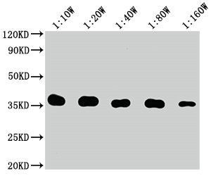
Western Blot Positive WB detected in: 10ng Flag Tag fusion protein Flag Tag antibody at 1:100000, 1:200000, 1:400000, 1:800000, 1:1600000 Secondary Goat polyclonal to mouse IgG at 1/50000 dilution Predicted band size: 35 kDa Observed band size: 35 kDa

CSB-MA000021M0M Image
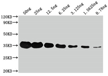
Western Blot Positive WB detected in: Flag Tag fusion protein at 50ng, 25ng, 12.5ng, 6.25ng, 3.625ng, 1.5625ng, 0.78ng All lanes: Flag Tag antibody at 1:1000 Secondary Goat polyclonal to mouse IgG at 1/50000 dilution Predicted band size: 35 kDa Observed band size: 35 kDa

CSB-MA000021M0M Image
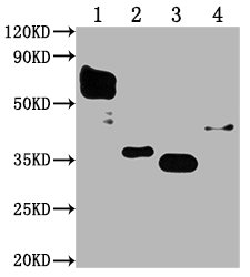
Western Blot Positive WB detected in: Flag Tag fusion protein1, 2, 3, 4 at 50ng All lanes: Flag Tag antibody at 1:1000 Secondary Goat polyclonal to mouse IgG at 1/50000 dilution Predicted band size: 59.6, 36, 35, 42 kDa Observed band size: 59.6, 36, 35, 42 kDa

CSB-MA000021M0M Image
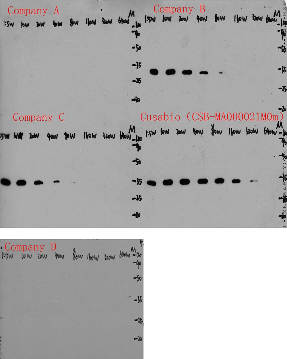
Western Blot Positive WB detected in: Flag Tag fusion protein at 10ng All lanes: Company A, Company B, Company C, Company D, CSB-MA000021M0m antibody at 1:50000, 1:100000, 1:200000, 1:400000, 1:800000, 1:1600000, 1:3200000, 1:6400000 Secondary Goat polyclonal to Mouse IgG at 1/50000 dilution Predicted band size: 35 kDa Observed band size: 35 kDa

CSB-MA000021M0M Image
Western Blot Positive WB detected in: Flag Tag fusion protein at 50ng, 25ng, 12.5ng, 6.25ng, 3.125ng, 1.5625ng, 0.78ng, 0.39ng All lanes: Company A, Company B, Company C, Company D, CSB-MA000021M0m antibody at 1:1000 Secondary Goat polyclonal to Mouse IgG at 1/50000 dilution Predicted band size: 35 kDa Observed band size: 35 kDa

CSB-MA000021M0M Image
Western Blot Positive WB detected in: 5 different recombinant proteins with Flag Tag All lanes: Company A, Company B, Company C, Company D, CSB-MA000021M0m antibody at 1:5000 Secondary Goat polyclonal to Mouse IgG at 1/50000 dilution

CSB-MA000021M0M Image
Immunoprecipitating Flag Tag in Transfected HEK-293 cell whole cell lysate Lane 1: Mouse control IgG instead of CSB-MA000021M0m in Transfected HEK-293 cell whole cell lysate Lane 2: CSB-MA000021M0m (1µl) + Transfected HEK-293 cell whole cell lysate (500µg) Lane 3: Transfected HEK-293 cell whole cell lysate (20µg) For western blotting, the blot was detected with CSB-MA000021M0m at 1:5000, and a HRP-conjugated Protein G antibody was used as the secondary antibody at 1:2000

CSB-MA000021M0M Image
Immunoprecipitating Flag Tag in 293F transfected whole cell lysate Lane 1: Mouse control IgG instead of CSB-MA000021M0m in Flag Tag in 293F transfected whole cell lysate. For western blotting, a HRP-conjugated Protein G antibody was used as the secondary antibody (1/2000) Lane 2: Company A (1µl), Company B (1µl), Company C (1µl), Company D (1µl), CSB-MA000021M0m (1µl) + Flag Tag in 293F transfected whole cell lysate (500µg) Lane 3: Flag Tag in 293F transfected whole cell lysate (20µg)

CSB-MA000021M0M Image
Immunofluorescence staining of 293T cells transfected with Flag tag by CSB-MA000021M0m at 1:300, counter-stained with DAPI. The cells were fixed in 4% formaldehyde and blocked in 10% normal Goat Serum. The cells were then incubated with the antibody overnight at 4°C. The secondary antibody was Alexa Fluor 488-congugated AffiniPure Goat Anti-Rabbit IgG(H+L). The image on the right is the 293T cells transfected without Flag tag.
CSB-MA000021M0M Image
Western Blot Positive WB detected in: 10ng Flag Tag fusion protein Flag Tag antibody at 1:100000, 1:200000, 1:400000, 1:800000, 1:1600000 Secondary Goat polyclonal to mouse IgG at 1/50000 dilution Predicted band size: 35 kDa Observed band size: 35 kDa
CSB-MA000021M0M Image
Western Blot Positive WB detected in: Flag Tag fusion protein at 50ng, 25ng, 12.5ng, 6.25ng, 3.625ng, 1.5625ng, 0.78ng All lanes: Flag Tag antibody at 1:1000 Secondary Goat polyclonal to mouse IgG at 1/50000 dilution Predicted band size: 35 kDa Observed band size: 35 kDa
CSB-MA000021M0M Image
Western Blot Positive WB detected in: Flag Tag fusion protein1, 2, 3, 4 at 50ng All lanes: Flag Tag antibody at 1:1000 Secondary Goat polyclonal to mouse IgG at 1/50000 dilution Predicted band size: 59.6, 36, 35, 42 kDa Observed band size: 59.6, 36, 35, 42 kDa
CSB-MA000021M0M Image
Western Blot Positive WB detected in: Flag Tag fusion protein at 10ng All lanes: Company A, Company B, Company C, Company D, CSB-MA000021M0m antibody at 1:50000, 1:100000, 1:200000, 1:400000, 1:800000, 1:1600000, 1:3200000, 1:6400000 Secondary Goat polyclonal to Mouse IgG at 1/50000 dilution Predicted band size: 35 kDa Observed band size: 35 kDa
CSB-MA000021M0M Image
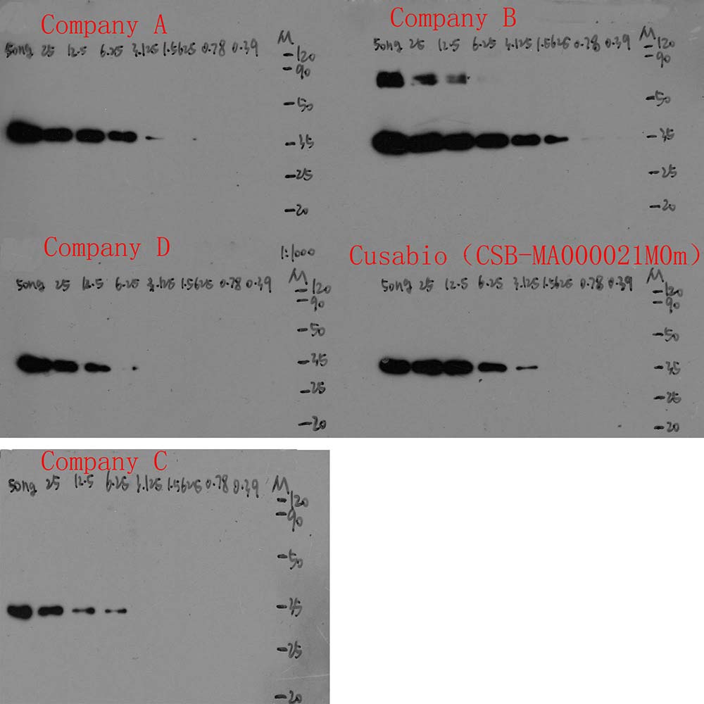
Western Blot Positive WB detected in: Flag Tag fusion protein at 50ng, 25ng, 12.5ng, 6.25ng, 3.125ng, 1.5625ng, 0.78ng, 0.39ng All lanes: Company A, Company B, Company C, Company D, CSB-MA000021M0m antibody at 1:1000 Secondary Goat polyclonal to Mouse IgG at 1/50000 dilution Predicted band size: 35 kDa Observed band size: 35 kDa
CSB-MA000021M0M Image
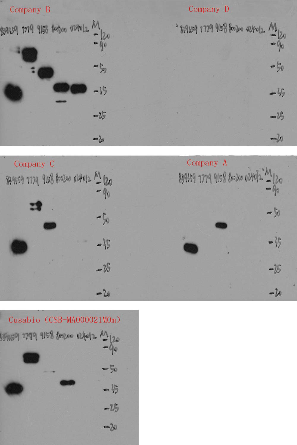
Western Blot Positive WB detected in: 5 different recombinant proteins with Flag Tag All lanes: Company A, Company B, Company C, Company D, CSB-MA000021M0m antibody at 1:5000 Secondary Goat polyclonal to Mouse IgG at 1/50000 dilution
CSB-MA000021M0M Image
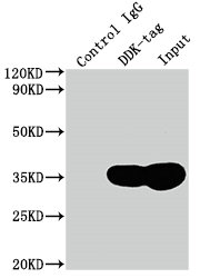
Immunoprecipitating Flag Tag in Transfected HEK-293 cell whole cell lysate Lane 1: Mouse control IgG instead of CSB-MA000021M0m in Transfected HEK-293 cell whole cell lysate Lane 2: CSB-MA000021M0m (1µl) + Transfected HEK-293 cell whole cell lysate (500µg) Lane 3: Transfected HEK-293 cell whole cell lysate (20µg) For western blotting, the blot was detected with CSB-MA000021M0m at 1:5000, and a HRP-conjugated Protein G antibody was used as the secondary antibody at 1:2000
CSB-MA000021M0M Image
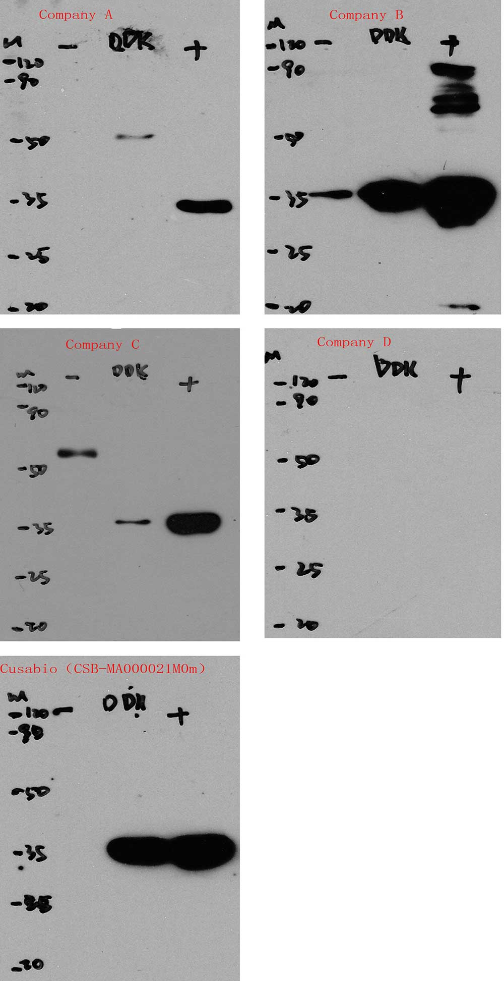
Immunoprecipitating Flag Tag in 293F transfected whole cell lysate Lane 1: Mouse control IgG instead of CSB-MA000021M0m in Flag Tag in 293F transfected whole cell lysate. For western blotting, a HRP-conjugated Protein G antibody was used as the secondary antibody (1/2000) Lane 2: Company A (1µl), Company B (1µl), Company C (1µl), Company D (1µl), CSB-MA000021M0m (1µl) + Flag Tag in 293F transfected whole cell lysate (500µg) Lane 3: Flag Tag in 293F transfected whole cell lysate (20µg)
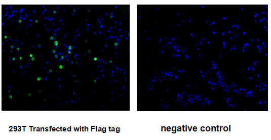
CSB-MA000021M0M Image
Immunofluorescence staining of 293T cells transfected with Flag tag by CSB-MA000021M0m at 1:300, counter-stained with DAPI. The cells were fixed in 4% formaldehyde and blocked in 10% normal Goat Serum. The cells were then incubated with the antibody overnight at 4°C. The secondary antibody was Alexa Fluor 488-congugated AffiniPure Goat Anti-Rabbit IgG(H+L). The image on the right is the 293T cells transfected without Flag tag.
| Manufacturer: | Cusabio Biotech |
| Preservative: | 0.03% ProClin™ 300 |
| Physical state: | Liquid |
| Type: | Monoclonal Primary Antibody - Unconjugated |
| Shipping Condition: | Blue Ice |
| Unit(s): | 100 ul, 50 ul |
| Host name: | Mouse |
| Clone: | 4G5C8 |
| Isotype: | IgG2a |
| Immunogen: | DYKDDDDK(Flag) synthetic peptide conjugate to KLH |
| Application: | ELISA, IF, IP, WB |
Description
Description: Flag-tag, or Flag octapeptide, is a polypeptide protein tag that can be added to a protein using recombinant DNA technology. It can be used for affinity chromatography, then used to separate recombinant, overexpressed protein from wild-type protein expressed by the host organism. It can also be used in the isolation of protein complexes with multiple subunits. A Flag-tag can be used in many different assays that require recognition by an antibody. If there is no antibody against the studied protein, adding a Flag-tag to this protein allows one to follow the protein with an antibody against the Flag sequence. Examples are cellular localization studies by immunofluorescence or detection by SDS PAGE protein electrophoresis.
Additional Text
Purification
Protein G purified
Antibody Clonality
Monoclonal
FAQs
SUPPORT
outstanding technical support
PRODUCT
we offer a full product guarantee
DELIVERY
we offer free delivery to UK universities and non profit organisations