GAPDH Monoclonal Antibody
Catalogue Number: CSB-MA000071M0M-CSB

CSB-MA000071M0M Image
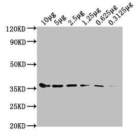
Western Blot Positive WB detected in: Hela whole cell lysate at 10µg, 5µg, 2.5µg, 1.25µg, 0.625µg, 0.3125µg All lanes: GAPDH antibody at 1:5000 Secondary Goat polyclonal to mouse IgG at 1/50000 dilution Predicted band size: 36 KDa Observed band size: 36 KDa Exposure time: 5min

CSB-MA000071M0M Image
Western Blot Positive WB detected in: Hela whole cell lysate, HepG2 whole cell lysate, Jurkat whole cell lysate, MCF-7 whole cell lysate All lanes: GAPDH antibody at 1:2000 Secondary Goat polyclonal to mouse IgG at 1/50000 dilution Predicted band size: 36 KDa Observed band size: 36 KDa Exposure time: 30s

CSB-MA000071M0M Image
IHC image of CSB-MA000071M0m diluted at 1:100 and staining in paraffin-embedded human breast cancer performed on a Leica BondTM system. After dewaxing and hydration, antigen retrieval was mediated by high pressure in a citrate buffer (pH 6.0). Section was blocked with 10% normal goat serum 30min at RT. Then primary antibody (1% BSA) was incubated at 4°C overnight. The primary is detected by a biotinylated secondary antibody and visualized using an HRP conjugated SP system.

CSB-MA000071M0M Image
IHC image of CSB-MA000071M0m diluted at 1:100 and staining in paraffin-embedded human kidney tissue performed on a Leica BondTM system. After dewaxing and hydration, antigen retrieval was mediated by high pressure in a citrate buffer (pH 6.0). Section was blocked with 10% normal goat serum 30min at RT. Then primary antibody (1% BSA) was incubated at 4°C overnight. The primary is detected by a biotinylated secondary antibody and visualized using an HRP conjugated SP system.

CSB-MA000071M0M Image
Immunofluorescence staining of Hela cells with CSB-MA000071M0m at 1:220, counter-stained with DAPI. The cells were fixed in 4% formaldehyde, permeabilized using 0.2% Triton X-100 and blocked in 10% normal Goat Serum. The cells were then incubated with the antibody overnight at 4°C. Nuclear DNA was labeled in blue with DAPI. The secondary antibody was FITC-conjugated AffiniPure Goat Anti-Mouse IgG(H+L).

CSB-MA000071M0M Image
Immunoprecipitating GAPDH in Hela whole cell lysate Lane 1: Mouse control IgG instead of CSB-MA000071M0m in Hela whole cell lysate. Lane 2: CSB-MA000071M0m (1µl) + Hela whole cell lysate (500µg) Lane 3: Hela whole cell lysate (20µg) For western blotting, the blot was detected with CSB-MA000071M0m at 1:5000, and a HRP-conjugated Protein G antibody was used as the secondary antibody at 1:2000

CSB-MA000071M0M Image
Overlay histogram showing Hela cells stained with CSB-MA000071M0m (red line). The cells were fixed with 70% Ethylalcohol (18h) and then permeabilized with 0.3% Triton X-100 for 2 min. The cells were then incubated in 10% normal goat serum to block non-specific protein-protein interactions followed by the antibody (1:200/1*106cells) for 1 h at 4°C. The secondary antibody used was FITC goat anti-mouse IgG(H+L) at 1/100 dilution for 30min at 4°C. Isotype control antibody (green line) was mouse IgG1 (1:200/1*106cells) used under the same conditions. Acquisition of >10,000 events was performed.

CSB-MA000071M0M Image
Western Blot Positive WB detected in: U87 whole cell lysate, PC3 whole cell lysate, 293 whole cell lysate, U251 whole cell lysate, A549 whole cell lysate, A375 whole cell lysate, MG-63 whole cell lysate All lanes GAPDH antibody at 1:5000 Secondary Goat polyclonal to mouse IgG at 1/50000 dilution Predicted band size: 36 KDa Observed band size: 36 KDa Exposure time : 30S

CSB-MA000071M0M Image
Western Blot Positive WB detected in: Rat heart tissue, Rat kidney tissue, Rat skeletal muscle tissue, Rat liver tissue, Rat brain tissue tissue, Rat spleen tissue All lanes GAPDH antibody at 1:1000 Secondary Goat polyclonal to mouse IgG at 1/50000 dilution Predicted band size: 36 KDa Observed band size: 36 KDa Exposure time : 1min

CSB-MA000071M0M Image
Western Blot Positive WB detected in: Rabbit heart tissue, Rabbit liver tissue,Rabbit spleen tissue, Rabbit lung tissue, Rabbit kidney tissue, Rabbit small intestine tissue, Rabbit skeletal muscle tissue All lanes GAPDH antibody at 1:5000 Secondary Goat polyclonal to mouse IgG at 1/50000 dilution Predicted band size: 36 KDa Observed band size: 36 KDa Exposure time : 5min

CSB-MA000071M0M Image
IHC image of CSB-MA000071M0m diluted at 1:100 and staining in paraffin-embedded human colon cancer performed on a Leica BondTM system. After dewaxing and hydration, antigen retrieval was mediated by high pressure in a citrate buffer (pH 6.0). Section was blocked with 10% normal goat serum 30min at RT. Then primary antibody (1% BSA) was incubated at 4°C overnight. The primary is detected by a biotinylated secondary antibody and visualized using an HRP conjugated SP system.

CSB-MA000071M0M Image
Immunofluorescence staining of HepG2 cells with CSB-MA000071M0m at 1:220, counter-stained with DAPI. The cells were fixed in 4% formaldehyde, permeabilized using 0.2% Triton X-100 and blocked in 10% normal Goat Serum. The cells were then incubated with the antibody overnight at 4°C. Nuclear DNA was labeled in blue with DAPI. The secondary antibody was FITC-conjugated AffiniPure Goat Anti-Mouse IgG(H+L).

CSB-MA000071M0M Image
Western Blot Positive WB detected in: 15µg hela whole cell lysate GAPDH antibody at 1:100000, 1:200000, 1:400000, 1:800000, 1:1600000 Secondary Goat polyclonal to mouse IgG at 1/50000 dilution Predicted band size: 36 KDa Observed band size: 36 KDa Exposure time: 5min

CSB-MA000071M0M Image
Overlay histogram showing Jurkat cells stained with CSB-MA000071M0m (red line). The cells were fixed with 70% Ethylalcohol (18h) and then permeabilized with 0.3% Triton X-100 for 2 min. The cells were then incubated in 10% normal goat serum to block non-specific protein-protein interactions followed by the antibody (1:200/1*106cells) for 1 h at 4°C. The secondary antibody used was FITC goat anti-mouse IgG(H+L) at 1/100 dilution for 30min at 4°C. Isotype control antibody (green line) was mouse IgG1 (1:200/1*106cells) used under the same conditions. Acquisition of >10,000 events was performed.

CSB-MA000071M0M Image
Western Blot Positive WB detected in: 15µg hela whole cell lysate GAPDH antibody at 1:100000, 1:200000, 1:400000, 1:800000, 1:1600000 Secondary Goat polyclonal to mouse IgG at 1/50000 dilution Predicted band size: 36 KDa Observed band size: 36 KDa Exposure time: 5min

CSB-MA000071M0M Image
Western Blot Positive WB detected in: Hela whole cell lysate at 10µg, 5µg, 2.5µg, 1.25µg, 0.625µg, 0.3125µg All lanes: GAPDH antibody at 1:5000 Secondary Goat polyclonal to mouse IgG at 1/50000 dilution Predicted band size: 36 KDa Observed band size: 36 KDa Exposure time: 5min

CSB-MA000071M0M Image
Western Blot Positive WB detected in: Hela whole cell lysate, HepG2 whole cell lysate, Jurkat whole cell lysate, MCF-7 whole cell lysate All lanes: GAPDH antibody at 1:2000 Secondary Goat polyclonal to mouse IgG at 1/50000 dilution Predicted band size: 36 KDa Observed band size: 36 KDa Exposure time: 30s

CSB-MA000071M0M Image
Western Blot Positive WB detected in: U87 whole cell lysate, PC3 whole cell lysate, 293 whole cell lysate, U251 whole cell lysate, A549 whole cell lysate, A375 whole cell lysate, MG-63 whole cell lysate All lanes GAPDH antibody at 1:5000 Secondary Goat polyclonal to mouse IgG at 1/50000 dilution Predicted band size: 36 KDa Observed band size: 36 KDa Exposure time : 30S

CSB-MA000071M0M Image
Western Blot Positive WB detected in: Rat heart tissue, Rat kidney tissue, Rat skeletal muscle tissue, Rat liver tissue, Rat brain tissue tissue, Rat spleen tissue All lanes GAPDH antibody at 1:1000 Secondary Goat polyclonal to mouse IgG at 1/50000 dilution Predicted band size: 36 KDa Observed band size: 36 KDa Exposure time : 1min

CSB-MA000071M0M Image
Western Blot Positive WB detected in: Rabbit heart tissue, Rabbit liver tissue,Rabbit spleen tissue, Rabbit lung tissue, Rabbit kidney tissue, Rabbit small intestine tissue, Rabbit skeletal muscle tissue All lanes GAPDH antibody at 1:5000 Secondary Goat polyclonal to mouse IgG at 1/50000 dilution Predicted band size: 36 KDa Observed band size: 36 KDa Exposure time : 5min

CSB-MA000071M0M Image
IHC image of CSB-MA000071M0m diluted at 1:100 and staining in paraffin-embedded human colon cancer performed on a Leica BondTM system. After dewaxing and hydration, antigen retrieval was mediated by high pressure in a citrate buffer (pH 6.0). Section was blocked with 10% normal goat serum 30min at RT. Then primary antibody (1% BSA) was incubated at 4°C overnight. The primary is detected by a biotinylated secondary antibody and visualized using an HRP conjugated SP system.

CSB-MA000071M0M Image
IHC image of CSB-MA000071M0m diluted at 1:100 and staining in paraffin-embedded human breast cancer performed on a Leica BondTM system. After dewaxing and hydration, antigen retrieval was mediated by high pressure in a citrate buffer (pH 6.0). Section was blocked with 10% normal goat serum 30min at RT. Then primary antibody (1% BSA) was incubated at 4°C overnight. The primary is detected by a biotinylated secondary antibody and visualized using an HRP conjugated SP system.

CSB-MA000071M0M Image
IHC image of CSB-MA000071M0m diluted at 1:100 and staining in paraffin-embedded human kidney tissue performed on a Leica BondTM system. After dewaxing and hydration, antigen retrieval was mediated by high pressure in a citrate buffer (pH 6.0). Section was blocked with 10% normal goat serum 30min at RT. Then primary antibody (1% BSA) was incubated at 4°C overnight. The primary is detected by a biotinylated secondary antibody and visualized using an HRP conjugated SP system.
| Manufacturer: | Cusabio Biotech |
| Preservative: | 0.03% ProClin™ 300 |
| Ph: | 7.4 |
| Physical state: | Liquid |
| Type: | Monoclonal Primary Antibody - Unconjugated |
| Shipping Condition: | Blue Ice |
| Unit(s): | 100 ul, 50 ul |
| Host name: | Mouse |
| Clone: | 14C2F11 |
| Isotype: | IgG1 |
| Immunogen: | Recombinant Human GAPDH protein (3-335AA) |
| Application: | ELISA, FC, IF, IP, WB, IHC |
Description
Description: Glyceraldehyde 3-phosphate dehydrogenase (GAPDH or G3PDH) is an enzyme of 37kDa that is consisdered as a cellular enzyme involved in glycolysis. Glyceraldehyde-3-phosphate dehydrogenase (GAPDH) is a pleiotropic enzyme that is overexpressed in apoptosis and in several human chronic pathologies. Its role as a mediator for cell death has also been highlighted. At the molecular level, sequential steps lead to nuclear translocation of GAPDH during cell death as follows: first, a catalytic cysteine in GAPDH (C150 in rat GAPDH) is S-nitrosylated by nitric oxide (NO) that is generated from inducible nitric oxide synthase (iNOS) and/or neuronal NOS (nNOS); second, the modified GAPDH becomes capable of binding with Siah1, an E3 ubiquitin ligase, and stabilizes it; third, the GAPDH-Siah protein complex translocates to the nucleus, dependent on Siah1's nuclear localization signal, and degrades Siah1's substrates in the nucleus, which results in cytotoxicity. A recent report suggests that GAPDH may be genetically associated with late-onset of Alzheimer's disease.-deprenyl, which has originally been used as a monoamine oxidase inhibitor for Parkinson's disease, binds to GAPDH and displays neuroprotective actions.
Additional Text
Gene Name
GAPDH
Gene ID
2597
Purification
Protein G purified
Uniprot ID
P04406
Antibody Clonality
Monoclonal
FAQs
SUPPORT
outstanding technical support
PRODUCT
we offer a full product guarantee
DELIVERY
we offer free delivery to UK universities and non profit organisations