TUBA1A Monoclonal Antibody
Catalogue Number: CSB-MA754656A0M-CSB

CSB-MA754656A0M Image

CSB-MA754656A0M Image

CSB-MA754656A0M Image

CSB-MA754656A0M Image

CSB-MA754656A0M Image

CSB-MA754656A0M Image

CSB-MA754656A0M Image

CSB-MA754656A0M Image

CSB-MA754656A0M Image

CSB-MA754656A0M Image

CSB-MA754656A0M Image

CSB-MA754656A0M Image

CSB-MA754656A0M Image

CSB-MA754656A0M Image

CSB-MA754656A0M Image
CSB-MA754656A0M Image
CSB-MA754656A0M Image
CSB-MA754656A0M Image
CSB-MA754656A0M Image
CSB-MA754656A0M Image
CSB-MA754656A0M Image
CSB-MA754656A0M Image
CSB-MA754656A0M Image
CSB-MA754656A0M Image
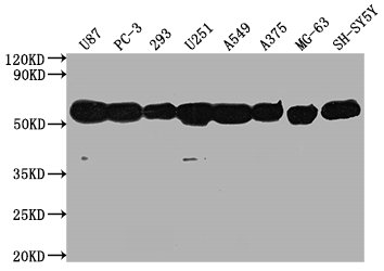
Western Blot Positive WB detected in: U87 whole cell lysate, PC-3 whole cell lysate, 293 whole cell lysate, U251 whole cell lysate, A549 whole cell lysate, A375 whole cell lysate, MG-63 whole cell lysate, SH-SY5Y whole cell lysate, All lanes: TUBA1A antibody at 1:5000 Secondary Goat polyclonal to Mouse IgG at 1/10000 dilution Predicted band size: 52 kDa Observed band size: 52 kDa
CSB-MA754656A0M Image
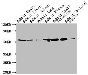
Western Blot Positive WB detected in: Rabbit heatrt tissue, Rabbit liver tissue, Rabbit spleen tissue, Rabbit lung tissue, Rabbit kidney tissue, Rabbit small intestine tissue, Rabbit skeletal muscle tissue All lanes: TUBA1A antibody at 1:5000 Secondary Goat polyclonal to Mouse IgG at 1/10000 dilution Predicted band size: 52 kDa Observed band size: 52 kDa
CSB-MA754656A0M Image
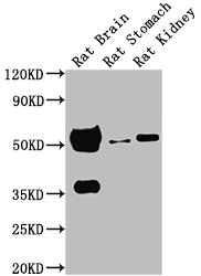
Western Blot Positive WB detected in: Rat brain tissue, Rat stomach tissue, Rat kidney tissue All lanes: TUBA1A antibody at 1:5000 Secondary Goat polyclonal to Mouse IgG at 1/10000 dilution Predicted band size: 52 kDa Observed band size: 52 kDa
CSB-MA754656A0M Image
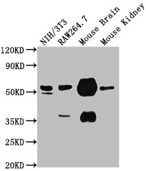
Western Blot Positive WB detected in: NIH/3T3 whole cell lysate, RAW264.7 whole cell lysate, Mouse brain tissue, Mouse kidney tissue All lanes: TUBA1A antibody at 1:5000 Secondary Goat polyclonal to Mouse IgG at 1/10000 dilution Predicted band size: 52 kDa Observed band size: 52 kDa
CSB-MA754656A0M Image
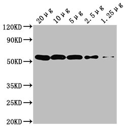
Western Blot Positive WB detected in: Hela whole cell lysate at 20µg, 10µg, 5µg, 2.5µg, 1.25µg All lanes: TUBA1A antibody at 1:5000 Secondary Goat polyclonal to Mouse IgG at 1/10000 dilution Predicted band size: 52 kDa Observed band size: 52 kDa
CSB-MA754656A0M Image
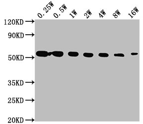
Western Blot Positive WB detected in: Hela whole cell lysate All lanes: TUBA1A antibody at 1:2500, 1:5000, 1:10000, 1:20000, 1:40000, 1:80000, 1:160000 Secondary Goat polyclonal to Mouse IgG at 1/10000 dilution Predicted band size: 52 kDa Observed band size: 52 kDa
CSB-MA754656A0M Image
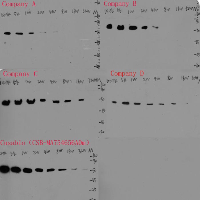
Western Blot Positive WB detected in: Hela whole cell lysate All lanes: Company A, Company B, Company C, Company D, CSB-MA754656A0m antibody at 1:2500, 1:5000, 1:10000, 1:20000, 1:40000, 1:80000, 1:160000, 320000 Secondary Goat polyclonal to Mouse IgG at 1/10000 dilution Predicted band size: 52 kDa Observed band size: 52 kDa
CSB-MA754656A0M Image
IHC image of CSB-MA754656A0m diluted at 1:150 and staining in paraffin-embedded human tonsil tissue performed on a Leica BondTM system. After dewaxing and hydration, antigen retrieval was mediated by high pressure in a citrate buffer (pH 6.0). Section was blocked with 10% normal goat serum 30min at RT. Then primary antibody (1% BSA) was incubated at 4°C overnight. The primary is detected by a biotinylated secondary antibody and visualized using an HRP conjugated SP system.
CSB-MA754656A0M Image
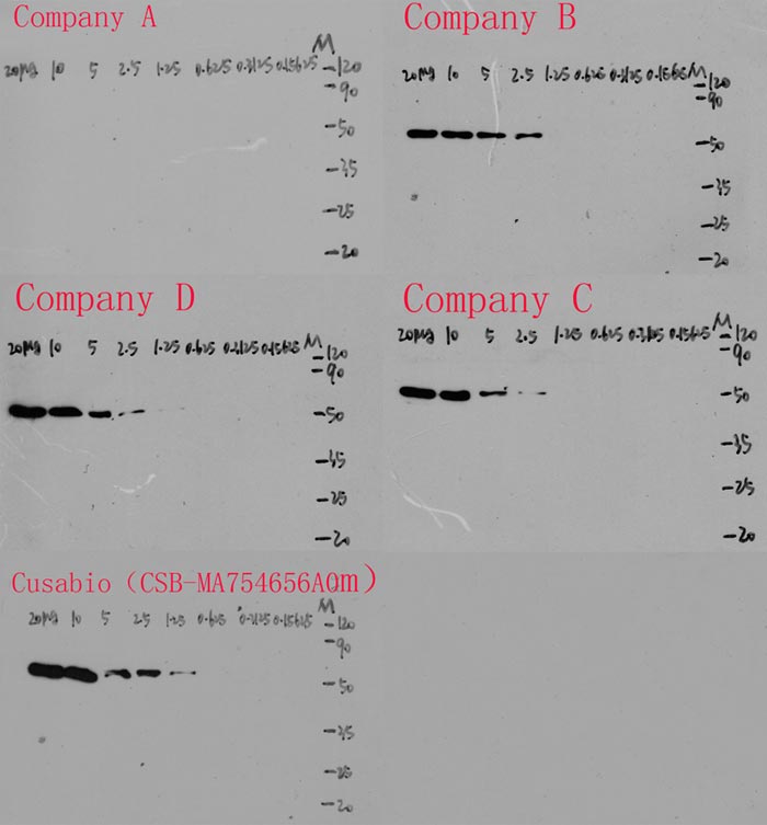
Western Blot Positive WB detected in: Hela whole cell lysate at 20µg, 10µg, 5µg, 2.5µg, 1.25µg, 0.625µg, 0.3125µg, 0.15625µg All lanes: Company A, Company B, Company C, Company D, CSB-MA754656A0m antibody at 1:5000 Secondary Goat polyclonal to Mouse IgG at 1/10000 dilution Predicted band size: 52 kDa Observed band size: 52 kDa
CSB-MA754656A0M Image
IHC image of CSB-MA754656A0m diluted at 1:150 and staining in paraffin-embedded human breast cancer performed on a Leica BondTM system. After dewaxing and hydration, antigen retrieval was mediated by high pressure in a citrate buffer (pH 6.0). Section was blocked with 10% normal goat serum 30min at RT. Then primary antibody (1% BSA) was incubated at 4°C overnight. The primary is detected by a biotinylated secondary antibody and visualized using an HRP conjugated SP system.
CSB-MA754656A0M Image
Overlay histogram showing Hela cells stained with Company A (5µg), Company B (5µg), Company C (5µg), Company D (5µg), CSB-MA754656A0m (5µg) (red line) at 1:150. The cells were incubated in 1x PBS /10% normal goat serum to block non-specific protein-protein interactions followed by primary antibody for 1 h at 4°C. The secondary antibody used was FITC goat anti-mouse IgG(H+L) at 1/200 dilution for 1 h at 4°C. Isotype control antibody (green line) was used under the same conditions. Acquisition of >10,000 events was performed.
CSB-MA754656A0M Image
Overlay histogram showing Hela cells stained with CSB-MA754656A0m (red line) at 1:150. The cells were incubated in 1x PBS /10% normal goat serum to block non-specific protein-protein interactions followed by primary antibody for 1 h at 4°C. The secondary antibody used was FITC goat anti-mouse IgG(H+L) at 1/200 dilution for 1 h at 4°C. Isotype control antibody (green line) was used under the same conditions. Acquisition of >10,000 events was performed.
CSB-MA754656A0M Image
Immunoprecipitating TUBA1A in Hela whole cell lysate Lane 1: Mouse control IgG (1µg) instead of CSB-MA754656A0m in Hela whole cell lysate. For western blotting, a HRP-conjugated Protein G antibody was used as the secondary antibody (1/2000) Lane 2: Company A (5µg), Company B (5µg), Company C (5µg), Company D (5µg), CSB-MA754656A0m (5µg) + Hela whole cell lysate (500µg) Lane 3: Hela whole cell lysate (20µg)
CSB-MA754656A0M Image
Immunoprecipitating TUBA1A in Hela whole cell lysate Lane 1: Mouse control IgG (1µg) instead of CSB-MA754656A0m in Hela whole cell lysate. For western blotting, a HRP-conjugated Protein G antibody was used as the secondary antibody (1/2000) Lane 2: CSB-MA754656A0m (5µg) + Hela whole cell lysate (500µg) Lane 3: Hela whole cell lysate (20µg)
CSB-MA754656A0M Image
Immunofluorescence staining of Hela cells with CSB-MA754656A0m at 1:75, counter-stained with DAPI. The cells were blocked in 10% normal Goat Serum and then incubated with the primary antibody overnight at 4°C. The secondary antibody was Alexa Fluor 488-congugated AffiniPure Goat Anti-Mouse IgG(H+L).
CSB-MA754656A0M Image
Western Blot Positive WB detected in: U87 whole cell lysate, PC-3 whole cell lysate, 293 whole cell lysate, U251 whole cell lysate, A549 whole cell lysate, A375 whole cell lysate, MG-63 whole cell lysate, SH-SY5Y whole cell lysate, All lanes: TUBA1A antibody at 1:5000 Secondary Goat polyclonal to Mouse IgG at 1/10000 dilution Predicted band size: 52 kDa Observed band size: 52 kDa
CSB-MA754656A0M Image
Western Blot Positive WB detected in: Rabbit heatrt tissue, Rabbit liver tissue, Rabbit spleen tissue, Rabbit lung tissue, Rabbit kidney tissue, Rabbit small intestine tissue, Rabbit skeletal muscle tissue All lanes: TUBA1A antibody at 1:5000 Secondary Goat polyclonal to Mouse IgG at 1/10000 dilution Predicted band size: 52 kDa Observed band size: 52 kDa
CSB-MA754656A0M Image
Western Blot Positive WB detected in: Rat brain tissue, Rat stomach tissue, Rat kidney tissue All lanes: TUBA1A antibody at 1:5000 Secondary Goat polyclonal to Mouse IgG at 1/10000 dilution Predicted band size: 52 kDa Observed band size: 52 kDa
CSB-MA754656A0M Image
Western Blot Positive WB detected in: NIH/3T3 whole cell lysate, RAW264.7 whole cell lysate, Mouse brain tissue, Mouse kidney tissue All lanes: TUBA1A antibody at 1:5000 Secondary Goat polyclonal to Mouse IgG at 1/10000 dilution Predicted band size: 52 kDa Observed band size: 52 kDa
CSB-MA754656A0M Image
Western Blot Positive WB detected in: Hela whole cell lysate at 20µg, 10µg, 5µg, 2.5µg, 1.25µg All lanes: TUBA1A antibody at 1:5000 Secondary Goat polyclonal to Mouse IgG at 1/10000 dilution Predicted band size: 52 kDa Observed band size: 52 kDa
CSB-MA754656A0M Image
Western Blot Positive WB detected in: Hela whole cell lysate All lanes: TUBA1A antibody at 1:2500, 1:5000, 1:10000, 1:20000, 1:40000, 1:80000, 1:160000 Secondary Goat polyclonal to Mouse IgG at 1/10000 dilution Predicted band size: 52 kDa Observed band size: 52 kDa
CSB-MA754656A0M Image
Western Blot Positive WB detected in: Hela whole cell lysate at 20µg, 10µg, 5µg, 2.5µg, 1.25µg, 0.625µg, 0.3125µg, 0.15625µg All lanes: Company A, Company B, Company C, Company D, CSB-MA754656A0m antibody at 1:5000 Secondary Goat polyclonal to Mouse IgG at 1/10000 dilution Predicted band size: 52 kDa Observed band size: 52 kDa
CSB-MA754656A0M Image
Western Blot Positive WB detected in: Hela whole cell lysate All lanes: Company A, Company B, Company C, Company D, CSB-MA754656A0m antibody at 1:2500, 1:5000, 1:10000, 1:20000, 1:40000, 1:80000, 1:160000, 320000 Secondary Goat polyclonal to Mouse IgG at 1/10000 dilution Predicted band size: 52 kDa Observed band size: 52 kDa
| Manufacturer: | Cusabio Biotech |
| Preservative: | 0.03% ProClin™ 300 |
| Physical state: | Liquid |
| Type: | Monoclonal Primary Antibody - Unconjugated |
| Shipping Condition: | Blue Ice |
| Unit(s): | 100 ul, 50 ul |
| Host name: | Mouse |
| Clone: | 7E5C12 |
| Isotype: | IgG2a |
| Immunogen: | A synthesized peptide derived from human Tubulin alpha-1A chain (297-309) |
| Application: | ELISA, FC, IF, IP, WB, IHC |
Description
Description: Tubulin is the major constituent of microtubules. It binds two moles of GTP, one at an exchangeable site on the beta chain and one at a non-exchangeable site on the alpha chain.
Additional Text
Gene Name
TUBA1A
Uniprot ID
Q71U36
Gene ID
7846
Purification
Protein A purified
Antibody Clonality
Monoclonal
SUPPORT
outstanding technical support
PRODUCT
we offer a full product guarantee
DELIVERY
we offer free delivery to UK universities and non profit organisations