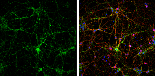
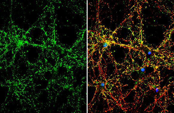
Synaptophysin antibody detects Synaptophysin protein at synaptic vesicles by immunofluorescent analysis. Sample: DIV9 rat E18 primary cortical neurons were fixed in 4% paraformaldehyde at RT for 15 min. Green: Synaptophysin protein stained by Synaptophysin antibody (GTX100865) diluted at 1:500. Red: beta Tubulin 3/ Tuj1, stained by beta Tubulin 3/ Tuj1 antibody [GT11710] (GTX631836) diluted at 1:500. Blue: Fluoroshield with DAPI (GTX30920).
GTX100865 Image
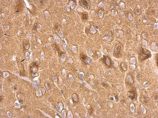
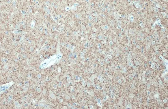
Synaptophysin antibody detects Synaptophysin protein at on rat fore brain by immunohistochemical analysis. Sample: Paraffin-embedded rat fore brain. Synaptophysin antibody (GTX100865) dilution: 1:500. Antigen Retrieval: Trilogy™ (EDTA based, pH 8.0) buffer, 15min
GTX100865 Image
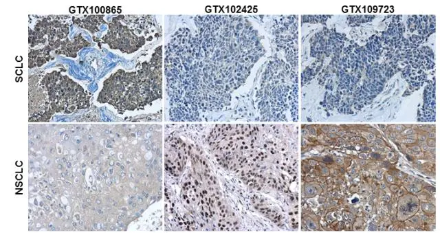
Immunohistochemical characterization of Synaptophysin (GTX100865), p63 (GTX102425) and Cytokeratin 7 (GTX109723) in human small cell lung cancer (SCLC) and non-small cell lung cancer (NSCLC) specimens. Sample: Paraffin-embedded human SCLC (upper panel) and NSCLC (lower panel). The section was pre-treated using heat mediated antigen retrieval with sodium citrate buffer (pH6) for 15 mins. The section was then incubated with primary antibody at 1:500 overnight at 4C and detected using an HRP conjugated avidin-biotin-peroxidase Complex system. DAB was used as the chromogen and counterstained with haematoxylin. Antigen Retrieval: Citrate buffer, pH 6.0, 15 min
GTX100865 Image
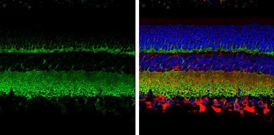
Synaptophysin antibody detects Synaptophysin protein expression by immunohistochemical analysis. Sample: Paraffin-Embedded adult mouse retina. Green: Synaptophysin protein stained by Synaptophysin antibody (GTX100865) diluted at 1:250. Red: beta Tubulin 3/ TUJ1, stained by beta Tubulin 3/ TUJ1 antibody [GT11710] (GTX631836) diluted at 1:250. Blue: Fluoroshield with DAPI (GTX30920). Antigen Retrieval: Citrate buffer, pH 6.0, 15 min
GTX100865 Image
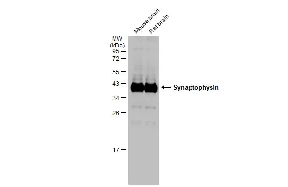
Various tissue extracts (50 µg) were separated by 12% SDS-PAGE, and the membrane was blotted with Synaptophysin antibody (GTX100865) diluted at 1:50000. The HRP-conjugated anti-rabbit IgG antibody (GTX213110-01) was used to detect the primary antibody.
GTX100865 Image
Synaptophysin antibody detects Synaptophysin protein by immunofluorescent analysis. Sample: DIV10 rat E18 primary hippocampal neuron cells were fixed in 4% paraformaldehyde at RT for 15 min. Green: Synaptophysin stained by Synaptophysin antibody (GTX100865) diluted at 1:500. Red: Tau, stained by Tau antibody [GT287] (GTX634809) diluted at 1:500. Blue: Fluoroshield with DAPI (GTX30920).
GTX100865 Image
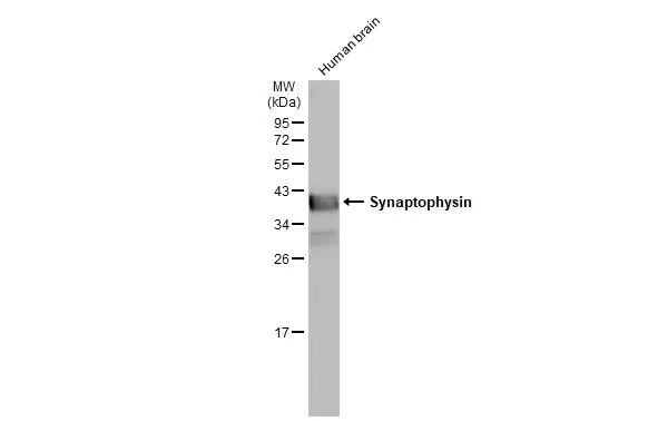
Human tissue extract (30 µg) was separated by 12% SDS-PAGE, and the membrane was blotted with Synaptophysin antibody (GTX100865) diluted at 1:50000. The HRP-conjugated anti-rabbit IgG antibody (GTX213110-01) was used to detect the primary antibody.
GTX100865 Image
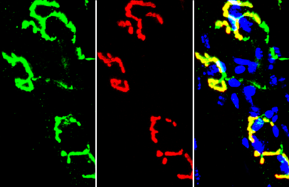
Synaptophysin antibody detects Synaptophysin protein by immunohistochemical analysis. Sample: Frozen-sectioned mouse muscle. Green: Synaptophysin stained by Synaptophysin antibody (GTX100865) diluted at 1:250. Red: α-Bungarotoxin, stained by α-Bungarotoxin, Alexa Fluor™ 594 conjugate (B13423) diluted at 1:5000. Blue: Hoechst 33342 staining.
GTX100865 Image
Synaptophysin antibody detects Synaptophysin protein at cell membrane and cytoplasm by immunohistochemical analysis. Sample: Paraffin-embedded mouse brain. Synaptophysin stained by Synaptophysin antibody (GTX100865) diluted at 1:500. Antigen Retrieval: Citrate buffer, pH 6.0, 15 min
GTX100865 Image
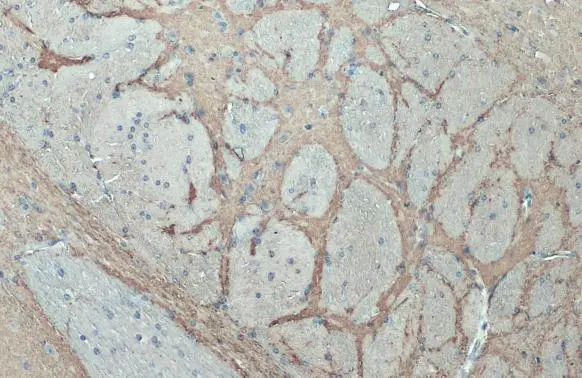
Synaptophysin antibody detects Synaptophysin protein at cell membrane and cytoplasm by immunohistochemical analysis. Sample: Paraffin-embedded rat brain. Synaptophysin stained by Synaptophysin antibody (GTX100865) diluted at 1:500. Antigen Retrieval: Citrate buffer, pH 6.0, 15 min
GTX100865 Image
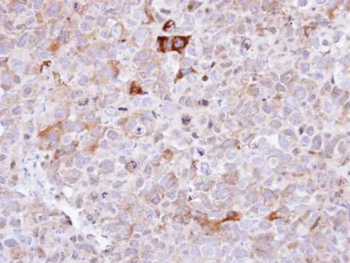
Immunohistochemical analysis of paraffin-embedded CL1-0 xenograft , using Synaptophysin(GTX100865) antibody at 1:100 dilution. Antigen Retrieval: Trilogy™ (EDTA based, pH 8.0) buffer, 15min
Description: Synaptophysin (p38) is an integral membrane protein of small synaptic vesicles in brain and endocrine cells.[supplied by OMIM]
Additional Text
Gene Name
SYP
Gene ID
6855
Uniprot ID
P08247
Concentration
0.65 mg/ml
Purification
Affinity Purified
Antibody Clonality
Polyclonal
Note
For In vitro laboratory use only. Not for any clinical, therapeutic, or diagnostic use in humans or animals. Not for animal or human consumption
Molecular Weight
34
Application Notes
WB: 1:500-1:50000. ICC/IF: 1:100-1:1000. IHC-P: 1:100-1:1000. *Optimal dilutions/concentrations should be determined by the researcher.Not tested in other applications.
Storage Note
Store as concentrated solution. Centrifuge briefly prior to opening vial. For short-term storage (1-2 weeks), store at 4C. For long-term storage, aliquot and store at -20C or below. Avoid multiple freeze-thaw cycles.