Benefits
- Highly specific – Specifically detect activation by binding sequence and with an antibody against the specific gene.
- High-throughput – With Whole Cell Lysis Buffer provided by Signosis, cell lysate can be prepared and high-through TF ELISA can be performed with limited number of cells from 96-well tissue plate.
- Highly sensitive – Detect with as little as 5ug nuclear extract using colorimetric-based kit and 0.5ug using chemiluminescent-based kit.
- Convenient and fast – Simple procedure with pre-coated plate included.
TF ELISA Strip Kit
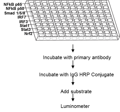 TF ELISA Strip kit is high sensitive and specific assay with a simple and optimized procedure. 12 of 8-well strips of white plate are pre-coated with 8 different consensus sequencing oligos respectively. The nuclear extract or the whole cell lysate is incubated on the well. The activated TF binds to the corresponding oligo on the well and is detected with a specific antibody and an HRP conjugated secondary antibody. The assay utilizes chemiluminescence detection method.
TF ELISA Kit (chemiluminescent)
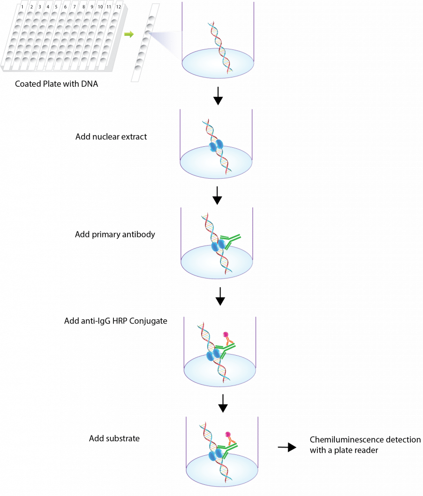 TF ELISA kit is high sensitive and specific assay with a simple and optimized procedure. The 96-well (8X12 strip) white plate is pre-immobilized with the TF consensus sequencing oligo. The activated TF in nuclear extract or the whole cell lysate is added in the well and binds to the oligo. The activated TF is detected with a specific antibody against the subunit and a HRP conjugated secondary antibody. After incubation, the wells are washed to remove unbound-labeled antibodies. The plate is further detected with HRP luminescent substrate. Luminescence is reported as relative light units (RLUs) on a microplate luminometer. The level of expression for each specific cytokine is directly proportional to the luminescent intensity.
TF ELISA Kit (Colourometric)
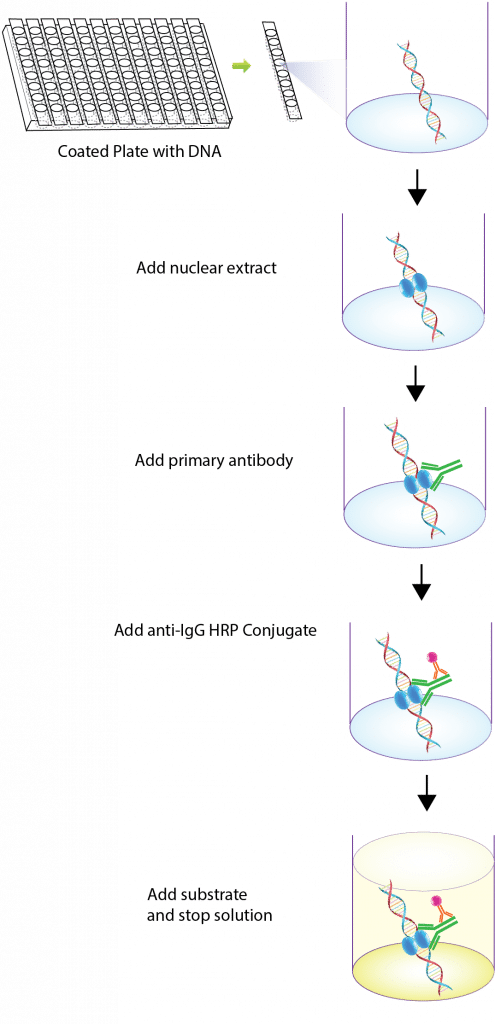 TF ELISA kit is high sensitive and specific assay with a simple and optimized procedure. The 96-well (8X12 strip) clear plate is pre-immobilized with the TF consensus sequencing oligo. The activated TF in nuclear extract or the whole cell lysate is added in the well and binds to the oligo. The activated TF is detected with a specific antibody against the subunit and a HRP conjugated secondary antibody. The assay utilizes colorimetric detection method, which can be easily measured by spectrophotometry.
CURRENT RANGE
NRF2 ELISA Kit
NRF2 plays a crucial role in cellular anti-oxidant defense, making it a therapeutic target for neurodegenerative diseases and cancer. Under unstressed conditions, NRF2 is kept in the cytoplasm by a cluster of proteins that degrade it quickly. Under oxidative stress, NRF2 is not degraded, but instead translocates to the nucleus where it binds to a DNA promoter and initiates gene expression. In the nucleus, NRF2 forms a heterodimer with a small Maf protein and binds to the Antioxidant Response Element in the upstream promoter region of many antioxidative genes, and initiates their transcription. Signosis has developed the NRF2 ELISA kit for the analysis of the NRF2 pathway and to facilitate studying activation of different NRF2-related pathways.
ELISA Kits Available
| NRF2 ELISA Kit (Colorimetric) TE-0009 |
| NRF2 ELISA Kit (Chemiluminescence) TE-0010 |
TFEB ELISA Kit
TFEB is a member of the basic Helix-Loop-Helix-Zipper family of transcription factors that specifically recognizes and binds E-box sequences (5′-CANNTG-3′) on DNA. TFEB is a master gene for lysosomal biogenesis, and therefore plays a central role in regulating expression of lysosomal genes. TFEB also acts as a positive regulator of autophagy by promoting expression of genes involved in autophagy. Under aberrant lysosomal storage conditions such as in lysosomal storage diseases, TFEB is translocated from the cytoplasm to the nucleus, resulting in the activation of its target genes. Signosis has developed the TFEB ELISA kits for sensitive and specific analysis of the activities of TFEB in a high throughput manner. The kit can be used for human, mouse and rat samples.
ELISA Kits Available
| TFEB ELISA Kit (Colorimetric) TE-0029 |
| TFEB ELISA Kit (Chemiluminescence) TE-0030 |
TF ELISA Strip Kit
Transcription factors (TFs) act as sensors to monitor cellular changes and signal transduction pathways and convert them to gene expression. Signosis TF ELISA Strip selects a number of TFs corresponding to most crucial pathways, such as NFkB, Stat, Smad, IRF and NRF2. Individual TF is captured and detected by a pair of sandwich assay reagent, one cis-element and one antibody to a TF. In addition, the strip kit targets a number of TFs that share the same cis-element, such as Stat1 and Stat3, p65 and p50, Smad2/3 and smad1/5/8, but they have distinctive biological functions and respond to different upstream signals or activation pathways. In the assay, the activation of these TFs can be specifically distinguished through specific antibodies. 8 TFs can be analyzed simultaneously, which the activation of the most important pathways can be determined accordingly.
List of Applicable Transcription Factors
| NFkB p65 | NFkB p50 | Smad 1/5/8 | IRF7 | IRF3 | Stat1 | Stat3 | Nrf2 |
ELISA Kits Available
| TF ELISA Strip Kit (Colorimetric) TE-1001 |
| TF ELISA Strip Kit (Chemiluminescence) TE-1002 |
NFkB ELISA Kits (p50 or p65)
NF-kappaB (NFkB) proteins comprise a family of eukaryotic transcription factors that are involved in the control of a large number of cellular and organismal processes. In addition, these transcription factors are associated with many diseases including cancer and arthritis. NFkB commonly refers specifically to a p50-RelA(p65) heterodimer, which is the major Rel/NF-kB complex in most cells. P65-p65 and p50-p50 heterodimers have been demonstrated to bind on DNA as well. NF-kB is present as a latent, inactive, IkB-bound complex in the cytoplasm. When a cell receives any of a multitude of extracellular signals, NF-kB rapidly enters the nucleus and activates gene expression. Signosis has developed the NFkB-p65 and NFkB p50 ELISA kits for sensitive and specific analysis of the activities of NFkB in a high throughput way. The kit can be used for human, mouse and rat samples.
NF-kB ELISA Kits Available
| NF-kB p65 ELISA Kit (Colorimetric) TE-0001 |
| NF-kB p65 ELISA Kit (Chemiluminescence) TE-0002 |
| NF-kB p50 ELISA Kit (Colorimetric) TE-0003 |
| NF-kB p50 ELISA Kit (Chemiluminescence) TE-0004 |
IRF ELISA Kits (IRF3 or IRF7)
IRF3 and IRF7 are members of the interferon regulatory transcription factor (IRF) family, involving in antiviral defence, cell growth regulation, and immune activation. Latent cytoplasmic IRF-3 and IRF7 are activated and phosphorylated following virus infection or treatment with dsRNA, and translocate to the nucleus and bind to their DNA binding sequences. Even though IRF3 and IRF7 are the key activators of the alpha/beta IFN genes, the recent studies have demonstrated that IRF3 and IRF7 have distinct DNA binding properties and induce preferentially on different promoters. Because of the common and distinct biological features of IRF-3 and IRF-7, Signosis have developed both IRF3 and IRF7 ELISA kits respectively and a combined kit (48 wells for IRF3 and 48 wells for IRF7) with specific DNA binding sites and antibodies to distinguish the activation of IRF3 and IRF7 in various biological conditions.
IRF ELISA Kits Available
| IRF3 ELISA Kit (Colorimetric) TE-0005 |
| IRF3 ELISA Kit (Chemiluminescence) TE-0017 |
| IRF7 ELISA Kit (Colorimetric) TE-0006 |
| IRF7 ELISA Kit (Chemiluminescence) TE-0018 |
| IRF3/IRF7 ELISA Kit (Colorimetric) TE-0007 |
| IRF3/IRF7 ELISA Kit (Chemiluminescence) TE-0019 |
PPAR gamma ELISA Kits
Peroxisome-proliferator-activated receptor gamma (PPAR gamma) is a ligand-activated transcription factor that plays an important role in the control of gene expression associated with a variety of physiological processes, particularly in metabolism and adipogenesis. With ligand binding, PPAR gamma forms a heterodimer with the retinoic X receptor, and binds to PPAR response elements in the promoter region of target genes, thus regulating their expression. Dysfunction of PPAR gamma leads to the pathological processes, such as metabolic diseases and cancer. Furthermore, modulation of receptor action in these diseases may be of therapeutic value. PPARgamma has been demonstrated to be the target of the thiazolidinediones (TZDs), which are widely used to treat type 2 diabetes. Signosis has developed PPARgamma ELISA, to help specific and sensitive measurement of the activation of PPARgamma.
ELISA Kits Available
| PPAR gamma ELISA Kit (Colorimetric) TE-0009 |
| PPAR gamma ELISA Kit (Chemiluminescence) TE-0010 |
STAT ELISA Kits (STAT3 and/or STAT1)
Stat3 (signal transducer and activator of transcription 3) promotes cell survival/proliferation, motility and immune tolerance and is considered as an oncogene. When signaled by cytokines and growth factors, such as oncostate, the activated Stat3 is translocated into the nucleus, where the gene expression is regulated by binding to the DNA recognition site. Signosis has developed a Stat3 ELISA kit to detect Stat3 activation. STAT1 exerts a wide spectrum of functions on both tumour cells and the immune system. Stat1 is usually considered as a tumour suppressor by enhancing inflammation and triggering anti-proliferative and pro-apoptotic responses in tumour cells. Upon activated by cytokines, such as INFgamma, stat1 forms homodimers or heterodimers with Stat3 or Stat2 and binds to the response element on the promoter region of target genes. Signosis has developed a Stat1 ELISA kit to monitor Stat1 activation.
While STAT3 promotes cell survival/proliferation, motility and immune tolerance and is considered as an oncogene, STAT1 enhances inflammation and innate and adaptive immunity, triggering in most instances anti-proliferative and pro-apoptotic responses in tumour cells. Despite being activated by common cytokines and growth factor receptor pathways, their activation is reciprocally regulated to redirect and balance cytokine/growth factor signals from proliferative to apoptotic, or from inflammatory to anti-inflammatory. Signosis has developed Stat3/Stat1 ELISA kit to distinguish the activation of Stat3 and Stat1.
STAT ELISA Kits Available
| STAT3 ELISA Kit (Colorimetric) TE-0020 |
| STAT3 ELISA Kit (Chemiluminescence) TE-0021 |
| STAT1 ELISA Kit (Colorimetric) TE-0023 |
| STAT1 ELISA Kit (Chemiluminescence) TE-0023 |
| STAT3/STAT1 ELISA Kit (Colorimetric) TE-0025 |
| STAT3/STAT1 ELISA Kit (Chemiluminescence) TE-0026 |
Smad ELISA Kits (2/3 or 1/5/8)
Smad transcription factors lie at the center of the transforming growth factor-beta (TGF-β) pathway, which is one of the most important cytokine signalling pathways. Members of the transforming growth factor-beta (TGF-β) superfamily bind to serine/threonine kinase receptors and specifically activate intracellular Smad proteins. Smads 2 and 3 are activated by activin/nodal and TGF-β, whereas Smads 1, 5 and 8 are activated by TGF-β-like BMP (Bone morphogenetic proteins). Smads family can be subsequently classified based on their activation by TGF-β or BMP cytokine family. These activated Smads form the complexes with co-Smads, translocate from cytoplasm into nucleus and bind to the distinctive consensus binding sequences on the target promoter region to regulate the transcription of genes. Signosis has developed the Smad2/3 ELISA kit for the analysis of TGF-β/Smad pathway, Smad1/5/8 ELISA kit for the analysis of BMP/Smad pathway, and a combined kit (48 wells for Smad2/3 and 48 wells for Smad1/5/8) to facilitate studying activation of different Smad-related pathways.
Smad ELISA Kits Available
| Smad2/3 ELISA Kit (Colorimetric) TE-0011 |
| Smad2/3 ELISA Kit (Chemiluminescence) TE-0012 |
| Smad1/5/8 ELISA Kit (Colorimetric) TE-0013 |
| Smad1/5/8 ELISA Kit (Chemiluminescence) TE-0014 |
| Smad2/3 and 1/5/8 ELISA Kit (Colorimetric) TE-0015 |
| Smad2/3 and 1/5/8 ELISA Kit (Chemiluminescence) TE-0016 |
SATB1 ELISA Kits
SATB1 is a nuclear protein which plays a crucial role in metastasis of breast cancer. It promotes tumour growth and metastasis by changing the expression of hundreds of genes, affecting cell adhesion, cell signalling, cell-cycle regulation, and other functions, including the epidermal growth factor gene Her2. SATB1 is not expressed in all cells. Only the metastatic cells express SATB1, and the most aggressive breast cancer cells show the highest levels of the protein. When SATB1 is detected in a breast tumour, the cancer is highly likely to progress or recur. Studies with human primary breast cancer tissue samples indicated that the highest levels of SATB1 were in samples from patients whose survival times had been shortest; patients whose tumour samples had no SATB1 expression generally had longer survival times. In addition, SATB1’s ability to regulate gene expression was identified as critical to T-cell development. Studies revealed that SATB1 interacts with HDAC1 or PCAF, and is regulated by phosphorylation and cleaved by Caspase 6. The expression is high in Jurkat and low in HeLa cells. Signosis offers SATB1 ELISA kit to facilitate the measurement of this important protein in cancer metastasis.
ELISA Kits Available
| SATB1 ELISA Kit (Colorimetric) TE-0008 |
| SATB1 ELISA Kit (Chemiluminescence) TE-0022 |
User Manuals
| NRF2 ELISA Kit (Colorimetric) | TE-0027 | |||
| NRF2 ELISA Kit (Chemiluminescence) | TE-0028 | |||
| STAT3/STAT1 ELISA Kit (Colorimetric) | TE-0025 | |||
| NFkB p65 ELISA Kit (Colorimetric) | TE-0001 | |||
| NFkB p65 ELISA Kit (Chemiluminescence) | TE-0003 | |||
| NFkB p50 ELISA Kit (Colorimetric) | TE-0002 | |||
| NFkB p50 ELISA Kit (Chemiluminescence) | TE-0004 | |||
| IRF3 ELISA Kit (Colorimetric) | TE-0005 | |||
| IRF7 ELISA Kit (Colorimetric) | TE-0006 | |||
| IRF7 ELISA Kit (Chemiluminescence) | TE-0018 | |||
| IRF3/IRF7 ELISA Kit (Colorimetric) | TE-0007 | |||
| PPAR gamma ELISA Kit (Colorimetric) | TE-0009 | |||
| PPAR gamma ELISA Kit (Chemiluminescence) | TE-0010 | |||
| Smad2/3 ELISA Kit (Colorimetric) | TE-0011 | |||
| Smad2/3 ELISA Kit (Chemiluminescence) | TE-0012 | |||
| Smad1/5/8 ELISA Kit (Colorimetric) | TE-0013 | |||
| Smad1/5/8 ELISA Kit (Chemiluminescence) | TE-0014 | |||
| Smad2/3 and 1/5/8 ELISA Kit (Colorimetric) | TE-0015 | |||
| SATB1 ELISA Kit (Colorimetric) | TE-1008 |
Citations
Ambroxol improves lysosomal biochemistry in glucocerebrosidase mutation-linked Parkinson disease cells. McNeill A, Magalhaes J, Shen C, Chau KY, Hughes D, Mehta A, Foltynie T, Cooper JM, Abramov AY, Gegg M, Schapira AH. Brain. 2014 Feb 25. (Epub ahead of print)