Validated Antibodies targeting NGFR and Melanopsin
NGFR (mu p75) Rabbit Polyclonal [AB-N01]
AB-N01 also known as mu p75, and the affinity-purified version, AB-N01AP recognize the p75 receptor in mouse.
Applications include immunohistochemistry, immunoprecipitation, immunoblotting, flow cytometry, and blocking the function of nerve growth factor receptor.
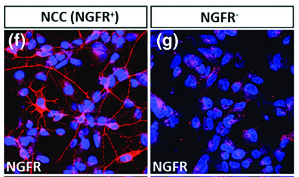
NGFR positive cells were only observed in the enriched population, thus confirming the specificity of the NGFR antibody for NCCs as has been previously documented. No NGFR positive cells were observed in the hESCs cultures. (Jones et al, 2018)
NGFR (ME20.4, p75) Mouse Monoclonal [AB-N07]
AB-N07 also known as ME20.4, recognizes the p75 receptor in human, cat, dog, pig, primate, and sheep.
Applications include flow cytometry, immunoprecipitation, immunohistochemistry, electron microscopy, immunocytochemistry, radioimmunoassay, and targeting.
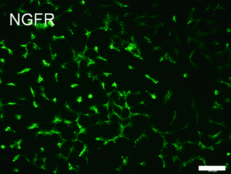
Expression of Neural Crest Cells (NCC) marker NGFR. Immunocytochemistry analysis shows that iPSC-NCCs are positive for NGFR. Cell nuclei were counterstained with DAPI. Scale bars, 100 µm. (Fujii et al, 2019)
NGFR (192-IgG, p75) Mouse Monoclonal [AB-N43]
AB-N43 also known as 192-IgG, recognizes the p75 receptor in rat.
Applications include flow cytometry, immunohistochemistry, and targeting.
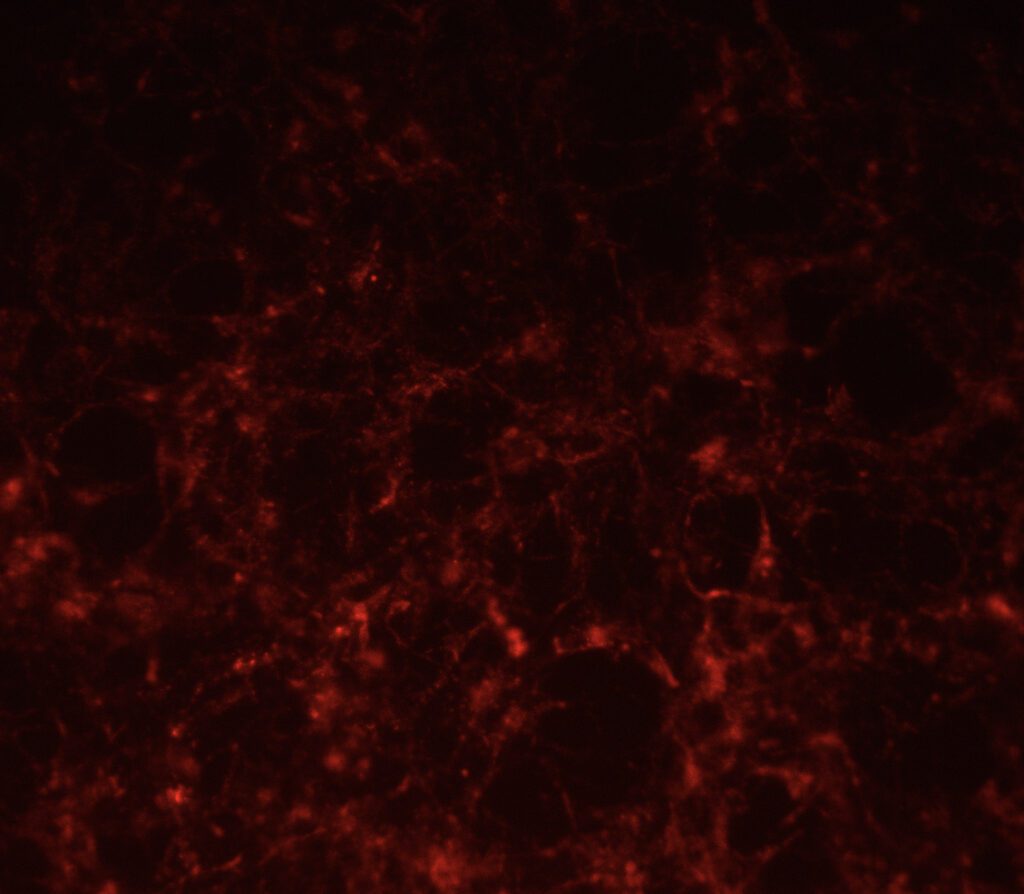
ICC image of Anti-NGFR (192-IgG) illuminated with Fab-pHast mouse.
Melanopsin Antibodies
I am text block. Click edit button to change this text. Lorem ipsum dolor sit amet, consectetur adipiscing elit. Ut elit tellus, luctus nec ullamcorper mattis, pulvinar dapibus leo.
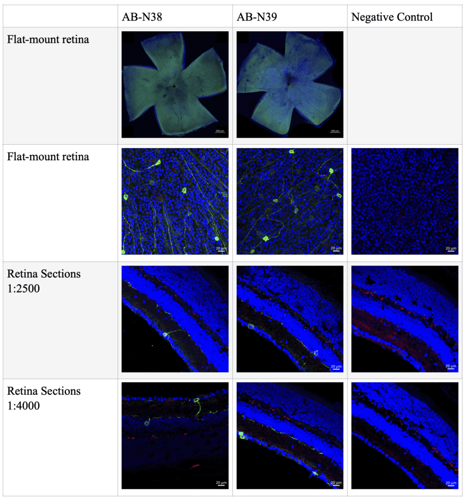
Staining of the antibody is shown in green. Blue is DAPI and tdTomato (red) signal from OPN4cre; R26Syp-tdT mouse line. Images supplied courtesy of Wenjin Xu, Xiarong Lu, and Ignacio Provencio at the University of Virginia.
Melanopsin Rabbit Polyclonal [AB-N38]
Anti-Melanopsin (UF006) recognizes a sequence representing the 15 most N-terminal amino acids of the mouse melanopsin extracellular domain. Anti-Melanopsin (UF006) does not cross-react with melanopsins of other species, it is very specific for mouse.
Applications include immunofluorescence.
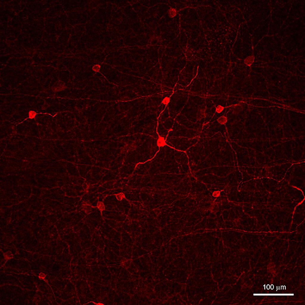
2-photon confocal microscope image from a single z-series using AB-N39 in flat-mount mouse retina at 1:4000 and an AlexaFluor-568 secondary antibody. This image is a single optic plane through the ganglion cell layer that also shows some proximal RGC dendrites. Image courtesy of Matthew Van Hook, Ph.D.
Melanopsin Rabbit Polyclonal, affinity-purified [AB-N39]
Affinity-purified anti-Melanopsin (UF008) was raised against the 15 N-terminal extracellular amino acids of mouse melanopsin. This antibody does not cross-react with melanopsins of other species, it is very specific for mouse.
Applications include immunofluorescence and targeting. This antibody works in both cross-section and flat-mount staining.
