Recombinant Antibodies

Section 1. What is a recombinant antibody?

A recombinant antibody (rAb) is generated from the cloned, in vitro-expressed heavy and light chains of a selected monoclonal antibody obtained through classical hybridoma methodology, display strategies alone or in combination, sorted single B cells from immunized animals, or by one of many other techniques. The fact that it is cloned means the rAb is defined by its primary sequence, which can be engineered to optimize affinity and be expressed in different binder formats. The numerous advantages of rAbs have been well-documented, with consistency of performance being of paramount significance.

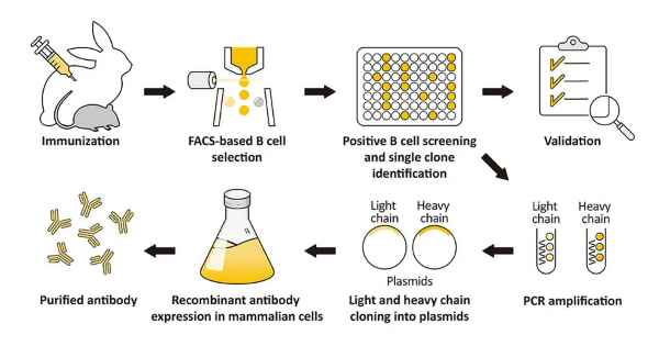
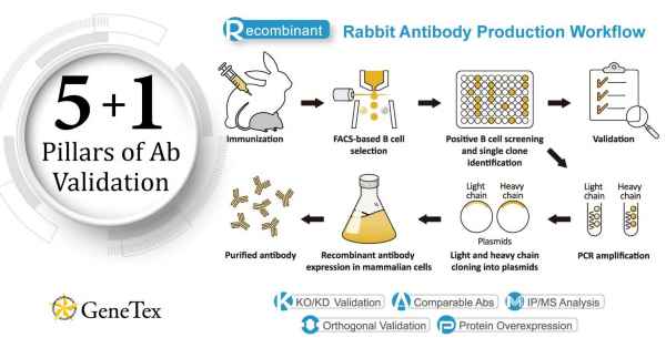
GeneTex’s rAb protocol employs a multi-parameter FACS-based approach to isolate antigen-specific IgG+ memory B cells from an immunized animal, with subsequent cloning of the antibody variable-region genes into an IgG backbone and expression in mammalian cells (Fig. 1) (1). This protocol is very rapid and can be completed in weeks, and also affords the opportunity to identify antibodies with diverse capabilities in various applications. Importantly, it allows cloning of the heavy and light chains from the same B cell, thereby preserving natural pairing. And once cloned, the supply of a given rAb is inexhaustible with exceptional reproducibility.

(A)

(B)

Figure 1. (A) The GeneTex recombinant rabbit monoclonal antibody workflow (Starkie et al. (2016)). (B) Advantages of recombinant antibodies.

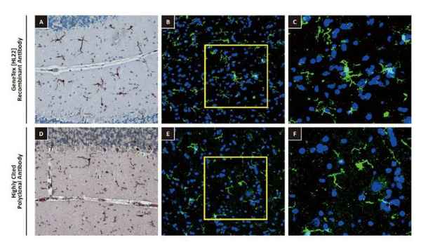
One outstanding advantage of this protocol is that the suitability of the individual clones for desired applications can be tested during screening. For example, GeneTex’s Iba1 rabbit recombinant antibody [HL22] (GTX635363) detects Iba1, a protein commonly used as an immunohistochemical marker of both quiescent and activated microglia. In its development and production, both paraffin- and frozen-IHC analyses (IHC-P, IHC-Fr, respectively) were conducted in the first screening process to identify the best clones for these applications, as shown below (Fig. 2). The clones were compared to a highly cited, market-leading commercial antibody to gauge their performance. This strategy provides us with valuable perspective on a clone’s market competitiveness during development. In addition, our recombinant antibody team has successfully expressed the antigen-binding regions of this antibody in the context of both mouse and rat IgG backbones, thus extending flexibility for multiplexed staining.

Figure 2. GeneTex’s recombinant rabbit monoclonal Iba1 antibody [HL22] (GTX635363) is superior to a competitor’s highly cited rabbit polyclonal antibody for both IHC-P (panels A vs. D) and IHC-Fr (panels B, C vs. E, F).

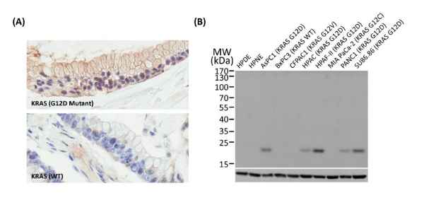
Another new recombinant rabbit monoclonal reagent is GeneTex’s RAS (G12D mutant) antibody [HL10] (GTX635362) (Fig. 3). It demonstrates a robust signal and specificity for the G12D mutation in wild-type (WT) and KRAS G12D mutant pancreatic tumor tissue sections (Fig. 3A) and in established cell line lysates (Fig. 3B), respectively. This RAS (G12D mutant) antibody (GTX635362) is the first commercial recombinant version that shows exceptional specificity by IHC-P for this key tumorigenic mutant protein on sequence-verified human pancreatic tumor samples.

Figure 3. (A) GeneTex’s recombinant rabbit RAS (G12D mutant) antibody [HL10] (GTX635362) is sensitive and specific for the RAS G12D mutation by IHC of a KRAS G12D mutant pancreatic tumor tissue section (top) compared to a wild-type KRAS section (bottom). (B) The antibody is sensitive and specific for the RAS G12D mutation by WB of extracts from wild-type and mutant KRAS-confirmed human pancreatic cell lines. GAPDH for the loading control was detected by GTX100118. Lane1: HPDE. Lane2: HPNE. Lane3: AsPC1 (KRAS G12D). Lane4: BxPC3 (KRAS WT). Lane5: CFPAC1 (KRAS G12V). Lane6: HPAC (KRAS G12D). Lane7: HPAF-II (KRAS G12D). Lane8: MIA PaCa-22 (KRAS G12C). Lane9: PANC1 (KRAS G12D). Lane10: SU86.86 (KRAS G12D).

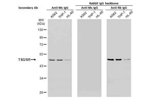
As mentioned above, the antigen-binding regions of a recombinant antibody can be inserted into various host IgG backbones or expressed in different binder formats (e.g., Fab fragments or scFvs). Here, GeneTex’s cloned TSG101 mouse monoclonal antibody (GTX70255) was converted to a rabbit IgG backbone (GTX635396) with preservation of performance. No cross-reaction was observed when the converted TSG101 rabbit IgG recombinant antibody was used in combination with an anti-mouse IgG secondary antibody (Fig. 4).scing elit. Ut elit tellus, luctus nec ullamcorper mattis, pulvinar dapibus leo.

Figure 4. Backbone switch from a cloned mouse IgG antibody to a rabbit IgG antibody.

Literature:

Section 2. GeneTex’s Recombinant Monoclonal Antibody Production Initiative

GeneTex’s production focus is the creation of novel recombinant rabbit monoclonal antibodies validated using enhanced protocols defined by the International Working Group for Antibody Validation (IWGAV).

Over the last decade, studies published in many of the top scientific journals have clearly described the significant impact of poorly validated antibody reagents on the reproducibility of biomedical research. The unfortunate reality is that the commercial antibody market is compromised by a substantial subset of products that have been demonstrated to be nonspecific. This means that there are two major issues that must be addressed in order to improve the overall quality of the commercial antibody market and therefore the integrity and reproducibility of the biomedical research dependent on antibodies. The first is more widespread adoption of production platforms that emphasize recombinant antibody technology (please see “Section 1: What is a recombinant antibody?”) while the second involves incorporation of improved and more coherent validation strategies. GeneTex is making major strides in both of these endeavors.

With the goal of providing better products for its customers through innovative technology, GeneTex has now shifted all of its new production to recombinant rabbit monoclonal antibodies (please see “Section 3: Why GeneTex’s rabbit-based HL clones are a better choice.”). Fully recombinant antibodies are defined by their primary sequence, their consistent performance and supply (issues that plague traditional polyclonals and hybridoma-based monoclonals with regard to lot-to-lot variability), and their ability to be engineered (i.e., backbone switching) to meet researcher requirements. These benefits may even evoke more favorable impressions by grant review and journal editorial committees. Other outstanding advantages of recombinant rabbit antibodies stem from the favorable biological characteristics of the rabbit immune system itself.

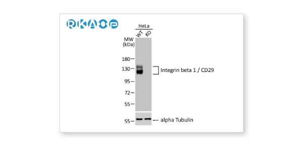
In conjunction with its pivot to recombinant antibody production, GeneTex’s second commitment to antibody reliability is its incorporation of enhanced validation protocols, based on the IWGAV proposal mentioned above, into its manufacturing and quality assurance (QA) workflow (1). This involves evaluation of each antibody using at least one, and preferably more, of five validation “pillars” defined in the IWGAV study. GeneTex’s version of the IWGAV plan follows the five standard strategies and includes (1) Knockout/Knockdown; (2) Comparable Antibodies; (3) Immunoprecipitation followed by Mass Spectrometry (IP/MS); (4) Biological and Orthogonal Validation; and (5) Recombinant Protein Expression (Fig. 1). The company refers to this as its “5+1” Pillar Plan when referencing new recombinant antibody production, with the “+1” designation referring to the pre-validation that is inherent in the application-specific assessment that occurs with clone selection during the recombinant antibody production process (please see “New Validated Recombinant Monoclonal Antibodies from GeneTex” below).

The new recombinant rabbit monoclonal antibodies that have been generated and evaluated using the production and validation approaches described above (these products are identified by “HL” in their clone designations) are just the beginning. GeneTex is also working to replace its successful polyclonals with new recombinants. The company envisions the “HL” clones representing the highest level of GeneTex validation and manufacturing capability.

Antibody fidelity, application-dependent reliability, and performance and supply consistency are crucial for ensuring research integrity. GeneTex joins other reputable companies in leveraging recombinant antibody technology and enhanced validation to create antibodies that researchers can trust.

Figure 1. GeneTex’s “5+1” Pillar Plan for recombinant antibody validation.

New Validated Recombinant Monoclonal Antibodies from GeneTex

Complete removal or significant reduction of the endogenous signal following genetic strategies involving genome editing or RNA interference, respectively.

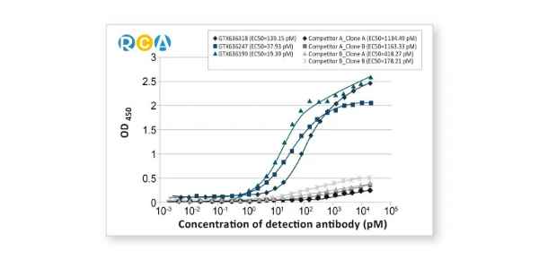
Comparable Antibodies

Independent antibodies against the same target but to different epitopes, often including distinct samples with different target protein expression levels.

Biological and Orthogonal Validation

Alterations in the endogenous level of the target protein in accordance with specific preparation conditions corresponding to defined biological characteristics, or comparison between antibody-dependent and -independent methods.

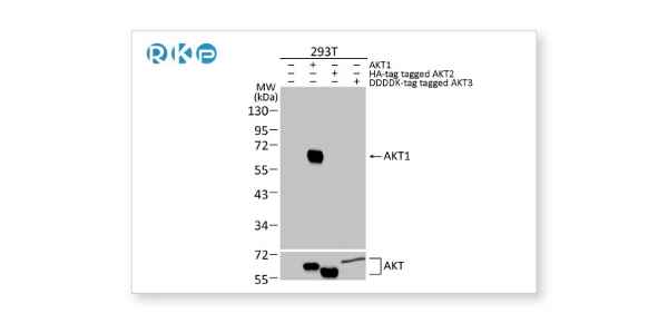
Protein Overexpression

Tagged target proteins overexpressed in transfected cells are used as a positive control for validation.

Recombinant Antibody

Antibody is generated using recombinant technology.

Section 3. Why GeneTex’s rabbit-based HL clones are a better choice.

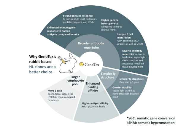
GeneTex’s recombinant rabbit monoclonal antibody production process is the platform for all new rabbit-based antibody generation (1). These products are distinguished by the “HL” designation in their respective clone numbers (e.g., “[HL1089]”). The rabbit, and specifically its immune system, is known to confer the following outstanding benefits for monoclonal antibody creation: broader antibody range, less complex immunoglobulin (Ig) structure, higher binding affinities, larger pool of lymphocytes to select antibodies (2). Each of these advantages is described in more detail below:

1. Broader antibody repertoire:

a) Rabbits belong to the order Lagomorpha, which is evolutionarily distinct from the order Rodentia. This means that human antigens present epitopes that are often more immunogenic in rabbits than in mice, expanding the number of generated antibodies that may in turn cross-react with murine protein orthologs (2-5).

b) The rabbit immune system is more successful in mounting an immune response to hapten or small molecule antigens, which is frequently not the case with the murine immune system (2-5).

c) Mice used to generate monoclonal antibodies are from inbred lines and offer less diversity in their immunogenic responses than do rabbits. In addition, mouse spleens are much smaller than rabbit spleens (2-5).

d) The rabbit immune system is different from those of mice and humans in that it utilizes somatic gene conversion (SGC) to expand its antibody range in addition to performing the same VDJ and VJ recombination and somatic hypermutation (SHM) mechanisms noted in mice and humans. Antibody diversity is further enhanced by other processes that include a kappa light chain variable gene structure distinctive for variability in the length of complementarity determining region 3 (LCDR3), as well as successive Ig development in the bone marrow, the gut-associated lymphoid tissues (GALT), and spleen and lymph node germinal centers (3, 5).

2. Simpler Ig structure:

a) The European rabbit (Oryctolagus cuniculus) is known to have single genes for IgE and IgM, and no genes for IgD. Interestingly, its genome contains 13 IGHA genes coding for at least 10 functional IgA subclasses. Importantly, though there are allelic variants, rabbits have only one IgG gene (and thus no subclass) and therefore differ from mice and humans (each with four IgG subclasses) (4, 5).

b) In addition to the disulfide bonds found in a human or mouse IgG (linking the heavy chain pair or one heavy chain to its associated light chain), rabbit IgG antibodies demonstrate a unique intrachain disulfide bond in their K1 light chain. This is significant as perhaps 90% of IgGs in New Zealand White rabbits are IgG-k (K1), and this extra bond is thought to confer stability onto the rabbit IgG (2, 5).

3. Enhanced binding affinity:

a)Rabbit monoclonal antibodies have very high affinities with Kd values characteristically in the picomolar range, with some possessing exceptionally low picomolar Kd values (2, 5).

b) Picomolar Kds translate to increased sensitivity with consistent specificity.

4. Larger lymphocyte pool:

a) Rabbits have a longer life span and their larger size (perhaps a 100-fold weight differential between a three-month-old rabbit and a six-week-old mouse) greatly facilitates blood and tissue sampling during antibody production (5).

b) Consistent with the statements above, rabbit spleens allow isolation of 50-fold more B cells compared to murine spleens, meaning more clones recognizing a wider selection of epitopes (2-5).

GeneTex hopes that the distinct advantages of rabbit monoclonal antibodies discussed above will encourage you to consider our HL clones for your research needs. As GeneTex has now shifted all of its new antibody production to the creation of these monoclonal antibodies, our product inventory will continue to expand rapidly moving forward.


GeneTex’s Well-validated HL Clone Antibodies

Section 4. IVD-related Applications for GeneTex’s Recombinant Antibodies

Advantages:

Superior Quality
Performance Consistency
Bulk Production (gram quantities)
International QMS Certifications:

Production in QMS IVD Laboratory
Validated Antibody Pairs for IVD
Product Development and Services

IVD-related Services:

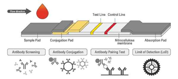
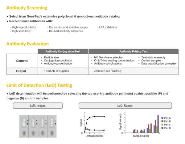
  1. Lateral Flow Assay (LFA) Development Service
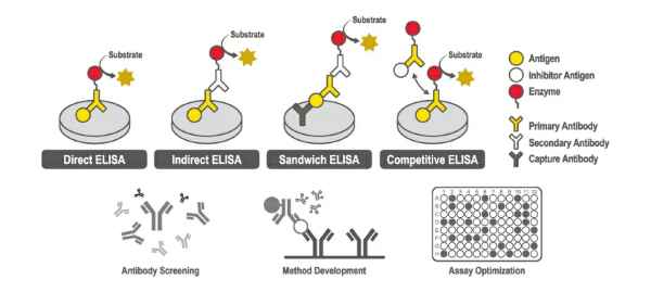
  2. Enzyme-Linked Immunosorbent Assay (ELISA) Development Service
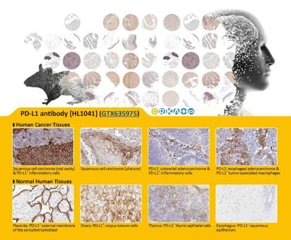
  3. Immunohistochemistry (IHC)-Validated Recombinant Antibodies


Lateral Flow Assay (LFA) Development Service

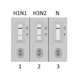
Influenza A Virus (FluA)

Capture:
Influenza A Virus Nucleoprotein antibody
[HL1078] (GTX636199)

Detection:
Influenza A Virus Nucleoprotein antibody
[HL1953] (GTX637790)

Samples:
1. FluA Nucleoprotein (H1N1) (GTX135904-pro)
2. FluA Nucleoprotein (H3N2) (GTX136317-pro)
3. Lysis buffer

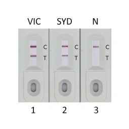
Influenza B Virus (FluB)

Capture:
Influenza B Virus Nucleoprotein antibody
[HL1069] (GTX636100)

Detection:
Influenza B Virus Nucleoprotein antibody
[HL1068] (GTX636099)

Samples:
1. Influenza B Virus Nucleoprotein (B/Victoria/02/1987) (GTX136277-pro)
2. Influenza B Virus Nucleoprotein (B/Sydney/3/2004) (GTX135867-pro)
3. Lysis buffer

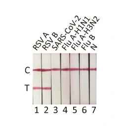
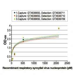
Respiratory Syncytial Virus (RSV)

Capture:
Respiratory Syncytial Virus Nucleoprotein
antibody [HL1248] (GTX636650)

Detection:
Respiratory Syncytial Virus Nucleoprotein
antibody [HL1246] (GTX636648)

Samples:
1. RSV type A Nucleoprotein (GTX136751-pro)
2. RSV type B Nucleoprotein (GTX136931-pro)
3. SARS-CoV-2 Nucleocapsid protein (GTX135592-pro)
4. FluA (H1N1) Nucleoprotein (GTX135904-pro)
5. FluA (H3N2) Nucleoprotein (GTX135903-pro)
6. FluB Nucleoprotein (GTX136277-pro)
7. Lysis buffer

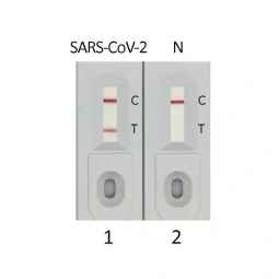
SARS-CoV-2 (COVID-19)

Capture:
SARS-CoV-2 (COVID-19) Nucleocapsid antibody
[HL455] (GTX635688)

Detection:
SARS-CoV-2 (COVID-19) Nucleocapsid antibody
[HL448] (GTX635686)

Samples:
1. SARS-CoV-2 (COVID-19) Nucleocapsid protein (GTX135592-pro)
2. Lysis buffer

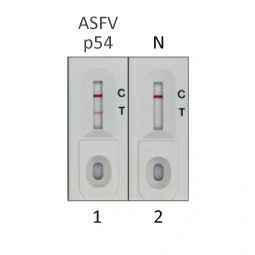
African Swine Fever Virus

Capture:
ASFV p54 antibody [HL1287]
(GTX636701)

Detection:
ASFV p54 antibody [GT1075]
(GTX635691)

Samples:
1. ASFV p54 (ECD) protein, His Tag (GTX135175-pro)
2. Lysis buffer

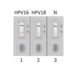
Human Papillomavirus type 16 E7

Capture:
Human Papillomavirus type 16 E7 antibody
[HL1647] (GTX637228)

Detection:
Human Papillomavirus type 16 E7 antibody
[HL1821] (GTX637546)

Samples:
1. HPV16 E7 protein (GTX133411-pro)
2. HPV18 E7 protein (GTX133412-pro)
3. Lysis buffer

Enzyme-Linked Immunosorbent Assay (ELISA) Development Service

Direct/Indirect ELISA

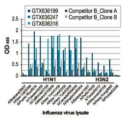
Virus subtype detection

Direct/Indirect ELISA

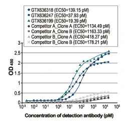
EC50 determination

Sandwich ELISA

Target protein detection and quantitation

Sandwich ELISA

Antibody pair screening

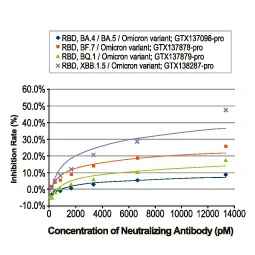
Competitive ELISA

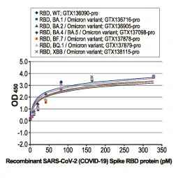
Neutralizing antibody evaluation

Immunohistochemistry (IHC)-Validated Recombinant Antibodies

Breast Cancer

Colon Cancer

Cervical Cancer

Hepatocellular Cancer

Lung Cancer

Esophageal Cancer

Glioblastoma

Ovarian Cancer

Pancreatic Cancer

Prostate  Cancer

Mouse/Rat Tissue Arrays

SUPPORT

outstanding technical support

PRODUCT

we offer a full product guarantee

DELIVERY

we offer free delivery to UK universities and non profit organisations