Polystyrene Microspheres

Polystyrene microspheres present a flexible platform for applications in diagnostics and bioseparations. They may be coated with recognition molecules, such as antibodies, antigens, peptides, or nucleic acid probes, and can be loaded with hydrophobic dyes and other compounds. Unmodified polymer spheres also find extensive use as standards for instrument set-up and calibration. Available in diameters ranging from 20nm to 200µm, products exhibit excellent size uniformity.

Plain polystyrene microspheres are ideal for protein adsorption, and have been utilized in a range of diagnostic tests and assays. Surface modified microspheres are available with carboxyl or primary amine groups for covalent ligand attachment. Affinity binding systems offer simple and efficient ligand attachment. Coatings of Fc binding proteins are able to orient antibodies for optimal activity, and streptavidin offers extremely stable attachment of biotinylated molecules, such as proteins, peptides, and oligonucleotides.

We offer uniform polystyrene latex microspheres that may be used as-is for standards or markers, or coated with proteins via adsorption for use in diagnostic tests and assays. See TechNote 204, Adsorption to Microspheres. Our spheres are synthesized via emulsion polymerization, and are available in diameters from ~20nm-90µm, with typical size CVs of 5-10%. For applications that require highly stringent CVs (e.g. 2-5%), call for product availability. Most products are supplied as a 10% solids suspension (100mg/mL) in de-ionized water with surfactant and sodium azide.

PS AND PS/DVB

Many proteins may be easily and stably absorbed to hydrophobic, non-functionalized microspheres. Divinylbenzene (DVB) confers additional solvent and heat resistance to our crosslinked spheres. Unless noted otherwise, PS and PS/DVB microspheres are available as ~10% solids (w/w) aqueous suspensions in these standard amounts: 0.5g, 1.0g, 1.5g, and 5.0g.

*Please note: If you are NOT ordering via website, please specify a lot number if multiple lots are available.
Catalog Number Nominal Diameter Specification Range
PS02001 0.025µm 0.015 – 0.035µm
PS02002 0.050µm 0.040 – 0.060µm
PS02003 0.075µm 0.065 – 0.085µm
PS02004 0.100µm 0.090 – 0.110µm
PS02005 0.125µm 0.115 – 0.135µm
PS02006 0.150µm 0.140 – 0.160µm
PS02007 0.175µm 0.165 – 0.185µm
PS02008 0.200µm 0.190 – 0.210µm
PS02009 0.300µm 0.270 – 0.330µm
PS02010 0.400µm 0.370 – 0.430µm
PS03001 0.500µm 0.470 – 0.530µm
PS03002 0.600µm 0.570 – 0.630µm
PS03003 0.700µm 0.670 – 0.730µm
PS03004 0.800µm 0.770 – 0.830µm
PS03005 0.900µm 0.870 – 0.930µm
PS04001 1.00µm 0.95 – 1.05µm
PS05001 2.00µm 1.80 – 2.20µm
PS05002 3.00µm 2.80 – 3.20µm
PS05003 4.00µm 3.80 – 4.20µm
PS06001 5.00µm 4.80 – 5.20µm
PS06002 5.50µm 5.30 – 5.70µm
PS06003 6.00µm 5.80 – 6.20µm
PS06004 7.00µm 6.80 – 7.20µm
PS06005 7.50µm 7.30 – 7.70µm
PS07001 10.00µm 9.50 – 10.50µm
PS07002 15.00µm 14.50 – 15.50µm
PS07003    20.00µm 19.00 – 21.00µm
PS08001 >25.µm >25.00µm

REFERENCES

  • Joubert, M, Hokom, M, Eakin, C, Zhou, L, Deshpande, M, Baker,M, …Jawa, J (2012) Highly Aggregated Antibody Therapeutics Can Enhance the in Vitro Innate and Late-stage T-cell Immune Responses J Biol Chem 287:25266
  • Noyhouzer, T., Perry, S. C., Vicente-Luis, A., Hayes, P. L., & Mauzeroll, J. (2018). The Best of Both Worlds: Combining Ultramicroelectrode and Flow Cell Technologies. Journal of The Electrochemical Society, 165(2), H10-H15. (Cat. PS03005, 0.9µm PS nanoparticles)
  • Fernandez-Bravo A, Yao K, Barnard ES, Borys N, Levy E, Tian B, … Schuck JP. (2018) Continuous-wave upconverting nanoparticle microlasers.Nature Nanotech;06/18.1748-3395 (nanoparticle-coated 5µm PS beads)
  • Arifin, N, Yunus, M H, Nolan, TJ, Lok, JB, & Noordin, R. (2018). Identification and Preliminary Evaluation of a Novel Recombinant Protein for Serodiagnosis of Strongyloidiasis. (PS02002, 0.5µm PS microspheres to pre-adsorb serum samples for immunoscreening)
  • Choy C H, & Botelho RJ. (2017). Quantifying phagocytosis by immunofluorescence and microscopy. In Phagocytosis and Phagosomes (pp. 43-53). Humana Press, New York, NY.(PS05003, 3.87µm PS, IgG opsonized with fluorescent secondary Ab)
  • Gigault C, Dalnoki-Veress K, & Dutcher JR. (2001) Changes in the morphology of self-assembled polystyrene microsphere monolayers produced by annealing. Journal of colloid and interface science, 243(1), 143-155.(1µm PS used to fabricate self-assembled monolayers)

Our carboxylate-modified polystyrene latex microspheres are suitable for the covalent immobilization of proteins, peptides, and nucleic acids. They are synthesized via emulsion polymerization, and are available in diameters up to ~20µm, with typical size CVs of 10%. Most products are supplied as 10% solids suspension (w/w) in de-ionized water with surfactant and sodium azide.

CARBOXYL POLYSTYRENE

EDAC-mediated coupling is often used for the covalent immobilization of amine-terminated proteins, DNA, or other molecules on carboxyl-functionalized PS and P(S/DVB) microspheres. Carboxyl polymer microspheres are available as ~10% solids (w/w) aqueous suspensions in these standard amounts: 0.5g, 1.0g, 1.5g, and 5.0g.

New to beads? See our PolyLink Protein Coupling Kit and TechNote 205, Covalent Coupling, for a protocol and tips on coating optimization and QC.

Catalog Number Nominal Diameter Specification Range
PC02001 0.025µm 0.015 – 0.035µm
PC02002 0.050µm 0.040 – 0.060µm
PC02003 0.070µm 0.061 – 0.080µm
PC02004 0.100µm 0.090 – 0.110µm
PC02005 0.125µm 0.115 – 0.135µm
PC02006 0.150µm 0.140 – 0.160µm
PC02007 0.175µm 0.165 – 0.185µm
PC02008 0.200µm 0.190-0.210µm
PC02009 0.300µm 0.270-0.330µm
PC02010 0.350µm 0.340 – 0.360µm
PC02011 0.400µm 0.370 – 0.430µm
PC03001 0.500µm 0.470 – 0.530µm
PC03002 0.600µm 0.570 – 0.630µm
PC03003 0.800µm 0.770 – 0.830µm
PC03004 0.900µm 0.870 – 0.930µm
PC04001 1.0µm 0.95 – 1.05µm
PC05001 2.00µm 1.80 – 2.20µm
PC05002 2.50µm 2.30 – 2.70µm
PC05003 3.00µm 2.80 – 3.20µm
PC05004 4.00µm 3.80 – 4.20µm
PC05005 4.50µm 4.30 – 4.70µm
PC06001 5.00µm 4.80 – 5.20µm
PC06002 5.50µm 5.30 – 5.70µm
PC06003 6.00µm 5.80 – 6.20µm
PC06004 7.00µm 6.80 – 7.20µm
PC07001 10.00µm 9.50 – 10.50µm
PC07002 15.00µm 14.50µm – 15.50µm
PC07003 20.00µm 19.00µm – 21.00µm
PC08001 >25.µm >25.00µm
*Please note: If you are NOT ordering via website, please specify a lot number if multiple lots are available.

REFERENCES

  • Park, MK, Briles, DE, Nahm, MH (2000) A Latex Bead-Based Flow Cytometric Immunoassay Capable of Simultaneous Typing of Multiple Pneumococcal Serotypes (Multibead Assay) Clin Vaccine Immunol v7(3) 486-489
  • Blanchette C, Lacayo CI, Fischer NO, Hwang M, Thelen MP (2012) Enhanced Cellulose Degradation Using Cellulase-Nanosphere Complexes. PLoS ONE 7(8): e42116
  • Rodrigues OR, & Monard S, (2016) A rapid method to verify single-cell deposition setup for cell sorters. Cytometry 89: 594–600
  • Borque L, Bellod L, Rus A, Seco L, Galisteo-Gonzàles F (2000) Development and Validation of an Automated and Ultrasensitive Immunoturbidimetric Assay for C-Reactive Protein. Clin Chem 46(11) 1839-1842
  • Perez-Amodio S, Holownia P, Davey C, Price C (2001) Effects of the Ionic Environment, Charge, and Particle Surface Chemistry for Enhancing a Latex Homogeneous Immunoassay of C-Reactive Protein. Anal. Chem.73 (14) 3417–3425
  • Lucas L, Han J, Yoon J (2006) Using highly carboxylated microspheres to simplify immunoassays and enhance diffusional mixing in a microfluidic device. Colloids and Surf. B: Biointerfaces 49(2) 106-111
  • Fu L, Kang C, Zhang P, Kang X, Li X, Wang H, …Qiu Y (2015) A modified quick PETIA for detecting anti-CCP antibodies in human serum, Anal Methods Issue 7
  • Deegan O, Walshe K, Kavanagh K, Doyle S. (2003) Quantitative detection of C-reactive protein using phosphocholine-labelled enzyme or microspheres. Anal. Biochem. 312 p175–181 (turbidimetric assay, 0.21µm PS-COOH)
  • Koskinen JM, Soukka JM, Meltola NJ, & Koskinen JO. (2018). Microbial identification from faces and urine in one step by two-photon excitation assay technique. Journal of immunological methods, 460, 113-118.(Cat. PC05003, 3.2µm PS-COOH microspheres, coated with anti-Legionella or anti-adenovirus Ab in ArcDia’s TPX assay.)
  • Nanavaty N, Lin L, Hinckley SH, & Kuret J. (2017) Detection and quantification methods for fibrillar products of in vitro tau aggregation assays. In Tau Protein (pp. 101-111). Humana Press, New York, NY.(Anionic PS-COOH, 40-90nm as tau aggregation inducers)

AMINE POLYSTYRENE

Amine-functionalized microspheres offer further options for coupling of biomolecules. Glutaraldehyde is commonly used for the immobilization of amine-containing compounds, and specialized linkers are available for targeted coupling of thiol or carbohydrate moieties. See TechNote 205, Covalent Coupling. Unless otherwise noted amine polymer microspheres are available as ~10% solids (w/w) aqueous suspensions in these standard amounts: 0.5g, 1.0g, 1.5g, and 5.0g. For microspheres larger than 1.00µm, please call for availability.

*Please note: If you are NOT ordering via website, please specify a lot number if multiple lots are available.
Catalog Number Nominal Diameter Specification Range
PA02001 0.200µm 0.190 – 0.210µm
PA03001 0.500µm 0.470 – 0.530µm
PA03002 0.750µm 0.720 – 0.780µm
PA04001 1.0µm 0.95 – 1.05µm

REFERENCES

  • Schleicher KD, Dettmer SL, Kapinos LE, Pagliara S, Keyser UF, Jeney S, LimRoderick YH (2014) Selective transport control on molecular velcro made from intrinsically disordered proteins. Nat Nano 9, 525-530
  • Bandura DR, Baranov VI, Ornatsky OI, Antonov A, Kinach R, Lou X… Tanner S.(2009) Mass Cytometry: Technique for Real Time Single Cell Multitarget Immunoassay Based on Inductively Coupled Plasma Time-of-Flight Mass Spectrometry. Anal Chem 81,6813–6822
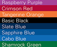
Visibly dyed microspheres are frequently used in lateral flow tests and “latex” agglutination tests. Our polystyrene microspheres are impregnated with vibrant dyes for optimal visualization. The colorpalette for a representation of our standard colors. Non-functionalized and functionalized versions are available to support adsorption and covalent binding strategies.

DYED POLYSTYRENE

Our non-functionalized microspheres are suitable for coating with antibody or other large proteins via adsorption. See TechNote 204, Adsorption to Microspheres, for a general adsorption protocol. Unless noted otherwise, these visibly dyed microspheres are supplied as a 5% solids suspension (w/v) and are available in these standard amounts: 0.5g, 1.0g, 1.5g, and 5.0g.

*Please note: If you are NOT ordering via website, please specify a lot number if multiple lots are available.
Catalog Number Dye Nominal Diameter Specification Range Product Data Sheet
DSCR001 Crimson Red 0.050µm 0.040 – 0.060µm PDS 717.pdf
DSCB002 Cabo Blue 0.200µm 0.190 – 0.210µm PDS 717.pdf
DSCR002 Crimson Red 0.200µm 0.190 – 0.210µm PDS 717.pdf
DSSG002 Shamrock Green 0.200µm 0.190 – 0.210µm PDS 717.pdf
DSBK002 Basic Black 0.200µm 0.190 – 0.210µm PDS 717.pdf
DSCR003 Crimson Red 0.300µm 0.270 – 0.330µm PDS 717.pdf
DSCR004 Crimson Red 0.400µm 0.370 – 0.430µm PDS 717.pdf
DSCB005 Cabo Blue 0.80µm 0.770 – 0.830µm PDS 717.pdf
DSCR005 Crimson Red 0.80µm 0.770 – 0.830µm PDS 717.pdf
DSSG005 Shamrock Green 0.80µm 0.770 – 0.830µm PDS 717.pdf
DSBK005 Basic Black 0.80µm 0.770 – 0.830µm PDS 717.pdf
DSCR006 Crimson Red 5.0µm 4.80 – 5.20µm PDS 717.pdf

References

Henderson, K., & Stewart, J. (2002). Factors influencing the measurement of oestrone sulphate by dipstick particle capture immunoassay. Journal of immunological methods, 270(1), 77-84.(0.3, 0.5, and 0.8µm dyed blue PS)

Henderson, K., & Stewart, J. (2000). A dipstick immunoassay to rapidly measure serum oestrone sulfate concentrations in horses. Reproduction, Fertility and Development, 12(4), 183-189.(0.31µm dyed blue PS)

Campbell, K., Fodey, T., Flint, J., Danks, C., Danaher, M., O’Keeffe, M., … & Elliott, C. (2007). Development and validation of a lateral flow device for the detection of nicarbazin contamination in poultry feeds. Journal of agricultural and food chemistry, 55(6), 2497-2503.(0.43µm dyed blue PS)

DYED CARBOXYL POLYSTYRENE

Biomolecules may be covalently immobilized to carboxyl-functionalized microspheres. A general coupling protocol is provided within TechNote 205, Covalent Coupling. Our dyed carboxyl microspheres are impregnated with vibrant dyes for optimal visualization. See our color palette for a representation of our standard colors. Unless noted otherwise, they are supplied as a 5% solids suspension (w/v) and are available in four standard volumes: 0.5g, 1.0g, 1.5g, and 5.0g.

*Please note: If you are NOT ordering via website, please specify a lot number if multiple lots are available.
Catalog Number Dye Nominal Diameter Specification Range Product Data Sheet
DCCB001 Cabo Blue 0.20µm 0.190 – 0.210µm PDS 717.pdf
DCCR001 Crimson Red 0.20µm 0.190 – 0.210µm PDS 717.pdf
DCSG001 Shamrock Green 0.20µm 0.190 – 0.210µm PDS 717.pdf
DCBK001 Basic Black 0.20µm 0.190 – 0.210µm PDS 717.pdf
DCCB002 Cabo Blue 0.50µm 0.470 – 0.530µm PDS 717.pdf
DCCR002 Crimson Red 0.50µm 0.470 – 0.530µm PDS 717.pdf
DCCB004 Cabo Blue 1.00µm 0.95 – 1.05µm PDS 717.pdf
DCCR004 Crimson Red 1.00µm 0.95 – 1.05µm PDS 717.pdf
DCTA004 Tangerine Orange 1.00µm 0.95 – 1.05µm PDS 717.pdf
DCSG004 Shamrock Green 1.00µm 0.95 – 1.05µm PDS 717.pdf
DCCB005 Cabo Blue 5.00µm 4.80 – 5.20µm PDS 717.pdf
DCCR005 Crimson Red 5.00µm 4.80 – 5.20µm PDS 717.pdf

REFERENCES

Terao, Y, Takeshita, K, Nishiyama, Y, Morishita, N, Matsumoto, T, & Morimatsu, F. (2015) Promising Nucleic Acid Lateral Flow Assay Plus PCR for Shiga Toxin–Producing Escherichia coli. Journ of food protection, 78(8), 1560-1568.

Sheng, W, Li, S, Liu, Y, Wang, J, Zhang, Y, & Wang, S. (2017). Visual and rapid lateral flow immunochromatographic assay for enrofloxacin using dyed polymer microspheres and quantum dots. Microchimica Acta, 184(11), 4313-4321.(Dyed Black carboxyl microspheres)

DYED PROTEIN-COATED POLYSTYRENE

At times, our inventory includes streptavidin-coated dyed microspheres supplied as ~1% solids (w/v) aqueous suspensions.

Catalog Number Coating / Dye Nominal Diameter Specification Range
CDCR001 SA / Crimson Red 0.20µm 0.190 – 0.210µm
CDCB001 SA / Cabo Blue 0.20µm 0.190 – 0.210µm

REFERENCES

  • Li S, Liu Y, Wang Y, Wang M, Liu C, & Wang Y. (2019) Rapid detection of Brucella spp. and elimination of carryover using multiple cross displacement amplification coupled with nanoparticles-based lateral flow biosensor. Frontiers in Cellular and Infection Microbiology, 9, 78. (Streptavidin Crimson Red nanoparticles [CP01R] for binding of biotinylated Brucella spp. amplicons in lateral flow biosensor.)

Fluorescent microspheres are a mainstay in diagnostics and life sciences research, finding use as fluorescent markers, assay substrates, and instrument standards. In order to meet the unique performance criteria required of microparticles in these different applications we stock an extensive inventory of fluorescent microspheres and offer custom dyeing services.

FLUORESCENT POLYSTYRENE

Our non-functionalized microspheres are suitable for coating via adsorption. See spectra for available fluorescent dyes. See TechNote 204, Adsorption to Microspheres, for a general adsorption protocol. Fluorescent polymer microspheres are available in these standard volumes: 1mL, 5mL, 10mL, and 100mL.

*Please note: If you are NOT ordering via website, please specify a lot number if multiple lots are available.
Catalog Number Fluorescence Color Nominal Diameter Specification Range
FSDG001 Dragon Green 0.05µm 0.040 – 0.060µm
FSFR001 Flash Red 0.05µm 0.040 – 0.060µm
FSDG002 Dragon Green 0.20µm 0.190 – 0.210µm
FSSY002 Suncoast Yellow 0.20µm 0.190 – 0.210µm
FSFR002 Flash Red 0.20µm 0.190 – 0.210µm
FSDG003 Dragon Green 0.50µm 0.470 – 0.530µm
FSPP003 Plum Purple 0.50µm 0.470 – 0.530µm
FSFR003 Flash Red 0.50µm 0.470 – 0.530µm
FSDG004 Dragon Green 1.00µm 0.95 – 1.05µm
FSEG004 Envy Green 1.00µm 0.95 – 1.05µm
FSPP004 Plum Purple 1.00µm 0.95 – 1.05µm
FSFR004 Flash Red 1.00µm 0.95 – 1.05µm
FSDG005 Dragon Green 2.00µm 1.80 – 2.20µm
FSPP005 Plum Purple 2.00µm 1.80 – 2.20µm
FSFR005 Flash Red 2.00µm 1.80 – 2.20µm
FSDG006 Dragon Green 4.00µm 3.80 – 4.20µm
FSEG006 Envy Green 4.00µm 3.80 – 4.20µm
FSFR006 Flash Red 4.00µm 3.80 – 4.20µm
FSDG007 Dragon Green 7.5µm 7.30 – 7.70µm
FSSY007 Suncoast Yellow 7.5µm 7.30 – 7.70µm
FSFR007 Flash Red 7.5µm 7.30 – 7.70µm
FSEG008 Envy Green 10.0µm 9.50 – 10.50µm
FSDG009 Dragon Green 15.00µm 14.5 – 15.50µm
FSDG011 Dragon Green >25.00µm >25.00µm

FLUORESCENT CARBOXYL POLYSTYRENE

Biomolecules may be covalently immobilized to carboxyl-functionalized microspheres. A general coupling protocol is provided in TechNote 205, Covalent Coupling. Fluorescent carboxyl microspheres and Europium chelate microspheres are available in these standard volumes: 1mL, 5mL, 10mL, and 100mL. See spectra for available fluorescent dyes. See also our Quantumplex™ kits for suspension array development.

*Please note: If you are NOT ordering via website, please specify a lot number if multiple lots are available.
Catalog Number Fluorescence Color Nominal Diameter Specification Range
FCDG001 Dragon Green 0.05µm 0.040 – 0.060µm
FCFR001 Flash Red 0.05µm 0.040 – 0.060µm
FCDG002 Dragon Green 0.10µm 0.090 – 0.110µm
FCDG003 Dragon Green 0.20µm 0.190 – 0.210µm
FCSG003 Surf Green 0.20µm 0.190 – 0.210µm
FCFR003 Flash Red 0.20µm 0.190 – 0.210µm
FCGB003 Glacial Blue 0.20µm 0.190 – 0.210µm
FCDG004 Dragon Green 0.40µm 0.370 -0.430µm
FCFR004 Flash Red 0.40µm 0.370 -0.430µm
FCDG005 Dragon Green 0.50µm 0.470 – 0.530µm
FCFR005 Flash Red 0.50µm 0.470 – 0.530µm
FCDG006 Dragon Green 1.0µm 0.95 – 1.05µm
FCEG006 Envy Green 1.0µm 0.95 – 1.05µm
FCSY006 Suncoast Yellow 1.0µm 0.95 – 1.05µm
FCGB006 Glacial Blue 1.0µm 0.95 – 1.05µm
FCFR006 Flash Red 1.0µm 0.95 – 1.05µm
FCSY007 Suncoast Yellow 2.0µm 1.80 – 2.20µm
FCDG008 Dragon Green 5.0µm 4.80 – 5.20µm
FCEG008 Envy Green 5.0µm 4.80 – 5.20µm
FCGB008 Glacial Blue 5.0µm 4.80 – 5.20µm
FCFR008 Flash Red    5.0µm 4.80 – 5.20µm
FCDG009 Dragon Green 10.0µm 9.50 – 10.50µm

EUROPIUM CHELATE

Our europium products offer extremely bright fluorescence and exceptional stability, in addition to well functionalized carboxylated surfaces for the covalent attachment of ligand. See PDS 741.pdf. See spectra for available fluorescent dyes.

Catalog Number Fluorescence Color Nominal Diameter Specification Range
FCEU001 Europium Chelate 0.10µm 0.090 – 0.110µm
FCEU002 Europium Chelate 0.20µm 0.190 – 0.210µm
FCEU003 Europium Chelate 0.30µm 0.270 – 0.330µm

FLUORESCENT MAGNETIC POLYMER

Both Encapsulated (ME catalog codes) and Classical (MC catalog codes) superparamagnetic microspheres are synthesized using a process that results in the distribution of iron oxide crystals throughout the polymer matrix. For specific information on the use of fluorescent magnetic particles for cell labeling & tracking, see TSD 0748. The surfaces of Encapsulated magnetic microspheres are expected to be free of iron oxide, while the Classical version will have some exposed iron oxide. COMPEL microspheres (UMC catalog codes) are highly uniform and spherical. Fluorescent magnetic microspheres are available in these standard volumes: 1mL, 5mL, 10mL, and 100mL. See spectra for available fluorescent dyes.

*Please note: If you are NOT ordering via website, please specify a lot number if multiple lots are available.
Catalog Number Description Fluorescence Color Nominal Diameter Specification Range
MCDG001 Classical Magnetic Polymer Dragon Green 0.9µm 0.50 – 2.00µm
MCFR001 Classical Magnetic Polymer Flash Red 0.9µm 0.50 – 2.00µm
MEDG001 Encapsulated Magnetic Polymer Dragon Green .86µm 0.50 – 2.00µm
MEFR001 Encapsulated Magnetic Polymer Flash Red .86µm 0.50 – 2.00µm
MEDG002 Encapsulated Magnetic Polymer Dragon Green 1.63µm 0.50 – 2.00µm
MESY002 Encapsulated Magnetic Polymer Suncoast Yellow 1.63µm 0.50 – 2.00µm
MEGB002 Encapsulated Magnetic Polymer Glacial Blue 1.63µm 0.50 – 2.00µm
MEFR002 Encapsulated Magnetic Polymer Flash Red 1.63µm 0.50 – 2.00µm
UMGB001 COMPEL™ Magnetic COOH modified Glacial Blue 3µm 2.50 – 3.50µm
UMDG001 COMPEL™ Magnetic COOH modified Dragon Green 3µm 2.50 – 3.50µm
UMEG001 COMPEL™ Magnetic COOH modified Envy Green 3µm 2.50 – 3.50µm
UMFR001 COMPEL™ Magnetic COOH modified Flash Red 3µm 2.50 – 3.50µm
UMGB002 COMPEL™ Magnetic COOH modified Glacial Blue 6µm 5.50 – 6.50µm
UMDG002 COMPEL™ Magnetic COOH modified Dragon Green 6µm 5.50 – 6.50µm
UMFR002 COMPEL™ Magnetic COOH modified Flash Red 6µm 5.50 – 6.50µm
UMGB003 COMPEL™ Magnetic COOH modified Glacial Blue 8µm 7.50 – 8.50µm
UMDG003 COMPEL™ Magnetic COOH modified Dragon Green 8µm 7.50 – 8.50µm
UMFR003 COMPEL™ Magnetic COOH modified Flash Red 8µm 7.50 – 8.50µm

FLUORESCENT PROTEIN COATED POLYMER

Our inventory includes a variety of streptavidin coated fluorescent microspheres. Please select the catalog code link below or contact our Customer Service Department for specific availability. See spectra for available fluorescent dyes.

Catalog Number Coating / Fluorescence Color Nominal Diameter Specification Range
CFDG001 SA / Dragon Green 0.20µm 0.190 – 0.210µm
CFDG002 SA / Dragon Green 0.40µm 0.370 – 0.430µm
CFFR002 SA / Flash Red 0.40µm 0.370 – 0.430µm
CFDG003 SA / Dragon Green 0.50µm 0.470 – 0.530µm
CFDG004 SA / Dragon Green 1.00µm 0.95 – 1.05µm
CFFR004 SA / Flash Red 1.00µm 0.95 – 1.05µm

STARLIGHT™ CALIBRATION SLIDES

Our StarLight™ Calibration Slides feature vibrant ~6µm fluorescent microspheres dyed with a single fluorophore for basic imaging checks and calibration. Our four standard versions are appropriate for use with common microscope filter sets: Glacial Blue (360, 450), Dragon Green (480, 520), Envy Green (525, 565), and Flash Red (660, 690). The calibration slides are available individually or as the full 4-slide collection. See fluorophore spectra here.
Catalog Number Description
SL1GB StarLight™ Calibration Slide – Glacial Blue
SL1DG StarLight™ Calibration Slide – Dragon Green
SL1EG StarLight™ Calibration Slide – Envy Green
SL1FR StarLight™ Calibration Slide – Flash Red
SL04K StarLight™ Collection – Slide 4 -Pack

DRAGON GREEN INTENSITY STANDARD

The Dragon Green Intensity Standard is a kit consisting of five individual populations of ~8µm polystyrene-based microspheres dyed with increasing amounts of the Dragon Green fluorophore. Dragon Green is an excellent spectral surrogate for fluorescein, and is suitable for use with fluorescein filter sets. View excitation and emission spectra here.

Catalog Number Description
DG06M Dragon Green Intensity Standard (5 Intensities)

FLASH RED INTENSITY STANDARD

The Flash Red Intensity Standard is a kit consisting of five individual populations of ~8µm polystyrene-based microspheres dyed with increasing amounts of the Flash Red fluorophore. As Flash Red is spectrally similar to Cy™5, traditional red fluorophore filter sets (e.g. Cy™5/microscope, PE-Cy™5 or APC/cytometer) may be used with this standard. View excitation and emission spectra here.

Catalog Number Description
FR06M Flash Red Intensity Standard (5 Intensities)

When circumstances call for the rapid development of microsphere reagents, particles coated with affinity binding proteins offer a ready solution. Affinity binding protocols are straightforward, typically involving a simple incubation of the spheres with the appropriate ligand. Binding proteins also provide an opportunity to optimally orient the ligand, as with the end-point immobilization of biotinylated oligonucleotides to streptavidin-coated microspheres, or the Fc-specific immobilization of antibodies via Protein A or G.

Particles coated with binding proteins also find significant use in purifications. Streptavidin-coated microspheres are often used in the isolation of biotinylated PCR amplicons. Protein A and Protein G microspheres are utilized for purification of antibodies and Fc-tagged fusion proteins, as well as for the recovery of Ab/Ag complexes in immunoprecipitation.

STREPTAVIDIN

Our streptavidin and SuperAvidin™ coated microspheres offer simple and extremely stable binding of biotinylated molecules, such as antibodies, peptides, and oligonucleotides for use in diagnostics, screening, and affinity purifications. See TechNote 101, ProActive® Microspheres. Streptavidin coated microspheres are available as ~1% solids (w/v) aqueous suspensions in four standard volumes: 1mL, 2mL, 5mL, or 10mL.

STREPTAVIDIN POLYSTYRENE

Catalog Number Nominal Diameter Specification Range
CP01000 0.10µm 0.090 – 0.110µm
CP01001 0.20µm 0.190 – 0.210µm
CP01003 0.50µm 0.470 – 0.530µm
CP01004 1.00µm 0.95 – 1.05µm
CP01005 3.0µm 2.80 – 3.20µm
CP01006 5.00µm 4.80 – 5.20µm
CP01007 10.00µm 9.50 – 10.50µm
CP01008 15.0µm 14.50 – 15.50µm

REFERENCES

Kahanovitz L, Seker E, Marks RS, Yarmush ML, Konry T, & Russell SJ. (2016) Development of a microsphere-based system to facilitate real-time insulin monitoring. Journal of diabetes science and technology, 10(3), 689-696.(10µm SA microspheres coated with biotin-anti-insulin Ab for insulin monitoring in a microdevice)

STREPTAVIDIN MAGNETIC POLYMER

The surfaces of Encapsulated magnetic microspheres are expected to be free of iron oxide, while the Classical version will have some exposed iron oxide. COMPEL microspheres (UMC catalog codes) are highly uniform and spherical. Fluorescent magnetic microspheres are available as ~1% solids (w/v) aqueous suspensions in these standard volumes: 1mL, 5mL, 10mL, and 100mL. See spectra for available fluorescent dyes.

Catalog Number Description Nominal Diameter Specification Range
CME0101 SA / Encapsulated Magnetic Polymer 0.86µm 0.7 – 1.2µm
UMC0100 SA/ COMPEL 3µm 2.50 – 3.50µm
UMC0101 SA / COMPEL 6µm 5.50 – 6.50µm
UMC0102 SA / COMPEL 8µm 7.50 – 8.50µm


 STREPTAVIDIN FLUORESCENT POLYSTYRENE

Catalog Number Fluorescence Color Nominal Diameter Specification Range
CFDG001 SA / Dragon Green 0.20µm 0.190 – 0.210µm
CFDG002 SA / Dragon Green 0.40µm 0.370 – 0.430µm
CFFR002 SA / Flash Red 0.40µm 0.370 – 0.430µm
CFDG003 SA / Dragon Green 0.50µm 0.470 – 0.530µm
CFDG004 SA / Dragon Green 1.00µm 0.95 – 1.05µm
CFFR004 SA / Flash Red 1.00µm 0.95 – 1.05µm

STREPTAVIDIN DYED POLYSTYRENE

Streptavidin-coated dyed microspheres supplied as ~1% solids (w/v) aqueous suspensions.

Catalog Number Coating / Dye Nominal Diameter Specification Range
CDCR001 SA / Crimson Red 0.20µm 0.190 – 0.210µm
CDCB001 SA / Cabo Blue 0.20µm 0.190 – 0.210µm

REFERENCES

  • Sepp, A, Tawfik, D, Griffiths, D(2002) Microbead display by in vitro compartmentalisation: selection for binding using flow cytometry. FEBS Letters V532(3)P455–458
  • Kahanovitz L, Seker E, Marks RS, Yarmush ML, Konry T, Russell SJ (2016) Development of a Microsphere-Based System to Facilitate Real-Time Insulin Monitoring. J Diabetes Sci Technol 10(3):689-96
  • Tanga MYH, Shum HC (2016) One-step immunoassay of C-reactive protein using droplet microfluidics Lab Chip, 16, 4359-4365

BIOTIN

Our biotin-coated microspheres have been fully characterized in terms of their ability to bind free avidin, and therefore will require minimal optimization when determining the correct concentration of ligand to be bound. Biotin coated beads are supplied as ~1% solids (w/v) aqueous suspensions. See TechNote 101, ProActive® Microspheres, for sample applications and procedures.

Catalog Number Description Nominal Diameter Specification Range
CP10000 Polymeric / Biotin 10µm 9.50 – 10.50µm

PROTEIN A AND PROTEIN G

Protein A and Protein G are proteins that readily bind the Fc regions of many types of antibodies. For this reason, they are used extensively for affinity purification of antibodies from serum or cell culture supernatant, and for capture of immunoprecipitated protein complexes. When Protein A and Protein G microspheres are used as reagents in tests and assays, the immobilized antibodies may be crosslinked to the Protein A- or Protein G-coated support for long-term stability. See TechNote 101, ProActive® Microspheres. Protein A and Protein G coated microspheres are available as ~1% solids (w/v) aqueous suspensions in four standard volumes: 1mL, 2mL, 5mL, or 10mL.

Catalog Number Description Nominal Diameter Specification Range
CP02000 Polymer / Protein A 1.00µm 0.95-1.05µm
CP02001 Polymer / Protein A 5.00µm 4.80-5.20µm
CP02002 Polymer / Protein A 10.0µm 9.50-10.50µm
PMG3N ProMag™ (Magnetic) / Protein G 3µm 2.70 – 3.30µm

SUPPORT

outstanding technical support

PRODUCT

we offer a full product guarantee

DELIVERY

we offer free delivery to UK universities and non profit organisations