Brain Progenitor/Stem Cells (BPCs)
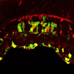
integrate and differentiate following transplantation into the mammalian retina. This figure illustrates green fluorescent protein expressing brain progenitor cells 28 days after transplantation into the eye of a Brazilian opossum, Monodelphis domestica, host. This cross-section of retinal tissue was stained with an antibody against calretinin, a marker used to identify subclasses of retinal neurons. To visualize the calretinin labeling pattern, we used Biotin-SP-Donkey Anti-Rabbit IgG (H+L) followed by Cy3-streptavidin. This image was created by merging confocal images of GFP fluorescence (green) with the calretinin antibody labeling pattern (red). Co-localization of the calretinin (red) in transplanted GFP-expressing progenitor cells (green) is indicated by the yellow color. Transplanted cells were located among calretinin-immunoreactive amacrine and ganglion cells.
Your first choice for scientific solutions
Find out moreSUPPORT
outstanding technical support
PRODUCT
we offer a full product guarantee
DELIVERY
we offer free delivery to UK universities and non profit organisations