Technical Articles

Technical articles consist of several parts of topics, including pathway research, cytokines, cancer, transmembrane proteins, et al. Among of these topics, such as pathway research, which are divided into various special topics. Click related links to the articles to help you plan and perform your experiment.

CYTOKINES

Cytokines are a large group of proteins that are important in cell signaling. Their release has an effect on the behavior of cells around them. It can be said that cytokines are involved in autocrine signaling, paracrine signaling and endocrine signaling as immunomodulating agents. Cytokines may include growth factors, chemokines, interferons, interleukins, colony-stimulating factors, and tumour necrosis factors(despite some overlap in the terminology). Cytokines are produced by a broad range of cells, including immune cells like macrophages, B lymphocytes, T lymphocytes and mast cells, as well as endothelial cells, fibroblasts, and various stromal cells; a given cytokine may be produced by more than one type of cell.

The Overview of Chemokines

Chemokines are a series of cytokines with small molecular weight whose main role is the recruitment of leukocyte subsets under homeostatic and pathological conditions. Their name is derived from their ability to induce directed chemotaxis in nearby responsive cells; they are chemotactic cytokines.

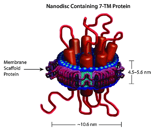
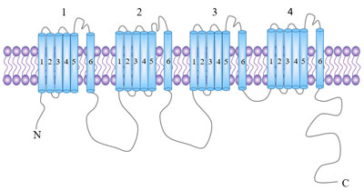
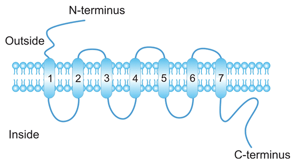
Trough interacting with chemokine receptors that are expressed on the cell surface as 7-transmembrane proteins coupled with G-protein for signaling transduction, chemokines can induce firm adhesion of targeted cells to the endothelium and direct the movement of targeted cells to their destination according to the concentration gradient of a given chemokine. In this article, we introduce the family of chemokines and receptors, its key role in the inflammation, and so on.

1. The Function of Chemokines

The major role of chemokines is to act as a chemoattractant to guide the migration of cells. Cells that are attracted by chemokines follow a signal of increasing chemokine concentration towards the source of the chemokine.

The function of chemokines are divided into two sections. One of them is homeostatic, which are constitutively produced in certain tissues and are responsible for basal leukocyte migration. These include: CCL14, CCL19, CCL20, CCL21, CCL25, CCL27, CXCL12 and CXCL13. This classification is not strict; for example, CCL20 can act also as pro-inflammatory chemokine. Another is inflammatory, which are formed under pathological conditions (on pro-inflammatory stimuli, such as IL-1, TNF-alpha, LPS, or viruses) and actively participate in the inflammatory response attracting immune cells to the site of inflammation. Examples are: CXCL-8, CCL2, CCL3, CCL4, CCL5, CCL11, CXCL10

2. The Types of Chemokines and Chemokine Receptors

Chemokines are a group of cytokines with small molecular weight whose main action is the recruitment of leukocyte subsets under homeostatic and pathological conditions.

Through interacting with chemokine receptors that are expressed on the cell surface as 7-transmembrane proteins coupled with G-protein for signaling transduction, chemokine can induce firm adhesion of targeted cells to the endothelium and direct the movement of targeted cells to their destination according to the concentration gradient of a given chemokine. According to behavior and structural characteristics, the chemokine family consists of 50 endogenous chemokine ligands in humans and mice (Table 1) [1][2][3][4][5][6].

Table 1a. CC chemokines

Name Protein Name Other Name(s) Receptor Uniprot ID
CCL1 C-C motif chemokine 1 I-309, TCA-3 CCR8 P22362
CCL2 C-C motif chemokine 2 MCP-1 CCR2 P13500
CCL3 C-C motif chemokine 3 MIP-1a CCR1 P10147
CCL4 C-C motif chemokine 4 MIP-1β CCR1CCR5 P13236
CCL5 C-C motif chemokine 5 RANTES CCR5 P13501
CCL6 C-C motif chemokine 6 C10, MRP-2 CCR1 P27784
CCL7 C-C motif chemokine 7 MARC, MCP-3 CCR2 P80098
CCL8 C-C motif chemokine 8 MCP-2 CCR1, CCR2B, CCR5 P80075
CCL9/CCL10 C-C motif chemokine 9 MRP-2, CCF18, MIP-1? CCR1 P51670
CCL11 Eotaxin C-C motif chemokine 11, Eosinophil chemotactic protein, Small-inducible cytokine A11 CCR2CCR3CCR5 P51671
CCL12 C-C motif chemokine 12 MCP-5 unknown Q62401
CCL13 C-C motif chemokine 13 MCP-4, NCC-1, Ckβ10 CCR2CCR3CCR5 Q99616
CCL14 C-C motif chemokine 14 HCC-1, MCIF, Ckβ1, NCC-2, CCL CCR1 Q16627
CCL15 C-C motif chemokine 15 Leukotactin-1, MIP-5, HCC-2, NCC-3 CCR1CCR3 Q16663
CCL16 C-C motif chemokine 16 LEC, NCC-4, LMC, Ckβ12 CCR1CCR2CCR5CCR8 O15467
CCL17 C-C motif chemokine 17 TARC, dendrokine, ABCD-2 CCR4 Q92583
CCL18 C-C motif chemokine 18 PARC, DC-CK1, AMAC-1, Ckβ7, MIP-4 unknown P55774
CCL19 C-C motif chemokine 19 ELC, Exodus-3, Ckβ11 CCR7 Q99731
CCL20 C-C motif chemokine 20 LARC, Exodus-1, Ckβ4 CCR6 P78556
CCL21 C-C motif chemokine 21 SLC, 6Ckine, Exodus-2, Ckβ9, TCA-4 CCR7 O00585
CCL22 C-C motif chemokine 22 MDC, DC/β-CK CCR4 O00626
CCL23 C-C motif chemokine 23 MPIF-1, Ckβ8, MIP-3, MPIF-1 CCR1 P55773
CCL24 C-C motif chemokine 24 Eotaxin-2, MPIF-2, Ckβ6 CCR3 O00175
CCL25 C-C motif chemokine 25 TECK, Ckβ15 CCR9 O15444
CCL26 C-C motif chemokine 26 Eotaxin-3, MIP-4a, IMAC, TSC-1 CCR3 Q9Y258
CCL27 C-C motif chemokine 27 CTACK, ILC, Eskine, PESKY, skinkine CCR10 Q9Y4X3
CCL28 C-C motif chemokine 28 MEC CCR3CCR10 Q9NRJ3

Table 1b. CXC chemokines

Name Protein Name Other Name(s) Receptor Uniprot ID
CXCL1 Growth-regulated alpha protein C-X-C motif chemokine 1, Gro-a, GRO1, NAP-3, KC CXCR2 P09341
CXCL2 C-X-C motif chemokine 2 Gro-β, GRO2, MIP-2a CXCR2 P19875
CXCL3 C-X-C motif chemokine 3 Gro-?, GRO3, MIP-2β CXCR2 P19876
CXCL4 Platelet factor 4 PF-4, C-X-C motif chemokine 4, roplact, Oncostatin-A CXCR3B P02776
CXCL5 C-X-C motif chemokine 5 ENA-78 CXCR2 P42830
CXCL6 C-X-C motif chemokine 6 GCP-2 CXCR1, CXCR2 P80162
CXCL7 Platelet basic protein NAP-2, CTAPIII, β-Ta, PEP unknown P02775
CXCL8 Interleukin-8 IL-8, NAP-1, MDNCF, GCP-1 CXCR1CXCR2 P10145
CXCL9 C-X-C motif chemokine 9 MIG, CRG-10 CXCR3 Q07325
CXCL10 C-X-C motif chemokine 10 IP-10, CRG-2 CXCR3 P02778
CXCL11 C-X-C motif chemokine 11 I-TAC, β-R1, IP-9 CXCR3CXCR7 O14625
CXCL12 Stromal cell-derived factor 1 SDF-1, PBSF CXCR4CXCR7 P48061
CXCL13 C-X-C motif chemokine 13 BCA-1, BLC CXCR5 O43927
CXCL14 C-X-C motif chemokine 14 BRAK, bolekine unknown O95715
CXCL15 C-X-C motif chemokine 15 Lungkine, WECHE unknown Q9WVL7
CXCL16 C-X-C motif chemokine 16 SRPSOX CXCR6 Q9H2A7
CXCL17 C-X-C motif chemokine 17 DMC, VCC-1 unknown Q6UXB2

Table 1c. C chemokines

Name Protein Name Other Name(s) Receptor Uniprot ID
XCL1 Lymphotactin Lymphotactin a, SCM-1a, ATAC XCR1 P47992
XCL2 Cytokine SCM-1 beta Lymphotactin β, SCM-1β XCR1 Q9UBD3


Table 1d CX3C chemokines

Name Protein Name Other Name(s) Receptor Uniprot ID
CX3CL1 Fractalkine Fractalkine, Neurotactin, ABCD-3 CX3CR1 P78423

3. Chemokine Signaling Pathway

Chemokines are a group of cytokines with small molecular weight whose main action is the recruitment of leukocyte subsets under homeostatic and pathological conditions.

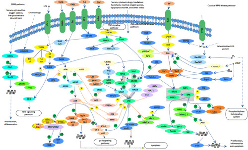
Through interacting with chemokine receptors that are expressed on the cell surface as 7-transmembrane proteins coupled with G-protein to transmit cell signals following ligand binding. Activation of G proteins, by chemokine receptors, causes the subsequent activation of an enzyme known as phospholipase C (PLC). PLC cleaves a molecule called phosphatidylinositol (4,5)-bisphosphate (PIP2) into two second messenger molecules known as Inositol triphosphate (IP3) and diacylglycerol (DAG) that trigger intracellular signaling events; DAG activates another enzyme called protein kinase C (PKC), and IP3 triggers the release of calcium from intracellular stores. These events promote many signaling cascades (such as the MAP kinase pathway) that generate responses like chemotaxis, degranulation, release of superoxide anions and changes in the avidity of cell adhesion molecules called integrin’s within the cell harbouring the chemokine receptor[7].

Fig.1. The picture of chemokine signaling pathway

4. Chemokine and Inflammation

Chemokines are chemotactic cytokines that direct the movement of circulating leukocytes to sites of inflammation or injury. Originally studied because of their role in inflammation, chemokines and their receptors are now known to play a crucial part in directing the movement of mononuclear cells throughout the body, engendering the adaptive immune response and contributing to the pathogenesis of a variety of diseases. Chemokine receptors are some of the most tractable drug targets in the huge battery of molecules that regulate inflammation and immunity[8][9]. Here, we survey the properties of chemokines and their receptors and highlight the roles of these chemoattractants in selected clinical disorders.

The chemokine system in innate immune cell homeostasis

Maintenance of hematopoietic stem cells and developing innate immune cells takes place largely in the bone marrow (BM) and is dependent on CXCL12/CXCR4 interactions[10]. As immune cell development progresses past the hematopoietic stem cell, CXCL12/CXCR4 interactions remain essential for BM retention and normal development of multiple immune lineages, including B cells, monocytes, macrophages, neutrophils, natural killer (NK) cells, and plasmacytoid dendritic cells[11]. CXCR4-mediated signaling plays a major role in promoting BM retention of many immune cells. However, exit from the BM may not be entirely passive. In studies examining monocyte development and release from the BM, blockade of CXCR4 induces only a small increase in the number of peripheral blood monocytes[12].

The chemokine system in acute inflammation

Acute Inflammation is a general pattern of immune response to cell injury characterized by rapid accumulation of immune cells at the site of injury. The acute inflammatory response is initiated by both immune and parenchymal cells at the site of injury and is coordinated by a wide variety of soluble mediators.

This coordination starts with the homeostatic prepositioning of innate immune cells throughout the periphery, where they act as local sensors of infection and inflammation through the activation of pattern recognition receptors (PRRs), the inflammasome, and/or RNA and DNA sensors. Neutrophils, monocytes, and basophils, almost all innate immune cells are present to some extent in the periphery under homeostatic conditions. There they lie in wait as sensors of pathogen invasion, via PRRs, or tissue damage, via the interleukin (IL)-33 pathway as one example. MCs and macrophages are classically described as essential immune sensors, based on their expression of a wide variety of PRRs and their broad localization throughout all vascularized tissues. MCs are uniquely capable of responding immediately to infectious or inflammatory stimuli. Lipopolysaccharide (LPS) stimulation of murine peritoneal MCs leads to immediate release of CXCL1 and CXCL2-containing granules, but not histamine-containing granules, as well as transcriptional activation of CXCL1 and CXCL2[13].

Inflammatory Diseases


Chemokines have been implicated in a wide range of diseases with prominent inflammatory components. For example, elevated levels of CC chemokines, particularly CCL2, CCL3, and CCL5, in the joints of patients with rheumatoid arthritis coincide with the recruitment of monocytes and T cells into synovial tissues. Inflammation is also a key factor in asthma, in which the chemokine CCL11 (eotaxin) and its receptor, CCR3, contribute to the recruitment of eosinophils to the lung. Psoriasis is another example of chemokine-mediated local cell recruitment and inflammation. Infiltrating effector T cells express CCR4, and its ligands (CCL17 and CCL22) are produced by cutaneous cells. CXCR3 has also been implicated in the recruitment of T cells to inflamed skin[14].

5. The Most Popular Questions about Chemokines

In this part, we enumerate several popular questions about chemokines and hope that may give some help for you.

What is the difference between chemokine and cytokine?

We can differentiate chemokine and cytokines from several aspects. With respect to definition, cytokines are immune-modulating agents which are made up from proteins, while chemokines are a super family of cytokines which mediate chemotaxis. With respect to function, cytokines are involved in both cellular and antibody-mediated immunity in the body, while chemokines are involved in the guiding of cells in the immune system to the site of infection. With respect to types, cytokines include chemokines, ILs, INFs, CSFs, TNFs and TGFs in the body, while chemokines involve CC chemokines, CXC chemokines, C chemokines and CX3C chemokines in the body.

What cells release chemokines?

Some chemokines have roles in development; they promote angiogenesis (the growth of new blood vessels), or guide cells to tissues that provide specific signals critical for cellular maturation. Other chemokines are inflammatory and are released from a wide variety of cells in response to bacterial infection, viruses and agents that cause physical damage such as silica or the urate crystals that occur in gout.

References

[1] Laing KJ, Secombes CJ. Chemokines[J]. Developmental and Comparative Immunology. 2004, 28 (5): 443–60

[2] Villeda SA, Luo J, Mosher KI, et al. The ageing systemic milieu negatively regulates neurogenesis and cognitive function[J]. Nature. 2011, 477 (7362): 90–4

[3] Murdoch C, Finn A. Chemokine receptors and their role in inflammation and infectious diseases[J]. Blood. 2000, 95 (10): 3032–43

[4] Cocchi F, DeVico AL, Garzino-Demo A, et al. Identification of RANTES, MIP-1 alpha, and MIP-1 beta as the major HIV-suppressive factors produced by CD8+ T cells[J]. Science. 1995, 270 (5243): 1811–5

[5] von Recum HA, Pokorski JK. Peptide and protein-based inhibitors of HIV-1 co-receptors[J]. Experimental Biology and Medicine. 2013, 238 (5): 442–9

[6] Garzino-Demo A, Moss RB, Margolick JB, et al. Spontaneous and antigen-induced production of HIV-inhibitory beta-chemokines are associated with AIDS-free status[J]. Proceedings of the National Academy of Sciences of the United States of America. 1999, 96 (21): 11986–91

[7] R. M. Ransohoff. Chemokines and chemokine receptors: standing at the crossroads of immunobiology and neurobiology[J]. Immunity. 2009, 5(31): 711–721

[8] Luster AD. Chemokines — chemotactic cytokines that mediate inflammation[J]. N Engl J Med. 1998, 338:436-45

[9] Israel F. Charo, and Richard M. Ransohoff. The Many Roles of Chemokines and Chemokine Receptors in Inflammation[J]. N Engl J Med. 2006, 354:610-21

[10] Ara T, Tokoyoda K, Sugiyama T, Egawa T, Kawabata K, Nagasawa T. Long-term hematopoietic stem cells require stromal cell-derived factor-1 for colonizing bone marrow during ontogeny[J]. Immunity. 2003, 19: 257–267

[11] Mercier FE, Ragu C, Scadden DT. The bone marrow at the crossroads of blood and immunity[J]. Nat Rev Immunol. 2012, 12: 49–60

[12] Wang Y, Cui L, Gonsiorek W, et al. CCR2 and CXCR4 regulate peripheral blood monocyte pharmacodynamics and link to efficacy in experimental autoimmune encephalomyelitis[J]. J Inflamm (Lond). 2009, 6: 32

[13] Caroline L. Sokol and Andrew D. Luster. The Chemokine System in Innate Immunity[J]. Cold Spring Harb Perspect Biol. 2015, 29:(5)

[14] Flier J, Boorsma DM, van Beek PJ, et al. Differential expression of CXCR3 targeting chemokines CXCL10, CXCL9, and CXCL11 in different types of skin inflammation[J]. J Pathol. 2001, 194:398-405

The Overview of Tumour Necrosis Factor (TNF)

Tumor necrosis factor was reported firstly by Lloyd J. Old as one kind of cytotoxic factor produced by macrophages, from Memorial Sloan-Kettering Cancer Center, New York, in 1975. TNF, also known as TNFα, is a cytokine that can directly kill tumor cells without obvious cytotoxicity to normal cells. It is a 17kDa protein consisting of 157 amino acids and a homotrimer in solution. It is produced chiefly by activated macrophages.

1. Tumor Necrosis Factor function

TNFα is mainly produced by activated macrophages, T lymphocytes, and natural killer (NK) cells, but is also expressed at lower levels by fibroblasts, smooth muscle cells, and tumor cells. The primary role of TNF is in the regulation of immune cells. TNF, being an endogenous pyrogen, is able to induce fever, apoptotic cell death, cachexia, inflammation and to inhibit tumorigenesis and viral replication and respond to sepsis via IL1 & IL6 producing cells.

2. Members

In this section, we present the members of TNF superfamily and the receptors in the table 1(table 1a and 1b).

Tumor Necrosis Factor superfamily

The tumor necrosis factor (TNF) superfamily refers to a superfamily of cytokines that are involved in modulating cell death, survival, proliferation, and differentiation. Nineteen proteins have been identified as part of the TNF family on the basis of sequence, function, and structural similarities (as shown in table 1a).

Table 1a TNF Superfamily-chromosomal location

Chromosomal Location mRNA Accession NO. Uniprot ID
Gene Name/alias Human Human Human Ligand Symbol
TNF/TNFα 6p21.3 NM_000594 P01375 TNFSF1A
Lymphotoxin-alpha/LTα 6p21.3 NM_000595 P01374 TNFSF1B
LTβ 6p21.3 NM_002341 Q06643 TNFSF3
OX40-L 1q25 NM_003326 P23510 TNFSF4
CD40-L, CD154 Xq26 NM_000074 P29965 TNFSF5
Fas-L 1q23 NM_000639 P48023 TNFSF6
CD27-L, CD70 19p13 NM_001252 P32970 TNFSF7
CD30-L, CD153 9q33 NM_001244 P32971 TNFSF8
4-1BB-L 19p13 NM_003811 P41273 TNFSF9
TRAIL 3q26 NM_003810 P50591 TNFSF10
RANK-L, TRANCE 13q14 NM_003701 O14788 TNFSF11
TWEAK 17p13 NM_003809 O43508 TNFSF12
APRIL/TALL2 17P13.1 NM_003808 O75888 TNFSF13
BAFF, BLYS, TALL1 13q32–q34 NM_006573 Q9Y275 TNFSF13B
LIGHT 19p13.3 NM_003807 O43557 TNFSF14
TL1A 9q33 NM_005118 O95150 TNFSF15
GITRL, AITRL 1q23 NM_005092 Q9UNG2 TNFSF18
EDA1 Xq12–q13.1 NM_001399 Q92838 unknown
EDA2 Xq12–q13.1 AF061189 unknown unknown


Tumor Necrosis Factor receptors

The portrait of all the receptors in the TNF Superfamily is large and complicated (Table 1b), including the official genome nomenclature site, which introduced the TNFSF and TNFRSF numbering system[1].

Table 1b TNF Receptor SuperFamily

Chromosomal Location mRNA Accession NO. Uniprot ID
Gene Name/alias Human Human Human Ligand Symbol
TNFR-1, p55–60 12p13.2 NM_001065 P19438 TNFRSF1A
TNFR2, p75–80 1p36.3-36.2 NM_001066 P20333 TNFRSF1B
LTβR 12p13 NM_002342 P36941 TNFRSF3
OX40 1p36 NM_003327 P43489 TNFRSF4
CD40 20q12–q13.2 NM_001250 P25942 TNFRSF5
FAS, CD95 10q24.1 NM_000043 P25445 TNFRSF6
DcR3 20q13 NM_003823 O95407 TNFRSF6B
CD27 12p13 NM_001242 P26842 TNFRSF7
CD30 1p36 NM_001243 P28908 TNFRSF8
4-1BB 1p36 NM_001561 Q07011 TNFRSF9
TRAILR-1, DR4 8p21 NM_003844 O00220 TNFRSF10A
TRAIL-R2, DR5 8p22-p21 NM_003842 O14763 TNFRSF10B
TRAILR3, DcR1 8p22-p21 NM_003841 O14798 TNFRSF10C
TRAILR4, DcR2 8p21 NM_003840 Q9UBN6 TNFRSF10D
RANK, TRANCE-R 18q22.1 NM_003839 Q9Y6Q6 TNFRSF11A
OPG, TR1 8q24 NM_002546 O00300 TNFRSF11B
FN14 16p13.3 NM_016639 Q9NP84 TNFRSF12A
TRAMP, DR3, LARD 1p36.3 NM_003790 Q93038 TNFRSF25
TACI 17p11.2 NM_012452 O14836 TNFRSF13B
BAFFR 22q13.1–q13.31 NM_052945 Q96RJ3 TNFRSF13C
HVEM, HveA, ATAR 1p36.3–p36.2 NM_003820 Q92956 TNFRSF14
p75NTR, NGFR 17q12–q22 NM_002507 P08138 TNFRSF16
BCMA 16p13.1 NM_001192 Q02223 TNFRSF17
AITR, GITR 1p36.3 NM_004195 Q9Y5U5 TNFRSF18
RELT 11q13.2 NM_152222 Q969Z4 TNFRSF19L
TROY, TAJ 13q12.11–q12.3 NM_018647 Q9NS68 TNFRSF19
EDAR 2q11–q13 NM_022336 Q9UNE0 unknown
DR6 6P12.2–21.1 NM_014452 O75509 TNFRSF21
EDA2R Xq11.1 NM_021783 Q9HAV5 TNFRSF27

3. Tumor Necrosis Factor Inhibitors

A tumor necrosis factor (TNF)-alpha inhibitor is a pharmaceutical drug that suppresses the physiologic response to tumor necrosis factor (TNF), which is part of the inflammatory response. TNF is involved in autoimmune and immune-mediated disorders such as rheumatoid arthritis, ankylosing spondylitis, inflammatory bowel disease, psoriasis, hidradenitis suppurativa and refractory asthma, so TNF inhibitors may be used in their treatment. The inhibitor, including etanercept, infliximab, adalimumab, certolizumab pegol, and golimumab, all bind to the cytokine TNF and inhibit its interaction with the TNF receptors[2].

As a cytokine, TNF is involved with the inflammatory and immune response and can bind to TNF receptor 1 (TNFR1) or TNF receptor 2 (TNFR2). It occurs in numerous forms, both monomeric and trimeric, as well as soluble and transmembrane[3]. Etanercept is a fusion protein of two TNFR2 receptor extracellular domains and the Fc fragment of human IgG1. It inhibits the binding of both TNF-alpha and TNF-beta to cell surface TNFRs. Infliximab is a chimeric monoclonal antibody that includes a murine variable region and constant human region. It binds to the soluble and transmembrane forms of TNF-alpha and inhibits the binding of TNF-alpha to TNFR[4].

The important side effects of TNF inhibitors include lymphomas, infections, congestive heart failure, demyelinating disease, a lupus-like syndrome, induction of auto-antibodies, injection site reactions, and systemic side effects[5].

4. Tumor Necrosis Factor Signaling Pathway

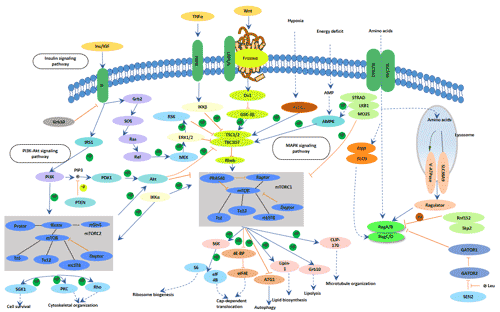
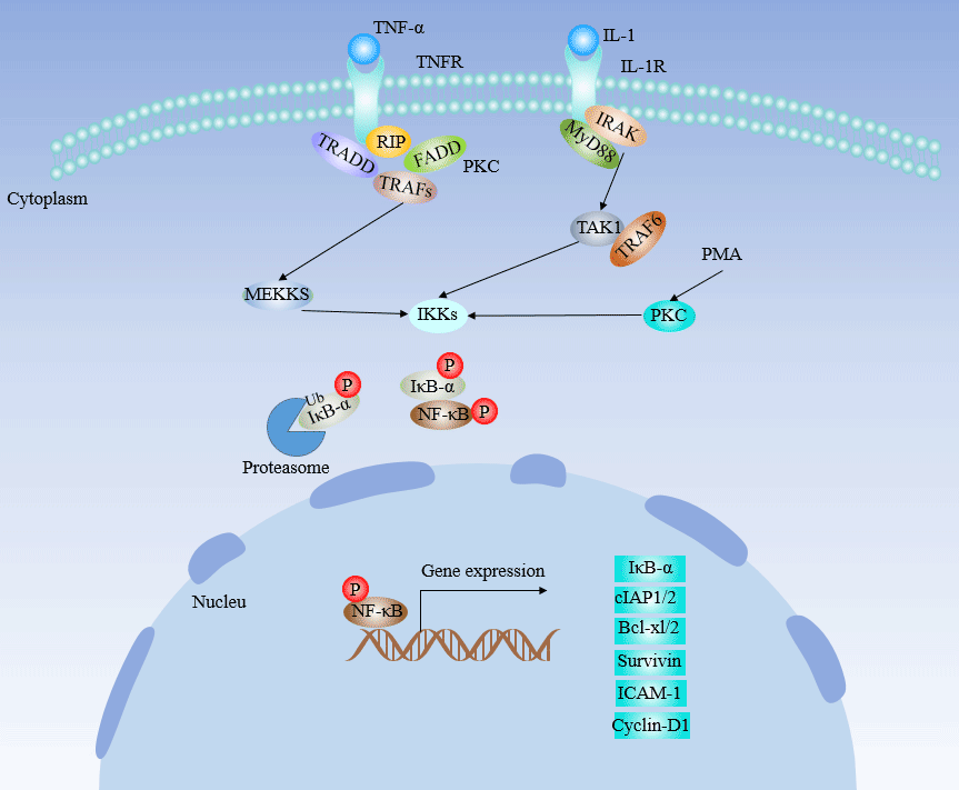
TNF can bind two receptors, TNFR1 (TNF receptor type 1) and TNFR2 (TNF receptor type 2) [6]. TNFR1 is expressed in most tissues, and can be fully activated by both the membrane-bound and soluble trimeric forms of TNF, whereas TNFR2 is found typically in cells of the immune system, and respond to the membrane-bound form of the TNF homotrimer. As most information regarding TNF signaling is derived from TNFR1, the role of TNFR2 is likely underestimated. Upon binding, TNF triggers the activation of numerous pathways including the NFκB and MAPK signaling pathway.

The picture of TNF signaling pathway

5. Tumor Necrosis Factor and Diseases

Accumulating evidences have revealed that dysregulation of TNF production was implicated in a variety of human diseases including Alzheimer’s disease[7], cancer[8], major depression[9], psoriasis[10] and inflammatory bowel disease (IBD). Its complex functions in the immune system include the stimulation of inflammation, cytotoxicity, the regulation of cell adhesion, and the induction of cachexia.

Tumor Necrosis Factor and Cancer

TNF-α is frequently detected in biopsies from human cancer, produced either by epithelial tumor cells, as for instance in ovarian and renal cancer; or stromal cells, as in breast cancer[11]. Cancer cell or stromal cell production of TNF-α is involved in the development of a range of experimental tumors, is partially responsible for NF-κB activation in initiated tumor cells and for the cytokine network in found in human cancer[12].

There are several known consequences of TNF-α production by cancer cells that begin to explain its role in cancer progression. As stated above ovarian cancer cell lines, but not normal ovarian surface epithelial cells, produce pg quantities of TNF-α protein and that there were differences in TNF-α mRNA stability between normal and malignant cells[13]. This endogenous TNF-α correlates with increased cancer cell expression of the chemokine receptor CXCR4 and its ligand CXCL12, in both cell lines and biopsies of disease[14]. TNF-α up-regulated CXCR4 expression, cancer cell migration and production of other inflammatory cytokines and chemokines by the ovarian epithelial cells. CXCR4 and its ligand CXCL12 are implicated in metastases and tumor cell survival in a wide range of cancers[15].

Anti-TNF therapy has shown only modest effects in cancer therapy. Treatment of renal cell carcinoma with infliximab resulted in prolonged disease stabilization in certain patients. Etanercept was tested for treating patients with breast cancer and ovarian cancer showing prolonged disease stabilization in certain patients via downregulation of IL-6 and CCL2. On the other hand, adding infliximab or etanercept to gemcitabine for treating patients with advanced pancreatic cancer was not associated with differences in efficacy when compared with placebo[16].

Tumor Necrosis Factor and Inflammation

As TNF plays a key role in the pathogenesis of chronic inflammatory diseases such as Crohn’s disease (CD) and rheumatoid arthritis (RA), a new class of drugs has been developed in an attempt to neutralise its biological activities.

Tumor Necrosis Factor and rheumatoid arthritis

The primary cellular elements in the RA inflammatory process that cause synovitis and other systemic manifestations are the CD4+ lymphocyte, macrophage, and synovial fibroblasts. The cells produce numerous cytokines, of which interleukin (IL)–1, IL-6, and TNF are arguably the most important in the RA inflammatory cascade. The important role of TNF-α has also been verified in clinical trials of TNF inhibitors. TNF-α, a homodimer produced primarily by macrophages/monocytes, is an excellent promoter of RA inflammation. Acting through a p55 or p75 receptor, it stimulates other cells to produce various inflammatory cytokines, for example, IL-2, IL-6, IL-8, IL-12, IL-18, and interferon (IFN)–γ[17].

Tumor Necrosis Factor and Crohn’s disease

Crohn’s disease (CD) is chronic inflammatory conditions characterized by episodes of remission and flare-ups that have a major impact on the patient’s physical, emotional and social well-being. Similar to its role in rheumatoid arthritis, TNFα has also been shown to be crucial in the pathogenesis of Crohn’s disease.

TNF is a crucial mediator in driving inflammatory processes in the gut. It is produced by a variety of mucosal cells, mainly macrophages and T cells, as a preform on the plasma membrane. In addition, Paneth cells in CD affected segments of the terminal ileum were shown to strongly express TNF RNA in contrast to Paneth cells in normal tissue, indicating an induction under pathogenic conditions. The transmembrane precursor form (mTNF), a homotrimer of 26 kDa subunits, is cleaved by the matrixmetalloproteinase TNF alpha converting enzyme (TACE/Adam17) into a soluble form. The expression of mTNF by CD14+ macrophages has been reported to be relevant in CD)[18].

Anti-tumor necrosis factor α (anti-TNFα) antibodies have been used as a potent therapy for CD. These particular agents are effective not only for CD patients who have never undergone surgery but also for those who have received surgery. In fact, fewer endoscopic recurrences were observed in patients who received anti-TNF agents after surgery than in those who did not.6 In the meanwhile, a report suggested that no significant difference in the endoscopic recurrence rate was observed between biological and conventional therapy after ileocecal resections in CD patients. In this context, the efficacy of anti-TNF agents for postoperative CD patients have been evaluated in patients who had no history of undergoing anti-TNFα antibody treatment prior to surgery. Because primary nonresponses or secondary loss of responses can frequently occur in CD patients who are administered anti-TNF agents, a substantial portion of patients who require anti-TNF agents after surgery receive the agents also prior to surgery in clinical practice[19][20].

References

[1] Carl F. Ware. TNF Superfamily 2008[J]. Cytokine Growth Factor Rev, 2008 (3-4): 183–186

[2] Valerie Gerriets1; Steve S. Bhimji2. Tumor Necrosis Factor (TNF), Inhibitors[J]. StatPearls Publishing, 2018 Jan

[3] Meroni PL, Valentini G, et al. New strategies to address the pharmacodynamics and pharmacokinetics of tumor necrosis factor (TNF) inhibitors: A systematic analysis[J]. Autoimmun Rev. 2015 (9): 812-29

[4] Komaki Y, Yamada A, et al. Efficacy, safety and pharmacokinetics of biosimilars of anti-tumor necrosis factor-α agents in rheumatic diseases; A systematic review and meta-analysis[J]. Autoimmun. 2017 (79): 4-16

[5] N Scheinfeld. A comprehensive review and evaluation of the side effects of the tumor necrosis factor alpha blockers etanercept, infliximab and adalimumab[J]. Journal of Dermatological Treatment, 2004 (15): 280–294

[6] Arianne L. Theiss, James G. Simmons, et al. Tumor Necrosis Factor (TNF) Increases Collagen Accumulation and Proliferation in Intestinal Myofibroblasts via TNF Receptor 2[J]. The journal of biological chemistry, 2005 (43): 36099 -36109

[7] Swardfager W, Lanctôt K, et al. A meta-analysis of cytokines in Alzheimer’s disease[J]. Biol Psychiatry, 2010 (68): 930–941

[8] Locksley RM, Killeen N, et al. “The TNF and TNF receptor superfamilies: integrating mammalian biology[J]. Cell, 2001 (104): 487–501

[9] Dowlati Y, Herrmann N, et al. A meta-analysis of cytokines in major depression[J]. Biol Psychiatry, 2010 (5): 446–457

[10] Victor FC, Gottlieb AB. TNF-alpha and apoptosis: implications for the pathogenesis and treatment of psoriasis[J]. Drugs Dermatol, 2002 (3): 264–75

[11] Balkwill, F. (2002). Tumor necrosis factor or tumor promoting factor[J]. Cytokine & Growth Factor Reviews, 2002 (13): 135–141

[12] Galban, S., Fan, J., et al. von Hippel-Lindau protein-mediatedrepression of tumor necrosis factor alpha translation revealed through use of cDNA arrays[J]. Molecular & Cellular Biology, 2003 (23): 2316–2328

[13] Tsan, M.-F. Toll-like receptors, inflammation and cancer[J]. Seminars in Cancer Biology, 2005 (16): 32–37

[14] Kulbe, H., Hagermann T, et al. The inflammatory cytokine TNF-a upregulates chemokine receptor expression on ovarian cancer cells[J]. Cancer Research, 2005 (65): 10355–10362

[15] Balkwill, F. The significance of cancer cell expression of CXCR4[J]. Seminars in Cancer Biology, 2004 (14): 171–179

[16] Korneev KV, Atretkhany KN, et al. TLR-signaling and proinflammatory cytokines as drivers of tumorigenesis[J]. Cytokine, 2017 (89): 127–135

[17] Ma X, Xu S. TNF inhibitor therapy for rheumatoid arthritis[J]. Biomed Rep, 2012 (2): 177–184.

[18] Narayanan Parameswaran, Sonika Patial. Tumor Necrosis Factor-α Signaling in Macrophages[J]. Crit Rev Eukaryot Gene Expr, 2010 (2): 87–103.

[19] Ulrike Billmeier, Walburga Dieterich, et al. Molecular mechanism of action of anti-tumor necrosis factor antibodies in inflammatory bowel diseases[J]. World J Gastroenterol, 2016 (42): 9300–9313

[20] Konstantinos Papamichael, Adam S Cheifetz. Defining and predicting deep remission in patients with perianal fistulizing Crohn’s disease on anti-tumor necrosis factor therapy[J]. World J Gastroenterol. 2017 (34): 6197–6200

The Overview of Interleukin

Interleukin is a kind of cytokines, which plays a critical role in immunological regulation and homeostasis. It is originally discovered from leukocytes. Currently, it is found to be produced by a lot of cells including macrophages, lymphocytic cells with a solid structure and function. In this article, we introduce interleukin briefly from the following aspects, involving family and receptor, signaling pathways, function and that role in inflammation.

1. Interleukin Family and Interleukin Receptors

The members of interleukin are wealthy which are named IL-1~IL-38.

The interleukin receptors can be classified as type I, type II and other type. The type II interleukin receptors include interleukin-10, 20, 22, 28 receptors, the other type include interleukin-1, 8 receptors, the rest of them are type I interleukin receptors.

Table 1. Interleukin Family and Receptors

Gene Name/Alias Uniprot ID Protein Name Receptors
IL-1 α P01583 Interleukin-1 alpha IL1R1, IL1R2
IL-1 β P01584 Interleukin-1 beta IL1R1IL1R2
IL-2 P60568 Interleukin-2 IL2RA, IL2RB, IL2RG
IL-3 P08700 Interleukin-3 IL3RA, IL3RB
IL-4 P05112 Interleukin-4 IL4R
IL-5 P05113 Interleukin-5 IL5RA, IL3RB
IL-6 P05231 Interleukin-6 IL6R
IL-7 P13232 Interleukin-7 IL7R
IL-8/CXCL8 P10145 Interleukin-8 IL-8, IL8RB
IL-9 P15248 Interleukin-9 IL9R
IL-10 P22301 Interleukin-10 IL10RA
IL-11 P20809 Interleukin-11 IL11RA
IL-12 α P29459 Interleukin-12 subunit alpha IL12RB1
IL-13 P35225 Interleukin-13 IL13RA1IL13RA2
IL-14/TXLNA P40222 Alpha-taxilin Unkown
IL-15 P40933 Interleukin-15 IL15RA
IL-16 Q14005 Pro-interleukin-16 CD4
IL-17 α Q16552 Interleukin-17A IL17RA
IL-18 Q14116 Interleukin-18 IL18R1
IL-19 Q9UHD0 Interleukin-19 IL20R
IL-20 Q9NYY1 Interleukin-20 IL20R
IL-21 Q9HBE4 Interleukin-21 IL21R
IL-22 Q9GZX6 Interleukin-22 IL22RA1
IL-23 Q9NPF7 Interleukin-23 subunit alpha IL23R
IL-24 Q13007 Interleukin-24 IL20R
IL-25 Q9H293 Interleukin-25 LY6E
IL-26 Q9NHP9 Interleukin-26 IL20R1
IL-27 α Q8NEV9 Interleukin-27 subunit alpha IL27RA
IL-27 β Q14213 Interleukin-27 subunit beta IL27RA
IL-28 Q8IU57 Interferon lambda receptor 1 IL28R
IL-29/IFNL1 Q8IU54 Interferon lambda-1 Unknown
IL-30 Q8NEV9 Interleukin-27 subunit alpha Unknown
IL-31 Q6EBC2 Interleukin-31 IL31RA
IL-32 P24001 Interleukin-32 Unknown
IL-33 O95760 Interleukin-33 Unknown
IL-35 Q14213 Consist of IL-12α and IL-27β chains Unknown
IL-36 α Q9UHA7 Interleukin-36 alpha Unknown
IL-36 β Q9NZH7 Interleukin-36 beta Unknown
IL-36 γ Q9NZH8 Interleukin-36 gamma Unknown

2. Common Signaling Pathway

In this part, we display several interleukin pathways, including IL-1, IL-10, IL-12, IL-17 and IL-7 signaling pathway.

IL-1 Signaling Pathway

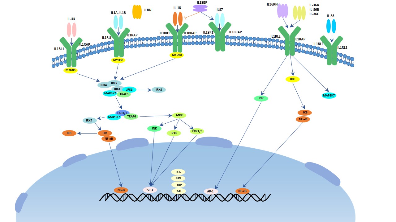
There are 11 members in this family. They (IL-1, 18, 33, 36, 37, 38) binds to IL1, 18 primary receptors. IL-1α, IL-1β, IL-18, IL-33, IL-36α, IL-36β, IL-36γ triggers MPKK and NF-κB leading to inflammatory response. While other members of IL-1 family have anti-inflammatory effects. IL-1 is one hallmark cytokine in this family which can excrete as co-stimulation of T helper cells, maturation and proliferation of B cells, activation of NK cells and is relation to inflammation.

The image of IL-1 signaling pathway

Fig.1. The image of IL-1 signaling pathway

IL-10 Pathway

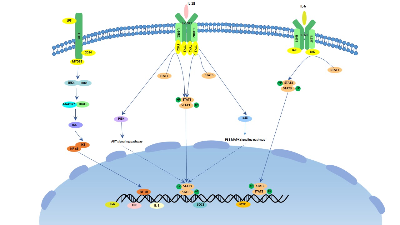
IL-10 is an important anti-inflammatory cytokine which is produced by activated T cells, B cells, macrophages and monocytes. It involves in cytokine production of macrophages, activation of B cells, stimulated Th2 cells, meanwhile, it inhibits cytokine production of Th1 cells. It can form a homodimer that binds to the tetrameric heterodimer IL-10 receptor and inhibit the IL-1, IL-6 and TNF signaling pathway. It is related with non-small cell lung cancers.

The image of IL-10 signaling pathway

Fig.2. The image of IL-10 signaling pathway

IL-12 Pathway

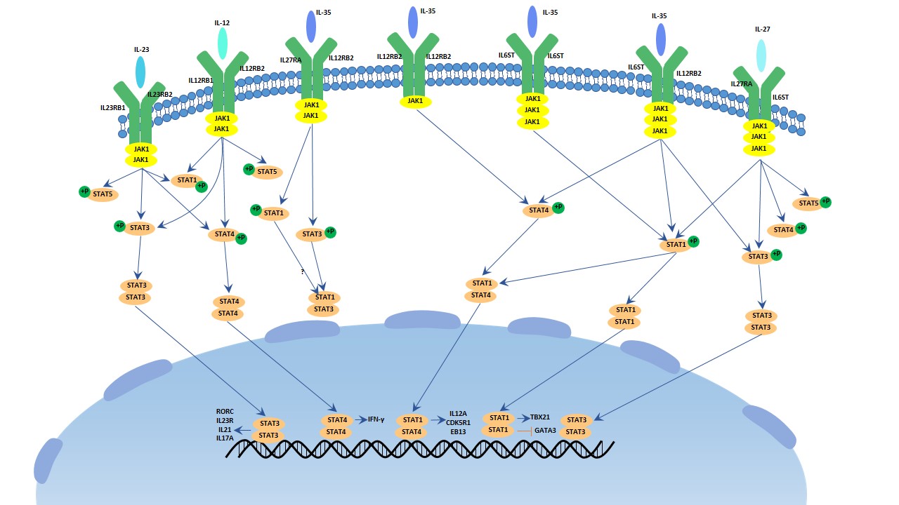
There are 4 members in this family. It (IL-12, 25, 27, 35) belonging to IL-6 superfamily is a special family as it is formed by heterodimers. It mediates phosphorylation of SAT1, 3, 4 by the JAK/STAT family. IL-12 involves in the stimulation and maintenance of Th1 cellular immune responses, including the normal host defend against various intracellular pathogens.

The image of IL-12 signaling pathway

Fig.3. The image of IL-12 signaling pathway

IL-17 Pathway

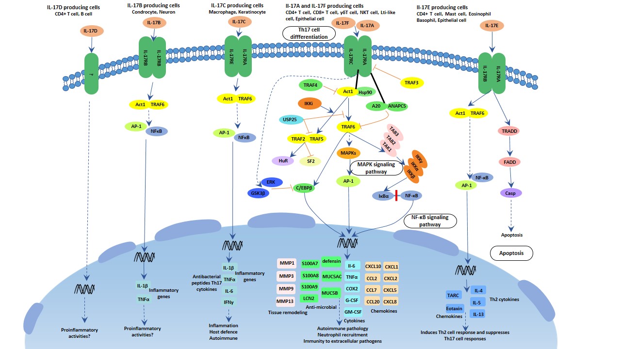
There are 6 members in the IL-17 family, and 5 members in its receptor family. It plays critical roles in host immunology. IL-17A is the hallmark in this family that it protects the host against extracellular pathogens, but also promotes inflammatory pathology in autoimmune disease, similar to IL-17C. While IL-17F involves in the mucosal host defense, IL-17E amplifies the Th2 immune response. The IL-17 family activates antibacterial cytokines and chemokines in MAPK, NF-κB and C/EBPs pathway. Act1 is considered as the master mediator in this pathway.

The image of IL-17 signaling pathway

Fig.4. The image of IL-17 signaling pathway

IL-7 Pathway

IL-7 is a kind of glycoprotein which is produced by bone marrows and weighs 25 kDa. It is important for the immunological development, but the mechanism is not clear. A recent research showed that: IL-7 downregulates the Scos3 (the inhibitor of cytokine signaling), thus much cytokines are stimulated and the T cells effectors are increased resulting the clearance of virus[1]. The article conducted by Marc Pellegrini is published in Cell in 2011.

IL-7 can increase the T effector numbers such as CD4 T cells, CD8 T cells, naive T cells and B cells, and then infiltrate organs like liver, kidney and brain. According to the assay in this research, it is T cells rather than B cells to exert the function of virus clearance. This result is consistent with the previous result that B cells contribute just a little to modulate T cells’ response. It also enlightens us to receive IL-7 therapy.

IL-7’s function depends on IL-6, meanwhile IL-7 augments many pro-inflammatory factors, particularly IL-17, IL-6 and IFNγ. But it limits the liver injury and cytotoxicity as it increases the IL-22, a cyto-protective cytokine.

Socs3 is one prominent repressor of IL-6 signaling pathway while IL-6 is increased with IL-7 treatment. It seems to be produced by T cells, and it is interfered by IL-7. Mice with socs3 deficiency can mimic the effect of virus elimination.

3. Interleukin Function

Interleukin is important for transmitting information, activating and regulating immune cells, mediates the activation, proliferation and differentiation of T cells and B cells.

Interleukin-1 contains IL-1α and IL-1β. While the former is produced by diverse cells, the later one is produced by some specific tissues. IL-1β is cleaved by caspase-1 which is formed by proteins termed as “the inflammasome”. It can lead to pain and short-time sleep, so that they study the relationship between fatigue and IL-1[2]. IL-1 also can simulate the nitric oxides, chemokines, cytokines and adhesion molecules that destroy the cartilage, while the IL-1 receptor antagonist can inhibit the destruction of IL-1 through the completion of the same receptor. The study helps us to understand the pathology of these diseases[3].

Interleukin-2 is popular object in the past 36 years as it is important for the immunotherapy of many diseases including cancers. But the adverse of taking it is fearful[4].Thus, timing of IL-2 administration and different T cells status are crucial to IL-2 therapies[5].

Interleukin-10 is an anti-inflammatory cytokine. In the meanwhile, IL-10 suppresses the immune responses by modulating the T cells and antigen-presenting cells. By controlling the receptor of IL-10, we can resolve the chronic viral infection[6]. The research was published in The Journal of Experimental Medicine in 2006.

Interleukin-18 is a regulator of cancer and autoimmune diseases. As a member of IL-1 family, it plays essential roles in inflammation process. It can cooperate with IL-12 to activate cytotoxic T cells (CTLs) and natural killer (NK) cells to produce IFNγ which may contribute to tumor immune. In addition, it can work with IL-23 to induce the secretion of IL-17. It has pros and cons effects in the treatment of many diseases[7].

4. Interleukin and Inflammation

Inflammation is a productive conduct to stimuli such as pathogens and damage cells. Its characters are heat, redness, swelling, pain and loss of function, sometimes accompanied with fever. Inflammasome activation leads to activation of caspase-1, resulting in the induction of apoptosis and the secretion of pro-inflammatory cytokines including IL-1β and IL-18[8]. Without inflammation, we can’t prevent harm from stimuli, but it can lead to chronic infection such as rheumatoid arthritis. Inflammation plays a key role in chronic inflammatory systemic diseases (CISD). There are overlaps between different CISD.

Interleukin-17 and Psoriasis

Psoriasis is a common, infammatory and chronic disease with the character of erythematous, scaly plaques. It is related to cardiovascular disease (CVD) such as artherosclerosis. As a result of immune dysregulation, its pathology refers to infammatory cytokines like tumor necrosis factor (TNF), interferon (IFN) and IL-1β, IL-6, IL-12, IL-17, IL-22, IL-23 produced from dendritic cells, T-helper cells and keratinocytes. Patients with psoriasis have more Th17 cells and higher level of IL-17A mRNA and protein. The recent study demonstrates over-expressing IL-17 causing increased inflammatory cells and levels of nitric oxide synthase (iNOS), cyclooxygenase-2 (COX-2), nitric oxide (NO) makes more severe psoriasis, and endothelial dysfunction. In the contrast, blocking of IL-7 or the genes acting downstream of IL7 improves the severity of psoriasis[8][9].

Interleukin and Rheumatoid Arthritis

Rheumatoid arthritis (RA) is a severe autoimmune disease accompanied with cartilage degradation, joint destruction and poor prognosis [8]. The study indicates that the polymorphism of inflammation genes leads to the susceptibility of RA[10][11][12]. Caspase-1 is a critical factor of RA, we can observe the reduction of RA in the caspase-1-/- mice mode[13]. Another member is NLRP1 which is related to the secretion of IL-1β and IL-18. Inhibiting the NLRP1 can ameliorate the symptom of RA[14].

This coordination starts with the homeostatic prepositioning of innate immune cells throughout the periphery, where they act as local sensors of infection and inflammation through the activation of pattern recognition receptors (PRRs), the inflammasome, and/or RNA and DNA sensors. Neutrophils, monocytes, and basophils, almost all innate immune cells are present to some extent in the periphery under homeostatic conditions. There they lie in wait as sensors of pathogen invasion, via PRRs, or tissue damage, via the interleukin (IL)-33 pathway as one example. MCs and macrophages are classically described as essential immune sensors, based on their expression of a wide variety of PRRs and their broad localization throughout all vascularized tissues. MCs are uniquely capable of responding immediately to infectious or inflammatory stimuli. Lipopolysaccharide (LPS) stimulation of murine peritoneal MCs leads to immediate release of CXCL1 and CXCL2-containing granules, but not histamine-containing granules, as well as transcriptional activation of CXCL1 and CXCL2[13].

Interleukin and Systemic Lupus Erythematosus

Systemic lupus erythematosus (SLE), accompanied with inflammatory joints, nephritis, mouth ulcer, swollen lymph nodes and butterfly-shaped red rash on the face, is a rare inflammatory rheumatic disease that attacks normal healthy tissues in the body. The major representation is lupus nephritis (LN) with severe kidney destruction. The study explains the relationship between the expression of inflammasome and LN[15] Tsai et al. reported inhibiting the expression of mRNA and production of IL-1β, IL-18, caspase-1 relieves the symptom of LN[16]. Ka et al. further reported that a main active ingredient in the herbal medicine Litsea cubeca, significantly inhibited activation of NLRP3 inflammasome and caspase-1 and the secretion of IL-1. Moreover, it effectively ameliorated LN symptoms in accelerated and severe LN mice[17].

Interleukin and Crohn’s Disease

Crohn’s disease (CD) is a chronic inflammatory condition of the GI tract with no known cure. It is demonstrated efficient to take antibody-based therapies directed against TNF or interleukin-12/interleukin-23 p40 subunit antibody or integrins in moderate-to-severe CD. However, many patients experience primary non-response or lose response over time to anti-TNF therapy, so that we require dose adjustment, or switch to a non-anti-TNF therapy. Interleukin-6 has multiple pro-inflammatory effects such as inhibition of apoptosis in T-cells and is a target for curing CD. A recent study of the IL-6 receptor inhibitor, tocilizumab, manifests a clinical benefit in moderate-to-severe CD[18].

References

[1]Pellegrini M, Calzascia T, et al. IL-7 Engages Multiple Mechanisms to Overcome Chronic Viral Infection and Limit Organ Pathology[J]. Cell, 2011(144): 601-603

[2]Megan E. Roerink, Marieke E. van der Schaaf, et al. Interleukin-1 as a mediator of fatigue in diseases: a narrative review[J]. Journal of Neuroinflammation, 2017(14): 16

[3]Claire J, Marjolaine G, et al. The role of IL-1 and IL-1Ra in joint inflammation and cartilage degradation[J]. Vitamins and Hormones, 2006(74): 371-403

[4]Dhupkar P, Gordon N. Interleukin-2: old and new approaches to enhance immune-therapeutic efficacy[J]. Adv Exp Med Biol, 2017(995): 33-51

[5]Blattman JN, Grayson JM, et al. Therapeutic use of IL2 to enhance antiviral T-cell responses in vivo[J]. Nat Med, 2003(9): 540-547

[6]Eirnaes M, Filippi CM, et al. Resolution of a chronic viral infection after interleukin-10 receptor blockade[J].J Exp Med, 2006(203): 2461-2472

[7]Esmailbeig M, Ghaderi A. Interleukin-18: a regulator of cancer and autoimmune diseases[J]. Eur Cytokine Netw, 2017(28): 127-140

[8]Yi YS. Role of inflammasomes in inflammatory autoimmune rheumatic diseases[J]. Korean J Physiol Pharmacol, 2018(1): 1-15

[9]Lockshin B, Balaqula Y, et al. Interleukin-17, inflammation, and cardiovascular risk in patients with psoriasis[J]. J Am Acad Dermatol, 2018, accepted

[10]Mathews RJ, Robinson JI, et al. Evidence of NLRP3-inflammasome activation in rheumatoid arthritis (RA); genetic variants within the NLRP3-inflammasome complex in relation to susceptibility to RA and response to anti-TNF treatment)[J]. Ann Rheum Dis, 2014(73): 1202-1210

[11]Jenko B, Praprotnik S, et al. NLRP3 and CARD8 polymorphisms influence higher disease activity in rheumatoid arthritis)[J]. J Med Biochem, 2016(35): 319-323

[12]Sui J, Li H. NLRP1 gene polymorphism influences gene transcription and is a risk factor for rheumatoid arthritis in han chinese. Arthritis Rheum, 2012(64): 647-654

[13]Joosten LA, Netea MG, et al. Inflammatory arthritis in caspase 1 gene-deficient mice: contribution of proteinase 3 to caspase 1-independent production of bioactive interleukin-1)[J]. Arthritis Rheum, 2009(60): 3651-3662

[14]Zhang L, Dong Y, et al. Hydroxysteroid dehydrogenase 1 inhibition attenuates collagen-induced arthritis)[J]. Int Immunopharmacol, 2013(17): 489-494

[15]Roberts TL, Idris A, et al. HIN-200 proteins regulate caspase activation in response to foreign cytoplasmic DNA)[J]. Science, 2009(323): 1057-1060

[16]Tsai PY, Ka SM, et al. Epigallocatechin-3-gallate prevents lupus nephritis development in mice via enhancing the Nrf2 antioxidant pathway and inhibiting NLRP3 inflammasome activation)[J]. Free Radic Biol Med, 2011(51): 744-754

[17]Ka SM, Lin JC, et al. Citral alleviates an accelerated and severe lupus nephritis model by inhibiting the activation signal of NLRP3 inflammasome and enhancing Nrf2 activation)[J]. Arthritis Res Ther. 2015(17): 331

[18]Danese S, Vermeire S, et al. Randomised trial and open-label extensive study of an anti-interleuki8n-6 antibody in crohn’s disease(ANDANTW I and II)[J]. Gut, 2017, doi:10.1136

The Overview of Colony-Stimulating Factor

Colony stimulating factors (CSFs) are glycoproteins that bind to receptor proteins on the surfaces of hemopoietic stem cells, activating intracellular signaling pathways, then promoting production of white blood cells (mainly granulocytes such as neutrophils), in response to infection. Administration of exogenous colony stimulating factors stimulates the stem cells in the bone marrow to produce more of the particular white blood cells. The new white blood cells migrate into the blood and fight the infection. In this article, we introduce the colony stimulating factors from several aspects briefly, including function, members, the relationship with inflammation and cancer, and the latest researches about that.

1. Colony-Stimulating Factors (CSFs) Function

Colony-stimulating factors (CSFs) activate intracellular signaling pathways that can cause the cells to proliferate and differentiate into a specific kind of blood cell (usually white blood cells. For red blood cell formation, see erythropoietin). CSFs circulate in the blood, acting as hormones, and are also secreted locally. General speaking, CSFs include macrophage colony-stimulating factor (CSF1), Granulocyte–macrophage colony-stimulating factor (CSF2), granulocyte colony-stimulating factor (CSF3) and interleukin-3 (IL-3; also known as multi-CSF). But in some extent, CSFs also involve erythropoictin (Epo) and thrombopoietin (Thpo) for their ability of stimulating the formation of red blood.

2. Members

In this section, we list the common colony-stimulating factors and corresponding receptors in the below Table 1.

Table 1. CSFs and receptors

Name Uniprot ID Species Receptor
(macrophage colony-stimulating factor(CSF1) P09603 Human CSF1R
macrophage colony-stimulating factor(CSF1) P07141 Mouse Csf1r
Granulocyte-macrophage colony-stimulating factor protein(GM-CSF) Q9GL44 Rhesus macaque CSF1R
Granulocyte-macrophage colony-stimulating factor protein(CSF2) P04141 Human CSF2RA
Granulocyte-macrophage colony-stimulating factor protein(CSF2) P01587 Mouse CSF2RA
Granulocyte colony-stimulating factor protein(CSF3) P09919 Human CSF3R
Granulocyte colony-stimulating factor protein(CSF3) P09920 Mouse Csf3r
Colony stimulating factor 3 F7H1Q6 Rhesus Macaque CSF3R
Erythropoietin(EPO) P01588 Human EpoR
Thrombopoietin(THPO) P40225 Human CD110

The Member of Colony-Stimulating Factors

CSF1, CSF2, CSF3 and multi-CSF were originally defined by their respective abilities to generate different types of myeloid populations from precursor bone marrow cells in vitro[1], M-CSF stimulated macrophages, G-CSF stimulated granulocytes, GM-CSF stimulated both granulocytes and macrophages, and multi-CSF induced the growth of all hematopoietic different cell types, respectively.

Unlike the other CSFs, CSF1 has the high levels in the blood and is ubiquitously and constitutively expressed in many tissues and by many cell types; the other CSFs are usually produced only after stimulation. For instance, GM-CSF and G-CSF are produced by stimulated haemopoietic and non-haemopoietic cells, and IL-3 is emerged by stimulated T cells and mast cells. The diverse receptor distribution and potential sources of the CSFs are key to the delineation of their separate biological functions and their potential roles in pathology.

Colony-Stimulating Factor Receptors

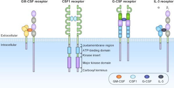
The receptors for the diverse CSFs (as shown in the Fig.1) are quite different and are expressed on different myeloid populations. General speaking, each receptor is stimulated by a specific CSF, except for CSF1R, which binds to both CSF1 and IL-34.

The CSF1 receptor, also known as CSF1R, is expressed on monocytes and macrophages, and is the only receptor tyrosine kinase that is activated by two ligands of unrelated sequence – CSF1 and IL-34. The biologically active regions of CSF1 and IL-34 have similar cytokine folds in spite of sharing low sequence similarity. However, although existed similarities, the IL-34–CSF1R complex differs from the CSF1–CSF1R complex in many structural ways.

The structures of the CSF receptors

Fig.1. The structures of the CSF receptors

The G-CSF receptor, also called G-CSFR, is a transmembrane protein that has the largest expression in neutrophils, but G-CSFR is also present in other types of cell. Thereby, we cannot affirm that direct interactions with G-CSF only occur in granulocyte lineage populations. Besides that, ligand-induced dimerization of G-CSFR rapidly triggers numerous downstream signal transduction pathways, such as MAPK and PI3K-AKT signaling pathway[2].

The GM-CSF receptor (GM-CSFR) is expressed on monocytes, macrophages, eosinophils and neutrophils. The fundamental GM-CSFR subunit, expressed mainly on myeloid populations, comprises of a unique ligand-binding α-chain that contains three extracellular domains and a signaling β-chain. And this β-common (βc) chain is also a component of the IL-3R. Emerging evidence revealed that GM-CSFR engagement can active JAK-STAT, MAPK, NF-κB and PI3K–AKT signaling pathway[3][4] .

Like GM-CSFR, IL-3 receptor (IL-3R) is a dodecamer, highly produced by many cell types, involving monocytes, macrophages, eosinophils, basophils, plasmacytoid dendritic cells (pDCs) and mast cells.

3. Colony Stimulating Factors (CSFs) and Inflammation

CSFs later became apparent that they could also affect more mature populations in these lineages, promoting their survival and/or proliferation, activation and differentiation, all of which could be relevant to inflammation. The effects of Csf gene deficiency in mice and of specific neutralizing monoclonal antibodies (mAbs) have spurred a reassessment of the roles that these CSFs have in steady-state haematopoiesis. Each of these CSFs can play a part in the host response to tissue injury and infection, which has potential implications for inflammatory and autoimmune diseases[5][6]. Roles for the different CSFs and their targeting, particularly GM-CSF and CSF1, are increasingly being investigated in preclinical models and clinical trials of inflammatory and autoimmune diseases. In this part, we will introduce the primary CSFs and inflammation.

Macrophage colony-stimulating factor (CSF1) and Inflammation

CSF1, also known as Macrophage colony-stimulating factor (M-CSF), unlike the other CSFs, is present at high levels in the blood and is ubiquitously and constitutively expressed in many tissues and by many cell types. CSF1 is one of the most important substances known to affect macrophage physiology. The binding of CSF1 to its sole specific receptor, CSF-1R, stimulates the survival, proliferation, and differentiation of mononuclear phagocytes[7][8]. Among the various candidates, CSF1 is considered an important cytokine. The up-regulation of CSF1 and its receptor CSF-1R has been reported in brain disease, as well as in diabetic complications. However, the mechanism is unclear. An elevated level of glycated albumin (GA) is a characteristic of diabetes, thus, it may be involved in monocyte/macrophage-associated diabetic complications[9].

Inflammation involving vessels and neural tissue occurs early in diabetic retinopathy, while excessive and persistent microglial activation is believed a major contributor to the inflammatory responses. Microglial activation occurs via a complex regulatory system involving multiple signals. Accumulating evidences showed M-CSF is a key cytokine in the regulation of the microglial activation, proliferation and migration in CNS[10][11]. Moreover, the high-affinity binding of M-CSF to CSF-1R plays a role in mononuclear/macrophage-associated diabetic complications, including diabetic retinopathy. Wei Liu previous study provided the evidence that the vigorous expression of M-CSF/CSF-1R occurred in the early diabetic retina and a robust induction of CSF-1R was observed on the activated microglia[12][13][14].

Granulocyte-macrophage colony-stimulating factor protein (CSF2) and Inflammation

Granulocyte–macrophage colony-stimulating factor(GM-CSF; also known as CSF2), particularly during inflammatory responses, can be produced by a number of both haemopoietic and non-haemopoietic cell types upon their stimulation. Moreover, the ligand can activate myeloid populations to produce inflammatory mediators. Therefore, GM-CSF is a multifunctional cytokine that acts at the interface between innate and adaptive immunity[15]. Adrian Achuthan et.al. have identified a new GM-CSF-driven pathway in monocytes/macrophages leading to CCL17 formation and with upstream involvement of JMJD3-regulated IRF4. This pathway was identified in both human and murine cells in vitro, and also in vivo, in the steady state and also during inflammatory responses. This pathway appears to be separate from the GM-CSF-driven pathway(s) in monocytes/macrophages, leading to expression of other key proinflammatory cytokines[16]. Moreover, Andrew D. Cook, et.al. previously identified a new GM-CSF→JMJD3→IRF4→CCL17 pathway that is active in monocytes/macrophages in vitro and important for the development in mice of inflammatory pain, as well as of arthritic pain and disease and suggested previously that CCL17 may be a therapeutic target in inflammatory conditions where GM-CSF is important[17].

Granulocyte colony-stimulating factor and Inflammation

Granulocyte colony-stimulating factor(G-CSF; also known as CSF3), is a glycoprotein that stimulates the bone marrow to produce granulocytes and stem cells and release them into the bloodstream. Like GM-CSF, G-CSF circulates at low concentrations, but its levels are elevated as part of the host response to infection or injury. In vitro, G-CSF can be produced by many cell types upon addition of pro-inflammatory stimuli such as TNF, IL-1 or lipopolysaccharide (LPS). G-CSF is considered to be a key regulator of granulopoiesis by inducing the development of neutrophils from progenitors and by promoting neutrophil release from the bone marrow. The role of neutrophils in inflammation, even in chronic situations, is becoming widely recognized. Given that G-CSF is a key ligand that controls neutrophil numbers (through its pro-survival and trafficking regulation functions) and activation, it is likely to be crucial for many aspects of neutrophil function, thereby providing clues as to potential therapeutic indications. An important role for an IL-17–G-CSF pathway in the regulation of neutrophil homeostasis has been proposed, as IL-17 can regulate G-CSF levels, which has potential implications for anti-IL-17 therapies in development[18][19][20][21][22] .

4. The latest researches about Colony Stimulating Factors

Colony stimulating factors are used in patients who are undergoing cancer treatment that causes low white blood cell counts (neutropenia) and puts the patient at risk of infection. Colony stimulating factors tend to reduce the time where patients are neutropenic. In this part, we numerate several latest researches about colony stimulating factors.

  • Several days ago, Baig H’s team have revealed that prophylactic granulocyte colony-stimulating factor use is appropriately highest for high-risk regimens and lowest for low-risk regimens yet still potentially underused in high risk regimens, overused in low-risk regimens, and not appropriately targeted in intermediate-risk regimens, indicating a need for further education on febrile neutropenia risk evaluation and appropriate granulocyte colony-stimulating factor use.
  • George DM, et al. have published one paper that named “Prodrugs for colon-restricted delivery: Design, synthesis, and in vivo evaluation of colony stimulating factor 1 receptor (CSF1R) inhibitors” in the journal of PLoS One, on 7 September 2018. The paper suggests that a suitable therapeutic index cannot be achieved with CSF1R inhibition by using GI-restricted delivery in mice. At the same time, these efforts provide a comprehensive frame-work in which to pursue additional gut-restricted delivery strategies for future GI targets.
  • Ashrafi F1, a researcher from department of internal medicine of Isfahan University of Medical Sciences in Isfahan, has demonstrated that G-CSF and biosimilar pegylated G-CSF are effective in reducing cytopenia in breast cancer patients treated with dose-dense chemotherapy, but side effects induced by pegylated G-CSF (Pegagen) are higher.

References

1] Burgess, A. W. & Metcalf, D. The nature and action of granulocyte-macrophage colony stimulating factors[J]. Blood. 1980(56)947–958

[2] Metcalf, D. Hematopoietic cytokines[J]. Blood. 2008, 111, 485–491

[3] Masek-Hammerman, K. et al. Monoclonal antibody against macrophage colony-stimulating factor suppresses circulating monocytes and tissue macrophage function but does not alter cell infiltration/activation in cutaneous lesions or clinical outcomes in patients with cutaneous lupus erythematosus[J]. Clin. Exp. Immunol. 2016, 183, 258–270

[4] Michaelson MD, Bieri PL, et al. CSF-1 deficiency in mice results in abnormal brain development[J]. Development. 1996, 122:2661-2672

[5] Théry C, Hétier E, et al. Expression of macrophage colonystimulating factor gene in the mouse brain during development[J]. J Neurosci Res. 1990, 26:129-133

[6] Ahmed N. Advanced glycation endproducts–role in pathology of diabetic complications[J]. Diabetes Res Clin Pract. 2005, 67:3-21

[7] Michaelson MD, Bieri PL, et al. CSF-1 deficiency in mice results in abnormal brain development[J]. Development. 1996, 122:2661-2672

[8] Chitu V, Stanley ER. Colony-stimulating factor-1 in immunity and inflammation[J]. Curr Opin Immunol. 2006, 18:39-48

[9] Chow F, Ozols E, et al. Macrophages in mouse type 2 diabetic nephropathy: correlation with diabetic state and progressive renal injury[J]. Kidney Int. 2004, 65:116-128

[10] Wautier MP, Boulanger E, et al. AGEs, macrophage colony stimulating factor and vascular adhesion molecule blood levels are increased in patients with diabetic microangiopathy[J]. Thromb Haemost. 2004, 91:879-885

[11] Wei Liu1, Ge Z Xu1, et al. Macrophage colony-stimulating factor and its receptor signaling augment glycated albumininduced retinal microglial inflammation in vitro[J]. BMC Cell Biology. 2011, 12:5

[12] John A. Hamilton, Andrew D. Cook, et al. Anti-colony-stimulating factor therapies for inflammatory and autoimmune diseases[J]. Nature. 2016;12(16):53-70

[13] Adrian Achuthan, Andrew D. Cook,, et al. Granulocyte macrophage colony-stimulating factor induces CCL17 production via IRF4 to mediate inflammation[J]. The Journal of Clinical Investigation. 2016;126(9):3453–3466

[14] Achuthan A. Granulocyte macrophage colony-stimulating factor induces CCL17 production via IRF4 to mediate inflammation[J]. J Clin Invest. 2016;126(9):3453–3466.

[15] Cornish, A. L., Campbell, I. K., et al. G-CSF and GM-CSF as therapeutic targets in rheumatoid arthritis[J]. Nat. Rev. Rheumatol. 2009, 5, 554–559.

[16] Eyles, J. L., Roberts, A. W., et al. Granulocyte colony-stimulating factor and neutrophils — forgotten mediators of inflammatory disease[J]. Nat. Clin. Pract. Rheumatol. 2006, 2, 500–510.

[17] Wright, H. L., Moots, R. J, et al. The multifactorial role of neutrophils in rheumatoid arthritis[J]. Nat. Rev. Rheumatol. 2014, 10, 593–601.

[18] Stark, M. A. Phagocytosis of apoptotic neutrophils regulates granulopoiesis via IL-23 and IL-17[J]. Immunity 2005, 22, 285–294.

[19] Joshi, A. Transcription factor, promoter, and enhancer utilization in human myeloid cells[J]. J. Leukoc. Biol. 2015, 97, 985–995.

The Overview of Interferons

Interferons are named for their ability to “interfere” with viral replication by protecting cells from virus infections. As an important class of CUSABIO cytokine proteins collection, we’ve got some questions from researchers doing the related research. Combining their questions and our exploration, we write this article.

In this article, we’ll cover these topics:

1. What is Interferon

Interferon, also known as IFN, is a kind of cytokines which plays important roles in viral defense, tumor inhibition and disease treatment through cytotoxic T cells, NK cells and DC cells, etc. Interferon, produced mainly by monocytes and macrophages and positioned at different cells’surface, regulates cell growth, cell differentiation, immunoediting. CUSABIO has produced various interferon related products, including proteins, antibodies and Elisa kits.

interferon-cytokines

2. Classification of Interferon

The IFN family is consist of three main classes of cytokines, type I IFNs, type II IFN and type III IFN. Among of them, type I IFN and type II IFN are well-studied.

Type I interferon

Type I interferon includes interferon α(which can be further divided into 13 different subtypes(IFN-α1, -α2, -α4, -α5, -α6, -α7, -α8, -α10, -α13, -α14, -α16, -α17 and -α21), β, ε, к, and ω in human. All type I IFNs bind a identical common cell-surface receptor, which is known as the type I IFN receptor. The receptor for type I interferons has multichain structures, which is composed of at least two different submits, IFNAR1 and IFNAR2. Amounting research results demonstrate that type I IFNs plays a key role in microbial infection. However, each member of type I IFNs also has its unique biologic functions. For the instance, although both IFN-α and IFN-β are elicited by Cl-13 infection, losing of IFN-β signaling is the primary factor that initiates early clearance of LCMV persistent infection by blocking IFNAR. in the stage of early viral clearance, the result of blocking IFN-β similar to that observed by blocking the IFNAR despite the presence of high levels of IFN-α; The study of Ng, C.T et al. reports that blockade of at least six forms of IFN-α can not accelerate viral clearance[1]. However, IFN-α was key to controlling viral spread, as only IFN-α blockade altered early viral dissemination, manifesting that the roles played by IFN-β as compared to IFN-α in controlling viral infection are different[2].

Type II Interferon

Type II interferon, only including interferon gamma, also known as IFN γ, is a cytokine that is critical for innate and adaptive immunity against viral, some protozoal and bacterial infections. Comparing with type I interferon, the protein does not exist marked structural homology. The receptor for type II interferons also has multichain structures, which is composed of two different submits, IFNGR1 and IFNGR2. IFN γ is Produced by lymphocytes, activated by specific antigens or mitogens, and has an antiviral activity and important immunoregulatory functions. It is a potent activator of macrophages, and has antiproliferative effects on transformed cells. IFN γ is an important mediator of immunity and inflammation that utilizes the JAK-STAT signaling pathway to activate the STAT1 transcription factor. In addition, emerging evidence shows that it can enforce the antiviral and antitumor effects of the type I interferons.

Type III Interferon

Type III interferon includes interferon λ, which can be further divided into 4 distinct subtypes, IFNλ1, IFNλ2, IFNλ3 and IFNλ4 in human. However, IFNλ1-λ4 are pseudogenes in mice, which prevents the function study of these cytokines in this animal model [3][4].

The members of interferon family are shown in the table 1.

Table 1. The member of IFNs family

Gene members Uniprot ID Protein Name Receptor&Description
IFNα1 P01562 Interferon alpha-1 IFNAR1: Interferon alpha/beta receptor 1 is a component of the receptor for type I interferons. Its function in general as heterodimer with IFNAR2. Type I interferon binding activates the JAK-STAT signaling cascade, and triggers tyrosine phosphorylation of a number of proteins including JAKs, TYK2, STAT proteins and the IFNR alpha- and beta-subunits themselves. Can form an active IFNB1 receptor by itself and activate a signaling cascade that does not involve activation of the JAK-STAT pathway.

IFNAR2: Interferon alpha/beta receptor 2, associated with IFNAR1 to form the type I interferon receptor, is receptor for interferons alpha and beta. It is Involved in IFN-mediated STAT1, STAT2 and STAT3 activation. Isoform 1 and isoform 2 are directly involved in signal transduction due to their association with the TYR kinase, JAK1. Isoform 3 is a potent inhibitor of type I IFN receptor activity.

IFNα2 P01563 Interferon alpha-2
IFNα4 P05014 Interferon alpha-4
IFNα5 P01569 Interferon alpha-5
IFNα6 P05013 Interferon alpha-6
IFNα7 P01567 Interferon alpha-7
IFNα8 P32881 Interferon alpha-8
IFNα10 P01566 Interferon alpha-10
IFNα13 P01562 Interferons alpha-13
IFNα14 P01570 Interferon alpha-14
IFNα16 P05015 Interferon alpha-16
IFNα17 P01571 Interferon alpha-17
IFNα21 P01568 Interferon alpha-21
IFNβ P01574 Interferon beta
IFNε Q86WN2 Interferon epsilon
IFNκ Q9P0W0 Interferon kappa
IFNω P05000 Interferon omega
IFNγ P01579 Interferon gamma IFNGR1: Interferon gamma receptor 1 associates with IFNGR2 to form a receptor for the cytokine interferon gamma.

IFNGR2: Interferon gamma receptor 2 associates with IFNGR1 to form a receptor for the cytokine interferon gamma. Ligand binding stimulates activation of the JAK/STAT signaling pathway.

IFNλ1 Q8IU54 IFNLR1: Interferon lambda receptor 1 is also known as IFNLR1. The IFNLR1/IL10RB dimer is a receptor for the cytokine ligands IFNL2 and IFNL3 and mediates their antiviral activity.

IL10RB: Interleukin-10 receptor subunit beta is shared cell surface receptor required for the activation of five class 2 cytokines: IL10, IL22, IL26, IL28, and IFNL1. The IFNLR1/IL10RB dimer is a receptor for the cytokine ligands IFNL2 and IFNL3 and mediates their antiviral activity.

IFNλ2 Q8IZJ0 Interferon lambda-2
IFNλ3 Q8IZI9 Interferon lambda-3
IFNλ4 K9M1U5 Interferon lambda-4

3. The Interferon Signaling Pathway

In the past two decades, accumulating evidences have revealed the mechanism of the interferon signaling pathway. Since the original discovery of the classical JAK-STAT signaling pathway, it has become clear that the connection and cooperation of multiple distinct signalling cascades are required for the generation of responses to interferons, including the mitogen-activated protein kinase p38 cascade and the phosphatidylinositol 3-kinase cascade. In this part, we focus on the most fundamental classical signaling pathway[5]. According to different type of interferon and corresponding receptors, we introduce the relevant signaling pathway, respectively.

Type I IFN Signaling Pathway

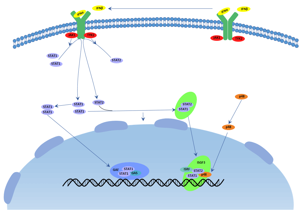
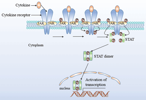
Almost all cell types particularly plasmacytoid dendritric cells(pDC) upon virus recognition can produce type I interferon. The receptors of type I interferon make up of IFNAR1 and IFNAR2. They stimulate the JAK-STAT pathway, resulting in the expression of IFN-stimulated genes(ISG), which are related to the antiviral host defense. An important transcriptional complex, induced by type I IFNs, is the ISG factor 3 (ISGF3) complex. The mature ISGF3 complex is consist of the phosphorylated STAT1 and STAT2, and combine with IRF9, which does not suffer tyrosine phosphorylation. This complex is the only complex that binds specific elements (known as IFN-stimulated response elements(ISREs)) that are present in the promoters of certain ISGs, then initiating their transcription. Overall, IFNα can be used to treat hepatitis B and C infections, while IFNβ can be used to treat multiple sclerosis. The receptors of type III interferon differ from the former one, but they have the common signaling pathway. Type III interferon plays critical roles in the antiviral host defense, predominantly in the epithelial tissues.

Type I IFN Signaling Pathway

Figure 1 Type I IFN Signaling Pathway

Type II IFN Signaling Pathway

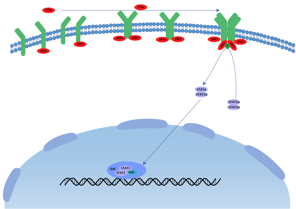
Type II interferon(interferon gamma) is produced by activating T cells, natural killer cells and macrophages et al. upon the stimuli of cytokines like interleukin 12. The receptors of type II interferon make up of IFNGR1 and IFNGR2. The transcription of type II IFN (IFN γ)-dependent genes is regulated by GAS elements, and STAT1 is the most important IFN γ-activated transcription factor for the regulation of these transcriptional responses. After binding to the type II IFN receptor by IFN γ, JAK1 and JAK2 are activated and then phosphorylate STAT1 on the tyrosine residue. Then phosphorylated STAT1 combines with another and forms STAT1–STAT1 homodimers , which translocate to the nucleus and bind GAS elements to initiate transcription. Comparing with type I IFNs, IFN γ does not induce to form ISGF3 complexes, and thereby cannot induce the transcription of genes which have only ISREs in their promoter. In addition to having antiviral activity, type II interferon has important immunoregulatory functions as it is a potent activator of macrophages and T helper 1 cells, at the same time, it has antiproliferative, antiviral and antitumor effects.

Type II IFN Signaling Pathway

Figure 2 Type II IFN Signaling Pathway

4. The Interferon and Diseases

Interferon is so important that it is related to many diseases and the application of it should be highlighted.

Interferon gamma and Tumor

The phases of tumor immunoediting are tumor elimination, tumor dormancy and tumor escape. IFNγ takes part in every phase and the pro-infammation. IFNγ helps the induction to the apoptosis or relapse and progression of tumor.

IFNγ can induce apoptosis and nonapoptosis directly and indirectly. IFNγ can activate the expression of IRF1, a tumor suppressor, in turn, it activates the expression of Bak and reduces the expression of BCL2. These materials stimulate cytochrome c and activate caspase leading to apoptosis of tumor cells. IFNγ simulates amounts of chemicals like ROS and RNI tending to undergo apoptosis. IFNγ can also induce autophagy in human cells. IFNγ can enhance p53-mediated apoptosis and TRAIL or FAS-mediated apoptosis by STAT signaling pathway.

IFNγ can induce dormancy leading to the arrest of cancer growth depend on STAT signaling pathway directly or indirectly. It is manifested that some breast cancer genes like HER2 are dormant in some patients for a long time.

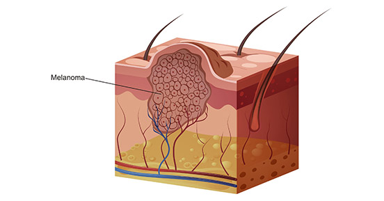
IFNγ facilitates tumor progression and relapse due to its inflammatory characteristic. Melanomas associates with macrophages which produce IFNγ. In turn, inhibiting IFNγ by UV exposure can abolish melanomas. High concentration of IFNγ may lead to NAFLD, subsequent NASH and HCC[6].

Interferon alpha and HIV

IFNα family has 13 members positioned on chromosome 9 in human. Different subtypes have high similarity in their sequence length identity as they have the same ancestor. While the affinity with their receptor, expression level and downstream signaling cascade are different from each subtype in different microenvironment. It is widely acknowledged that IFN has antiviral function. When it is induced by virus, the JAK-STAT signaling pathway is activated, the ISGs(IFN-stimulated genes) are upregulated to resist virus. However, some virus can evade from the IFN immunoregulation and the mechanism is not very clear, it is valuable to explore it deeply for the therapy.

So far, all reports on HIV or SIV infection showed different results in IFNα subtype gene expression, which strongly depended on the analyzed type of tissue, the cell or stimulus. Different IFNα subtypes induce different ISG expression patterns. Some IFNα subtypes(IFNα1, IFNα2, IFNα6, IFNα14, IFNα17 and IFNα21) strongly enhanced the mRNA expression of HIV restriction factors(Mx2, SAMHD1, Tetherin and Trim22), whereas all other IFNα subtypes only slightly increased the ISG expression compared to untreated controls in vitro. They observed that some IFNα subtypes(IFNα14, IFNα6, IFNα17 and IFNα21) potently decreased viral replication shown by cellular p24 levels and infectivity of cell culture supernatants. Interestingly, the clinically used subtype IFNα2 (for HBV therapy) only modestly suppressed HIV replication in vivo. What’s more, structural improvements of IFN might be a considerable option for future immunotherapy regimens in viral infections. It is well established that type I interferon efficiently induces antiviral restriction factors. Accumulating evidence suggests that other types of IFN, specific cytokines and other activators of the cell are also able to upregulate the expression of restriction factors and hence to establish an antiviral cellular state[7].

It was shown that the typical response to IFNα therapy for HCV-infected patients characterized by two phases of viral loads: a rapid decrease due to antiviral ISG expression and a slower decrease mediated by immune cells. Thus, the immunoregulatory activity of IFN may be required for a successful therapy of a chronic virus infection. However, type I interferon inducing immune activation can be either beneficial or harmful in HIV infection as IFNγ rather than IFNα mediates the persistent LCVM disease[2].

Interferon gamma and Tuberculosis

Tuberculosis is a serious infectious disease in our world. The defense depends on the cellular immune response mediated by T cells. IFNγ is produced by T cells, NK cells and macrophages with antiproliferation of transformed cell, antitumor and potentiating the antiviral effects of type I interferon. We obverved that mice with gko deficient could not produce IFNγ and survive well without the pathogen. After the infection of tuberculosis, mice with gko deficient live just 15±1d and have 10× to100× virus compared with the control. There are necrosis of spleen, liver and lung in the gko-knockout mice. After the injection of exogenous IFNγ can heighten the defense and prolong their survival without recovery absolutely. Macrophages activation by IFNγ is proved a resolution of tuberculosis. They have previously reported that NO, and its related gene RNI, are responsible for destruction of virulent tubercle bacilli by murine macrophages, a response that requires treatment in vitro with both IFNγ and TNFα. So that production of RNI, at least in the murine model, may be a necessary mechanism for the control of tuberculosis infection. What’s more, findings in the gko model may have relevance to tuberculosis in AIDS[8].

IFN gamma and Tolerance

IFNγ is one of the most important proinflammatory factor as it can induce the production of Th1 which initiates and maintains the inflammatory process in affected organs. To day, there are amounts of reports which associate IFNγ with immune tolerance induction, both in vitro and in vivo. It seems that these effects, either pro-inflammatory or tolerogenic, are largely dependent on the specific immunological setting and the timeline of the immune response. Dendritric cells(DCs) exerts atigen sampling and messages delivering to responding T cells originally. IFNγ can be produced by DCs and IFNγ can induce DCs activation and tolerance in some scenarios. Treatment of DCs with high doses IFNγ induces the expression of IDO which has an immunorepressive function leading to inhibiting effector T cells, and its long-term maintenance is shown to be supported by kynurenine-aryl hydrocarbon (AhR) pathway. IFNγ seems to be paradoxical that IFNγ sometimes aggravates the severity of autoimmune disease and attenuates it in others[9].

Interferon and Antiretroviraling Restriction Factors

Antiviral restriction factors is the vanguard in the defense line but it is constitutively expressed low in many cell types which should be enhanced encountering pathogens. IFN and some interleukin do this job and establish the antiviral state. The definition of restriction factors is fuzzy as it is diverse with solid function. Restriction factors like SERINC3 and SERINC5 has antiviral function without general inflammation and side effects. Restriction factors like PRRs and TLRs are receptors to restrict the pattern recognition of pathogen. High levels of IFN transiently suppresses viral replication during the chronic phase of HIV-1 infection and the induction of restriction factors contributes to this effect[4].

Interferon and Multiple Sclerosis

Multiple sclerosis(MS) is a chronic and dysregulatory disease of the central nervous system. IFNβ is the first approved and still the most widely used to treat MS. When produced in high amounts, as typically seen in acute viral infections, IFNβ up-regulates anti-inflammatory cytokines, and downregulates the pro-inflammatory ones. IFNα has similar effects of IFNβ. On the contrary, IFNγ has the opposite effects that it further induces Th1 immune response leading to the development and relapse of MS[10].

Interferon and Premature Birth

Pretern birth causes many severe health problems in children under 5 years old due to the infection and inflammation. The inflammation mediates by additional interleukin, tumor necrosis factor and IFNγ will probably cause fetal membrane damage, uterine contraction and biochemical and structural changes in the cervix. What’s more, newborns have weak and damaged innate and adaptive immune system with less IgG[11].

While IFNβ administration during early pregnancy seems to be pharmacologically safe and accumulating real-life evidence suggests that IFNβ therapies are not related to severe pregnancy problems. In consideration of these issues, IFNβ therapy might be continued in women with high risk of disease activity while trying to become pregnant. Additionally, IFNβ is not absorbed through the gastrointestinal tract, and available data suggests that the IFNβ milk levels attainable at usual doses are very low, hence, IFNβ administration seems to be safe for the baby[12][13][14].

Interferon and hepatitis C

Chronic hepatitis, caused by infection with hepatitis C virus C (HCV), also known as chronic hepatitis C (CHC), is parenterally transmitted and primarily through unsafe blood transfusions, the use of injected drugs and therapeutic injections. In humans, the only natural host for HCV is hepatocytes where the virus infects and replicates[15].

The type-1 IFNs include interferon α, β, ε, к, and ω in human. All type I IFNs have antiviral, antiproliferative and immunomodulatory activities, but their relative potencies are not different. Most forms of type I IFN have activity against HCV, yet few have been evaluated clinically[16]. Currently, the commercially available forms of IFNα used for hepatitis C (α2a, α2b and consensus IFN) have somewhat different potent in vitro but appear to yield similar response rates in treated patients. The type-1 IFNs might have similar clinical activities because they share the same, at least in part, cell-surface receptors and intracellular pathways of action. As a crucial mediator of the innate antiviral immune response, interferon alpha(IFNα) was a natural choice for treatment[17].

5. The latest research of Interferon

In this part, we are listing some latest researches about interferon.

#1 The research of Elitza S. Theel et al. suggested that the necessary transition to the QFT-Plus assay would be associated with a minimal difference in assay performance characteristics though comparison of the QuantiFERON-TB Gold Plus and QuantiFERON-TB Gold In-Tube Interferon-γ eelease assays in Patients at Risk for Tuberculosis and in healthcare workers. Please click here to obtain the article.

#2 Li F et al. demonstrated that mice deficient in E3 ligase gene Hectd3 remarkably increased host defense against infection by intracellular bacteria F. novicida, Mycobacterium, and Listeria by limiting bacterial dissemination. In the absence of HECTD3, type I IFN response was impaired during bacterial infection both in vivo and in vitro. These results indicated that HECTD3 mediated TRAF3 polyubiquitination and type I interferon induction during bacterial infection.

#3 Pulmonary, a researcher from critical care & sleep medicine of department of medicine in state university of New York, found that macrophages could be effectively immune-stimulated by aerosol therapy by repurposing IFN γ as an inhaled aerosol and targeting directly to the lung to treat a host of diseases affected by dysregulated immunity.

References

[1] Ng, C.T., Sullivan, B.M., et al. Blockade of Interferon Beta, but Not Interferon Alpha, Signaling Controls Persistent Viral Infection[J]. Cell Host Microbe.2015, 17, 653-661.

[2] Cherie T. Ng,1 Juan L. Mendoza, et al. Alpha and Beta Type 1 Interferon Signaling: Passage for Diverse Biologic Outcomes[J]. Cell. 2016, 164, 349-352.

[3] Nan Y, Wu C, et al. Interferon independent non-canonical STAT activation and virus induced inflammation[J]. Viruses, 2018(4): Apr.

[4] Kathrin S, Julia D, et al. Interferon α subtypes in HIV infection[J]. Cytokines Growth Factor Rev, 2018 Feb.

[5] Leonidas C. Platanias. Mechanisms of type I and type II interferon mediated signaling[J]. Nat Rev Immunol.2005 5(5):375-86.

[6] Aqbi HF, Wallace M, et al. IFN-γ orchestrates tumor elimination, tumor dormancy, tumor escape, and progression[J]. J Leukoc Biol, 2018 Feb.

[7] Hotter D, Kirchhoff F. Interferons and beyond: Induction of antiretroviral restriction factors[J]. J Leukoc Biol, 2018(3): 465-477.

[8] Flynn JL, Chan J, et al. An essential role for interferon γ in resistance to mycobacterium tuberculosis infection[J]. J Exp Med, 1993(6): 2249-2254.

[9] Rozman P, Svajger U. The tolerogenic role of IFN-γ[J]. Cytokines Growth Factor Rev, 2018 Apr

[10] Dumitrescu L, Constantinescu CS, et al. Recent developments in interferon-based therapies for multiple sclerosis[J]. Expert Opin Biol Ther, 2018 Apr.

[11] Helmo FR, Alves EAR, et al. Intrauterine infection, immune system and premature birth[J]. J Matern Fetal Neonatal Med, 2018(9): 1227-1233.

[12] Amato MP, Portaccio E. Fertility, pregnancy and childbirth in patients with multiple sclerosis: impact of disease-modifying drugs[J]. CNS Drugs, 2015(3): 207-220.

[13] Almas S, Vance J, et al. Management of multiple sclerosis in the breastfeeding Mother[J]. Mul Scler Int, 2016: 6527458.

[14] Hale TW, Siddiqui AA, et al.Transfer of interferon beta-1a into human breastmilk[J]. Breastfeed Med, 2012 (2):123-125.

[15] Shepard, C. W., et al. Global epidemiology of hepatitis C virus infection[J]. Lancet Infect. 2005, 5, 558-567.

[16] Robek, M. D., Boyd, et al. Lambda interferon inhibits hepatitis B and C virus replication[J]. J. Virol. 2005, 79, 3851-3854.

[17] Jordan J. Feld and Jay H. Hoofnagle. Mechanism of action of interferon and ribavirin in treatment of hepatitis C[J]. Nature.2005, 436(7053):967-72.

The Overview of Growth Factors

A growth factor, which generally considered as a series of cytokines, is a naturally occurring substance that stimulate cell growth, differentiation, survival, inflammation, and tissue repair. Usually it is a protein or a steroid hormone. Growth factors are important to regulate a variety of cellular processes, which can be secreted by neighboring cells, distant tissues and glands, or even tumor cells themselves. Normal cells show a demand for several growth factors to maintain proliferation and viability. Growth factors can exert their stimulation though endocrine, paracrine or autocrine mechanisms.

1. Growth Factors Classification

According to the database from Wikipedia, we conclude the classification of growth factors in the Table 1, which includes gene name, Uniprot ID and corresponding receptors.

Table 1. The Classification of Growth Factors

Uniprot ID Receptors
Epidermal growth factor (EGF) P01133 EGFR
Fibroblast growth factor (FGF) FGF1 P05230 FGFR1FGFR2[1]FGFR3[2]FGFR4[3]
FGF2 P09038 FGFR1FGFR2
FGF3 P11487 FGFR1FGFR2
FGF4 P08620 FGFR1FGFR2
FGF5 P12034 FGFR1
FGF6 P10767 FGFR1FGFR2
FGF7 P21781 FGFR2[4]
FGF8 P55075 FGFR1FGFR2
FGF9 P31371 FGFR2FGFR3[5]
FGF10 O15520 FGFR1FGFR2
FGF11 Q92914 None[6]
FGF12 P61328 None[6]
FGF13 Q92913 None[6]
FGF14 Q92915 None[6]
FGF15 O35622 To be supplemented
FGF16 O43320 To be supplemented
FGF17 O60258 FGFR1FGFR2
FGF18 O76093 FGFR3[7]
FGF19 O95750 FGFR1
FGF20 Q9NP95 To be supplemented
FGF21 Q9NSA1 FGFR1
FGF22 Q9HCT0 FGFR2
FGF23 FGFR1
Insulin P01308 insulin receptor (IR)
Insulin-like growth factors(IGF) IGF1 P05019 insulin receptor (IR)IGF1R
IGF2 P01344 insulin receptor (IR), GF1R, IGF2R[8]
Transforming growth factors TGF-α P01135 EGFR[9]
TGF-β1 P01137 TGFBR1
TGF-β2 P61812 TGFBR2
TGF-β3 P10600 TGFBR3
Vascular endothelial growth factor(VEGF) VEGF-A P15692 VEGFR-1, VEGFR-2
VEGF-B P49765 VEGFR-1
VEGF-C P49767 VEGFR-2VEGFR-3
VEGF-D O43915 VEGFR-3
PlGF P49763 VEGFR-1
Platelet-derived growth factor (PDGF) PDGFA P04085 PDGFRAPDGFRB
PDGFB P01127 PDGFRAPDGFRB
PDGFC Q9NRA1 PDGFRAPDGFRB
PDGFD Q9GZP0 PDGFRAPDGFRB
Neurotrophins Brain-derived neurotrophic factor (BDNF) P23560 TrkB, LNGFR
Nerve growth factor (NGF) P01138 TrkA
Neurotrophin-3 (NT-3) P20783 TrkC
Neurotrophin-4 (NT-4) P34130 TrkB
Growth differentiation factor-9 (GDF9) O60383 BMPRII, TGFBR1[9][10]
Hepatocyte growth factor (HGF) HGFR(MET)
Hepatoma-derived growth factor (HDGF) P51858 To be supplemented
Migration-stimulating factor (MSF) Q92954 CD44

2. Featured Growth Factors & Receptors

Fibroblast Growth Factors (FGFs) and Receptors

a. Fibroblast growth factors

The fibroblast growth factors are a family of cell signaling proteins, also known as potent regulators of cell proliferation, differentiation and function, which are critically important in normal development, tissue maintenance, wound repair and angiogenesis. FGFs are also associated with several pathological conditions. Mutations in FGF genes are associated with various diseases such as cardiovascular disease, osteoarthritis, hepatocellular carcinoma, intervertebral disc homeostasis and hypophosphatemia[11][12][13].

FGFs bind heparan sulfate glycosaminoglycans (HSGAGs), which facilitates dimerization (activation) of FGF receptors (FGFRs). There are 22 members of the FGF family in humans from FGF-1 to FGF-23 except for FGF-15 which exists in the mouse. All FGFs except for four members (FGF11-FGF14) bind to FGF Receptors. Because FGF11-FGF14, also known as fibroblast growth factor-homologous factors or FHF1-FHF4, share significant sequence and structural homology with FGFs, and the manner of binding HS is similar to the FGFs. In contrast, Olsen et al. And Mohammadi et al. Who analysed FHF-mediated FGFR activation showed that FHFs are incapable of activating any of the four FGFRs, likely as a result of the structural incompatibility of the FGFR interacting region[14][15]. According to the present stage of knowledge, FHFs act as intracellular signaling molecules that function independently of FGFRs, via interaction with islet brain-2 scaffold protein and voltage-gated sodium channels, as discussed in detail later[16]. Based on phylogenetic analysis, the human FGF family can be further divided into seven subfamilies; FGF1-FGF2, FGF4-FGF6, FGF 3/7/10/22, FGF 8/17/18, FGF 9/16/20, FGF 11-FGF14, and FGF 19/21/23. Some other research discoveries showed that FGF24 and 25 are exist in zebra fish, but no data is available about their mammalian counterparts[17][18].

b. Fibroblast growth factors receptors

The fibroblast growth factor receptors, also known as FGFRs, are receptors that bind to members of the fibroblast growth factor family of proteins. FGFRs are single-pass transmembrane receptors with extracellular ligand-binding domains and an intracellular tyrosine kinase domain that have intracellular tyrosine kinase activity, and belong to the family of receptor tyrosine kinases (RTK)[19]. Activation of RTKs by their respective ligands induces kinase activation that in turn initiates intracellular signaling networks that ultimately orchestrate key cellular processes, such as cell proliferation, growth, differentiation, migration, and survival. In this way, RTKs play pivotal biological roles during the development and adult life of multicellular organisms. FGFRs are associated with several pathological conditions, such as cancer cell migrations[20], tumor initiation and progression[21], carcinogenesis[22], congenital skeletal dysplasias[23], et al. Mutations in FGFR genes are associated with various diseases such as breast cancer[24], bladder cancer[25], gastric cancer[26], clear-cell renal cell carcinoma[27], developmental corruption and malignant disease[28]et al.

The FGFRs includes four genes encoding closely related transmembrane, tyrosine kinase receptors (termed FGFR1 to FGFR4). A typical FGFR consists of a signal peptide that is cleaved off, three immunoglobulin (Ig)–like domains, an acidic box, a transmembrane domain, and a split tyrosine kinase domain.

Transforming growth factors & receptors

a. Transforming growth factors

Transforming growth factor, also known as Tumor growth factor, or TGF, is used to refer to two classes of polypeptide growth factors, Transforming growth factor alpha (TGFα) and transforming growth factor beta (TGFβ). TGFα and TGFβ are autocrine/paracrine growth factors that are classically thought of as potent promoters and inhibitors of cell growth, respectively, in normal tissues[29][30][31].

TGFα is upregulated in some human cancers and acts through the EGF receptor. It is produced in macrophages, keratinocytes and brain cells, and induces epithelial development. TGFα is also thought to be invovlved in the pathogenesis process of hepatocarcinogenesis. TGFβ acts through the TGFβ receptor (TGFβR) and exists in three known isoforms in humans, TGFβ1, TGFβ2, and TGFβ3[32], which are upregulated in colorectal adenomas and cancer, and directly associated with more advanced tumor characteristics[33][34]. Moreover, they also play crucial roles in tissue regeneration, cell differentiation, embryonic development, and regulation of the immune system. Isoforms of transforming growth factor-beta (TGF-β1) are also involved in the pathogenesis of epithelial-mesenchymal transition via the NF-κB pathway[35]. TGFβ receptors are single pass serine/threonine kinase receptors[36].

b. Transforming growth factors receptors

In the last paragraph, we have introduced that the receptor of TGFα is epidermal growth factor receptor ( also known as EGFR), which is a transmembrane protein that is a receptor for members of the epidermal growth factor family (EGF family) of extracellular protein ligands, and the receptor of TGFβ also has three isoforms in humans, TGFRI, TGFRII and TGFRIII.

EGFR is a transmembrane protein that is activated by binding of its specific growth factor ligands, including TGFα and epidermal growth factor. Upon activation by its specific ligands, EGFR turns to be a transition from an inactive monomeric form to an active homodimer. Mutations that EGFR overexpression, also known as upregulation or amplification, have been associated with several cancers, including squamous-cell carcinoma of the lung, anal cancers, glioblastoma and epithelian tumors of the head and neck[37]. Aberrant EGFR signaling has been implicated in inflammatory disease and monogenic disease. Furthermore, EGFR also has been reported to play a critical role in TGFβ1 dependent fibroblast to myofibroblast differentiation[38].

The Transforming Growth Factor beta (TGFβ) receptors are a superfamily of serine/threonine kinase receptors. There are three isoforms receptors bind members of the TGFβ superfamily of growth factor, TGFRβ1, TGFRβ2, TGFRβ3, can be distinguished by their structural and functional properties. TGFβR1 (also known as ALK5) and TGFβR2 have similar ligand-binding affinities and can be differentiated from each other just by peptide mapping. Both TGFβR1 and TGFβR2 have a high affinity for TGFβ1 and low affinity for TGFβ2. TGFβR3 (also known as β-glycan) has a high affinity for both homodimeric TGFβ1 and TGFβ2 and the heterodimer TGF-β1.2. The TGFβ receptors also bind TGFβ3.

Vascular endothelial growths factor and Receptors

a. Vascular endothelial growth factors

Vascular endothelial growth factors, also known as vascular permeability factor (VPF), or VEGF, constitute a sub-family of growth factors produced by cells that stimulate the formation of blood vessels and a mitogen for vascular endothelial cells. VEGFs are important signaling proteins involved in both vasculogenesis, the de novo formation of the embryonic circulatory system, and angiogenesis, the growth of blood vessels from pre-existing vasculature. There are five members in the vascular growth factor family, include VEGF-A, VEGF-B, VEGF-C, VEGF-D and placenta growth factor (PlGF). VEGF-A is the first discovery member of the vascular endothelial growth factor family. Vascular endothelial growth factors are key mediators of angiogenesis that is crucial for development and metastasis of tumors. Overexpression of VEGF can contribute to disease. Solid cancers cannot grow beyond a limited size without an adequate blood supply; cancers that can express VEGF are able to grow and metastasize. When VEGF is overexpressed, it can cause vascular disease in the retina of the eye and other parts of the body. Drugs can inhibit VEGF and control or slow those diseases, such as aflibercept, bevacizumab, ranibizumab and pegaptanib sodium (Macugen).

b. Vascular endothelial growth factor Receptors

All members of the VEGF family stimulate cellular responses by binding to tyrosine kinase receptors (the VEGFRs) on the cell surface, causing them to be dimer and become activated through transphosphorylation. The VEGFRs have an extracellular portion, which is consist of 7 immunoglobulin-like domains, a single transmembrane spanning region and an intracellular portion containing a split tyrosine-kinase domain. There are three main subtypes of VEGFR, numbered 1, 2 and 3, also known as Flt-1, KDR/Flk-1 and Flt-4, respectively.

VEGF-A binds to VEGFR-1 (Flt-1) and VEGFR-2 (KDR/Flk-1). VEGFR-2 appears to mediate almost all of the known cellular responses to VEGF, which can combines with VEGF-A, VEGF-C, VEGF-D and VEGF-E[39]. Although VEGFR-1 is thought to modulate VEGFR-2 signaling, the function of VEGFR-1 is less well defined. Another function of VEGFR-1 is to act as a dummy/decoy receptor, sequestering VEGF from VEGFR-2 binding. In fact, an alternatively spliced form of VEGFR-1 (sFlt1) is not a membrane bound protein but is secreted and functions primarily as a decoy[40]. A third receptor has been discovered (VEGFR-3), however, VEGF-A is not a ligand for this receptor. VEGFR-3 mediates lymphangiogenesis by binding to VEGF-C and VEGF-D.

3. The Signaling Pathways, Most Popular with Researchers

TGF-β Signaling Pathway

In TGF-β signaling pathway, the family members of TGF-βs include TGF-betas, activins and bone morphogenetic proteins (BMPs), which are structurally related secreted cytokines found in various species ranging from insects to mammals. TGF-β signaling pathway is involved in many cellular processes, including cell growth, cell differentiation, apoptosis, cellular homeostasis, et al.

The image of TGF-β signaling pathway

Fig. 1. The image of TGF-β signaling pathway

The mechanism includes four parts, Ligand binding, Receptor recruitment and phosphorylation, CoSMAD binding, and Transcription. TGF-beta family ligands binds to the type II receptor and recruits Type I, whereby type I receptor is phosphorylated and activated by type II receptor. Then, the type I receptor phosphorylates receptor-activated Smads(R-Smads: Smad1, Smad2, Smad3, Smad5, and Smad8). Once phosphorylated, R-Smads combine with the co-mediator Smad, Smad4, and the heteromeric complex then translocates into the nucleus. In the nucleus, Smad complexes activate specific genes through cooperative interactions with other DNA-binding and coactivator (or co-repressor) proteins.

VEGF Signaling Pathway

When a VEGF binds to its receptor, the receptor can transiently exert its kinase activity and form a complex with an intracellular tyrosine or serine/threonine kinase. Then, the activated receptors result in the activation of other proteins in the signaling pathway and the production of various second messengers. Finally, these signals are transmitted into the nucleus and induce the expression of specific genes.

The image of VEGF signaling pathway

Fig. 2. The image of VEGF signaling pathway

Now, although the evidence that VEGFR-2 is the major mediator of VEGF-driven responses in endothelial cells also is not very much, it is considered to be a crucial signal transducer in both physiologic and pathologic angiogenesis. The binding of VEGF to VEGFR-2 leads to form receptor dimer, then, followed by intracellular activation of the PLCγ; Subsequently, PKC-Raf kinase-MEK-mitogen-activated protein kinase (MAPK) pathway and initiation of DNA synthesis and cell growth, whereas activation of the phosphatidylinositol 3′ -kinase (PI3K)-Akt pathway leads to increased endothelial-cell survival. Activation of PI3K, FAK, and p38 MAPK is implicated in cell migration signaling.

Wnt Signaling Pathway

The Wnt signaling pathway is a group of signal transduction pathway made of proteins that pass signals into a cell through cell surface receptors.

The image of Wnt signaling pathway

Fig. 3. The image of Wnt signaling pathway

Wnt signaling diversifies into three main branches: 1. The canonical Wnt pathway, also known as classical, activates target genes through stabilization of β-catenin in the nucleus. The function of this pathway during embryonic development has been originally elucidated by experimental analysis of axis development in the frog Xenopus laevis and of segment polarity and wing development in the fly Drosophila melanogaster. 2. The planar cell polarity pathway involves RhoA and Jun Kinase (JNK) and controls cytoskeletal rearrangements. Its main role is the temporal and spatial control of embryonic development, as exemplified in the polar arrangement of cuticular hairs in Drosophila or the convergent-extension movements in Xenopus embryos. 3. The Wnt/Ca2+ pathway is stimulated by Wnt 5a and Wnt 11 and involves an increase in intracellular Ca2+ and activation of Ca2+-sensitive signalling components, such as calmodulin-dependent kinase, the phosphatase calcineurin, and the transcription factor NF-AT.

4. The Latest Researches of Growth Factor

  • Komaki Y’s team have revealed that hepatocyte growth factor facilitates esophageal mucosal repair and inhibits the submucosal fibrosis in a rat model of esophageal ulcer. If you want to know more information, please click here to view the article.
  • Regeenes R1, Silva PN, et al. have demonstrated that fibroblast growth factor receptor 5 (FGFR5) is a co-receptor for FGFR1 that is up-regulated in beta-cells by cytokine-induced inflammation. If you want to know more information, please click here to view the article.
  • Breit A1, Miek L, et al. have found that insulin-like growth factor-1 acts as a zeitgeber on hypothalamic circadian clock gene expression via glycogen synthase kinase-3β signaling. If you want to know more information, please click here to view the article.

References

[1] Stauber DJ, DiGabriele AD, et al. Structural interactions of fibroblast growth factor receptor with its ligands[J]. Proceedings of the National Academy of Sciences of the United States of America. 2000, 97 (1): 49–54

[2] Santos-Ocampo S, Colvin JS, et al. Expression and biological activity of mouse fibroblast growth factor-9[J]. The Journal of Biological Chemistry. 1996, 271 (3): 1726–31

[3] Ornitz DM, Xu J, et al. Receptor specificity of the fibroblast growth factor family [J]. The Journal of Biological Chemistry. 1996, 271(25):15292-15297

[4] Duchesne L, Tissot B, et al. N-glycosylation of fibroblast growth factor receptor 1 regulates ligand and heparan sulfate co-receptor binding [J]. The Journal of Biological Chemistry. 2006, 281(37):27178-27189

[5] Davidson D, Blanc A, et al. Fibroblast growth factor (FGF) 18 signals through FGF receptor 3 to promote chondrogenesis[J]. The Journal of Biological Chemistry. 2005, 280(21):20509-20515

[6] Chellaiah A, Yuan W, et al. Mapping ligand binding domains in chimeric fibroblast growth factor receptor molecules. Multiple regions determine ligand binding specificity[J]. The Journal of Biological Chemistry. 1999, 274 (49): 34785–94

[7] Pavel Krejci, Jirina Prochazkoval, et al. Molecular pathology of the fibroblast growth factor family[J]. Hum Mutat. 2009, 30(9): 1245–1255

[8] Laureys G, Barton DE, et al. Chromosomal mapping of the gene for the type II insulin-like growth factor receptor/cation-independent mannose 6-phosphate receptor in man and mouse[J]. Genomics.1988, 3 (3): 224–9

[9] Ojeda, S. R.; Ma, Y. J., et al. The transforming growth factor alpha gene family is involved in the neuroendocrine control of mammalian puberty[J]. Molecular Psychiatry. 1997, 2 (5): 355–358

[10] Gilchrist, R., Lane, M., et al. Oocyte-secreted factors: regulators of cumulus cell function and oocyte quality[J]. Human Reproduction Update. 2008, 14(2), pp.159-177

[11] Yun, Y.-R., Won, J. E., et al. Fibroblast growth factors: biology, function, and application for tissue regeneration[J]. Tissue Eng. 2010, 218142

[12] Coleman SJ, Grose RP, et al. Fibroblast growth factor family as a potential target in the treatment of hepatocellular carcinoma[J]. J Hepatocell Carcinoma. 2014, 29;1:43-54

[13] Ellman MB, An HS, et al. Biological impact of the fibroblast growth factor family on articular cartilage and intervertebral disc homeostasis[J]. Gene. 2008, 15;420(1):82-9

[14] Olsen SK, Garbi M, et al. Fibroblast growth factor (FGF) homologous factors share structural but not functional homology with FGFs[J]. J Biol Chem. 2003, 278:34226–34236

[15] Mohammadi M, Dikic I, et al. Identification of six novel autophosphorylation sites on fibroblast growth factor receptor 1 and elucidation of their importance in receptor activation and signal transduction[J]. Mol Cell Biol. 1996; 16:977–989

[16] Goldfarb M. Fibroblast growth factor homologous factors: evolution, structure, and function[J]. Cytokine Growth Factor Rev. 2005; 16:215–220

[17] Roy NM, Sagerstrom GG. An early Fgf signal required for gene expression in the zebrafish hindbrain primordium[J]. Brain Res Dev Brain Res. 2004; 148(1): 27-42

[18] Katoh Y, Katoh M. Comparative genomics on FGF7, FGF10, FGF22, orthologs, and identification of fgf25[J]. Int J Mol Med. 2005; 16(4): 767-70

[19] Schlessinger J. Cell signaling by receptor tyrosine kinases[J]. Cell. 2000, 103:211–25

[20] Nguyen T, Mège RM. N-Cadherin and Fibroblast Growth Factor Receptors crosstalk in the control of developmental and cancer cell migrations[J]. Eur J Cell Biol. 2016, 95(11):415-426

[21] Feng S, Zhou L, et al. Fibroblast growth factor receptors: multifactorial-contributors to tumor initiation and progression[J]. Histol Histopathol. 2015, 30(1):13-31

[22] Haugsten EM, Wiedlocha A, et al. Roles of fibroblast growth factor receptors in carcinogenesis[J]. Mol Cancer Res. 2010, 8(11):1439-52

[23] Sanak M. Molecular genetics of congenital skeletal dysplasias related to mutations of fibroblast growth factor receptors[J]. Med Wieku Rozwoj. 1999, 3(1):67-82

[24] Wang S, Ding Z. Fibroblast growth factor receptors in breast cancer[J]. Tumour Biol. 2017, 39(5):1010428317698370

[25] Morales-Barrera R, Suárez C, et al. Targeting fibroblast growth factor receptors and immune checkpoint inhibitors for the treatment of advanced bladder cancer: New direction and New Hope[J]. Cancer Treat Rev. 2016, 50:208-216

[26] Inokuchi M, Fujimori Y, et al Therapeutic targeting of fibroblast growth factor receptors in gastric cancer[J]. Gastroenterol Res Pract. 2015, 2015:796380

[27] Sonpavde G, Willey CD, et al. Fibroblast growth factor receptors as therapeutic targets in clear-cell renal cell carcinoma[J]. Expert Opin Investig Drugs. 2014, 23(3):305-15

[28] Kelleher FC, O’Sullivan H, et al. Fibroblast growth factor receptors, developmental corruption and malignant disease[J]. Carcinogenesis. 2013, 34(10):2198-205

[29] Lemmon MA, Schlessinger J. Cell signaling by receptor tyrosine kinases[J]. Cell. 2010, 141:1117–1134

[30] Potter JD. Colorectal cancer: molecules and populations[J]. J Natl. Cancer Inst. 1999, 91:916–932

[31] Ikushima H, Miyazono K. TGFbeta signalling: a complex web in cancer progression[J]. Nat Rev Cancer 2010, 10:415-424

[32] Huakang Tu, Thomas U. Ahearn, et al. Transforming Growth Factors and Receptor as Potential Modifiable Pre-Neoplastic Biomarkers of Risk for Colorectal Neoplasms[J]. MOLECULAR CARCINOGENESIS. 2015, 54:821-830

[33] Bellone G, Gramigni C, et al. Abnormal expression of Endoglin and its receptor complex (TGF-beta1 and TGF-beta receptor II) as early angiogenic switch indicator in premalignant lesions of the colon mucosa[J]. Int J Oncol. 2010, 37:1153–1165

[34] Hawinkels LJ, Verspaget HW, et al. Active TGF-beta1 correlates with myofibroblasts and malignancy in the colorectal adenoma-carcinoma sequence[J]. Cancer Sci. 2009, 100:663–670

[35] Li Y, Zhu G, et al. Simultaneous stimulation with tumor necrosis factor-α and transforming growth factor-β1 induces epithelial-mesenchymal transition in colon cancer cells via the NF-κB pathway[J]. Oncol Lett. 2018, 15(5):6873-6880

[36] Badawy AA, El-Hindawi A, et al. Impact of epidermal growth factor receptor and transforming growth factor-α on hepatitis C virus-induced hepatocarcinogenesis[J]. APMIS. 2015, 123(10):823-31

[37] Walker F, Abramowitz L, et al. Growth factor receptor expression in anal squamous lesions: modifications associated with oncogenic human papillomavirus and human immunodeficiency virus[J]. Human Pathology. 2009, 40 (11): 1517–27

[38] Midgley AC, Rogers M, et al. Transforming growth factor-β1 (TGF-β1)-stimulated fibroblast to myofibroblast differentiation is mediated by hyaluronan (HA)-facilitated epidermal growth factor receptor (EGFR) and CD44 co-localization in lipid rafts[J]. The Journal of Biological Chemistry[J]. 2013, 288 (21): 14824–38

[39] Holmes K, Roberts OL, et al. Vascular endothelial growth factor receptor-2: structure, function, intracellular signalling and therapeutic inhibition[J]. Cell Signal. 2007, 19 (10): 2003–2012

[40] Zygmunt T, Gay CM, et al. Semaphorin-PlexinD1 Signaling Limits Angiogenic Potential via the VEGF Decoy Receptor sFlt1[J]. Dev Cell. 2011, 21 (2): 301–314
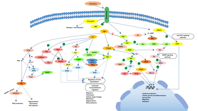
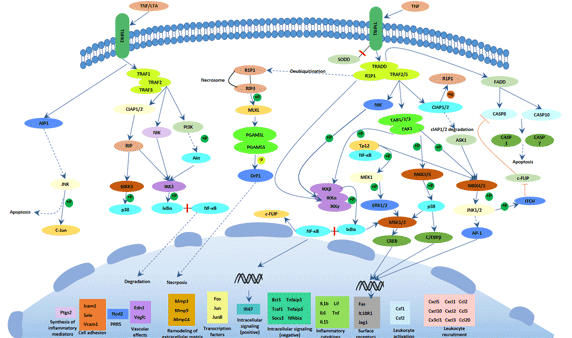
CANCER

Cancer is a group of diseases that involve abnormal cell growth with the potential to invade or spread to other parts of the body. They form a subset of neoplasms. Comparing with benign tumors, which do not spread to other parts of the body. A neoplasm or tumor is a group of cells that have undergone unregulated growth and will often form a mass or lump, but may be distributed diffusely.

Melanoma, the Most Serious Type of Skin Cancer

1. What is Melanoma?

Melanoma, usually referred to as malignant melanoma, is a highly malignant tumor derived from melanocytes which control the pigment in your skin. The figure 1 shows melanoma cells extending from the surface of the skin into the deeper skin layers. Melanoma occurs mostly in the skin, but also in the mucous membrane and viscera, accounting for about 3% of all tumors. Melanoma is the deadliest form of skin cancer and strikes tens of thousands of people around the world each year. The number of cases is rising faster than any other type of solid cancer.

a diagram of melanoma
Figure 1. a diagram of melanoma

In 2020, the American Cancer Society’s estimates that there will have approximately 100,350 new melanomas diagnosed (about 60,190 in men and 40,160 in women), and 6,850 deaths from melanoma (about 4,610 men and 2,240 women). The rates of melanoma have been rising rapidly over the past few decades, but this has varied by age. So how does melanoma develop?


2. How does Melanoma Develop?

Melanoma occurs when something goes wrong in melanocytes, the cells producing the pigment melanin. Normally, skin cells develop in a controlled and orderly way — healthy new cells push older cells toward to the surface of skin, where they die and eventually fall off. But when some cells undergo DNA damage, new cells may start to grow out of control and can eventually form a mass of cancerous cells. So what damages DNA in skin cells?

In fact, it isn’t still clear. It may be a combination of factors, including environmental and genetic factors, causes melanoma. Currently, doctors believe exposure to ultraviolet (UV) radiation from the sun and from tanning lamps and beds is the leading cause of melanoma. UV light doesn’t cause all melanomas, especially those that occur in places on your body that don’t receive exposure to sunlight. This suggests that other factors may contribute to your risk of melanoma.


3. What are the Risk Factors of Melanoma?

As mentioned previously, exposure to ultraviolet (UV) radiation and other sources of ultraviolet light, like tanning beds, is a very important risk factor. In addition to the ultraviolet light, factors that may increase risk of melanoma include:

  • Race. The American Cancer Society states that the lifetime risk of developing melanoma is about 2.6% for white people, 0.1% for Black people and 0.6% for Hispanic people. Accumulating evidence has revealed that melanoma in white people is 20 times more common than black people.
  • A family history of melanoma. If a close relative (such as a parent, child or sibling) has had melanoma, you may have a greater chance of developing melanoma.
  • Weakened immune system. People with weakened immune systems also have an increased risk of melanoma and other skin cancers. Your immune system may be impaired if you take medicine to suppress the immune system, such as after an organ transplant, or if you have a disease that impairs the immune system, such as AIDS.
  • Age. The risk of melanoma grows as you age. The average age at diagnosis is 65, even though it’s one of the most common cancers among young adults.


4. What are the Symptoms of Melanoma?

Studies reported that the origin of melanocytes is associated with the fetal period and the melanocyte precursors. The melanocyte precursors are produced in the neural ridge which migrate to various localizations in the body during fetal development, including the skin, meningeal coverings, mucous membrane, the upper part of esophagus, and the eyes. So melanoma can develop by malignant transformation of melanocytes anywhere on your body [2] [3]. The most likely areas, though, are chest and back for men, legs for women, neck and face, because these areas have more exposure to the sun than other parts of the body.

The first symptoms of melanoma often are changes in an existing mole and the development of a new pigmented or unusual growth on your skin. Clues that a mole might be melanoma are: irregular shape, irregular border, multicolored or uneven coloring, larger than a quarter of an inch, changes in size, shape, or color, itchiness or bleeding. Sometimes, the skin will appear normal even though melanoma has begun to develop.

As well as sun exposure, distinct genetic alterations have been identified as associated with melanoma [4]. In the next section, we illustrate the mechanisms of melanoma as brief.


5. What are the Mechanisms of Melanoma?

Melanoma caused by transformation of melanocytes requires a complex interaction of exogenous and endogenous events. There are tremendous progress has been reported to make in unravelling the genetic basis of melanoma [5] [6] [7]. As the figure 2a shows, under normal conditions, mitogen-activated protein kinase (MAPK) and phosphatidylinositol 3‑kinase (PI3K)–AKT signaling permit balanced control of basic cellular functions, including cell cycle regulation, survival, motility and metabolism.

However, in melanoma, several genetic alterations depicted in the figure 2b are frequently observed, leading to constitutive pathway activation (indicated by thick arrows) and loss of cellular homeostasis. Malignant transformation can require combinations of genetic defects. The functional consequences of genetic events determine whether mutations can coexist or remain mutually exclusive. For example, mutations in NRAS and BRAF occur very rarely in the same melanoma cell, whereas combined genetic alterations of BRAF and PTEN are common.

signaling pathways in melanoma
Figure 2. signaling pathways in melanoma
*this diagram is derived from publication on Nature Reviews.


6. How to Diagnose Melanoma?

There are a series of tests and procedures used to diagnose melanoma, including:

  • Physical examination. Your doctor will ask questions about your health history and examine your skin carefully to look for signs (as mentioned on section 4) that may indicate melanoma.
  • Blood chemistry studies. As the levels of lactate dehydrogenase (LDH) can be higher than normal when you have melanoma, you will be informed to check this enzyme in your blood. But note that LDH levels may not be checked for early stage disease.
  • Skin biopsy. It usually refers to removing a sample of tissue for testing, and is the only way to confirm melanoma. The sample removed from your skin is sent to a lab for examination. If at all possible, the entire suspected area should be removed.
  • Lymph node biopsy. If melanoma is diagnosed, your doctor may need to find out if cancer cells have spread, though they won’t do this for melanoma in situ. The first step is to perform a sentinel node biopsy.
  • Imaging tests. Imaging tests are used to see if cancer has spread beyond the skin to other parts of the body. These tests include CT scan, MRI and PET scan.


7. What are the Treatment of Melanoma?

If there’s a diagnosis of melanoma, it’s important to determine the stage. This will provide information on your overall outlook and help guide treatment. Treatment depends on the stage of melanoma. Melanoma is staged based on the size of the tumor and the extent to which the cancer has spread. The stages of melanoma are divided into five stages. We collect the stages and treatments of melanoma on the following table.

Description Treatments
Stage 0 The tumor is in the epidermis and has not spread elsewhere. The suspicious tissue of melanoma in stage 0 is possible to be completely removed during a biopsy. You may not need further treatment.
Stage 1 The tumor is less than 2 mm thick, is developing slowly and hasn’t spread to other organs. Very thin melanomas can be completely removed during biopsy. If not, they can be surgically removed later. This involves removing the cancer along with a margin of healthy skin and a layer of tissue underneath the skin. Early-stage melanoma doesn’t necessarily require additional treatment.
Stage 2 The tumor is from 1-4+ mm thick, may be ulcerated, and hasn’t spread to other organs.
Stage 3 The cancer has spread to nearby skin or lymph nodes, lymph nodes may be enlarged, and the developing tumor may be ulcerated. In stage 3, wide-excision surgery is used to remove the tumor and affected lymph nodes. In stage 4, The skin tumors and some enlarged lymph nodes can be removed by surgery. Moreover, you also need to have surgery to remove tumors on internal organs based on the number, size, and location of tumors.
Stage 4 The cancer has spread to distant organs or lymph nodes. At this stage the cancer may be referred to as a metastatic melanoma.

Besides, stages 3 and 4 generally require some additional treatments, involving:

  • Immunotherapy drugs. Currently, the immunotherapy drugs usually include interferon or interleukin-2 (IL-2) or checkpoint inhibitors, such as ipilimumab, nivolumab, and pembrolizumab.
  • Targeted therapy for those cancers related to mutations in the BRAF gene. These may include cobimetinib, dabrafenib, trametinib, and vemurafenib.
  • Targeted therapy for melanoma related to mutations in the C-KIT gene. These may include imatinib and nilotinib.
  • Vaccines. These may include Bacille Calmette-Guerin (BCG) and T-VEC (Imlygic).
  • Radiation therapy. This is used to shrink tumors and to kill cancer cells that may have been missed during surgery. Radiation can also help relieve symptoms of cancer that has metastasized.
  • Isolated limb perfusion. This involves infusing only the affected arm or leg with a heated solution of chemotherapy.
  • Systemic chemotherapy. This may include dacarbazine (DTIC) and temozolomide (Temodar), which may be used to kill cancer cells throughout your body.


References

[1] Karl Smart, Ian Pope, Robert Sullivan. MELANOMA [J]. Nature. 2014, 515: S109.

[2] Ribas A, Slingluff CL, Rosenberg SA. Cutaneous Melanoma [J]. Principles and Practice of Oncology. 2015. pp. 1346-94.

[3] Coricovac D, Dehelean C, Moaca EA, et al. Cutaneous melanoma long road from experimental models to clinical outcome: a review [J]. Int J Mol Sci. 2018, 19:1566.

[4] Dirk Schadendorf, David E. Fisher, Claus Garbe, et al. Melanoma [J]. NATURE REVIEWS. 2015.

[5] Bastian, B. C. The molecular pathology of melanoma: an integrated taxonomy of melanocytic neoplasia [J]. Annu. Rev. Pathol. 2014, 9, 239–271.

[6] Sheppard, K. E. & McArthur, G. A. The cell-cycle regulator CDK4: an emerging therapeutic target in melanoma [J]. Clin. Cancer Res. 2013, 19, 5320–5328.

[7] Shi, J. et al. Rare missense variants in POT1 predispose to familial cutaneous malignant melanoma [J]. Nat. Genet. 2014, 46, 482–486.

Leukemia, Why is it So Common?

Upon mentioned leukemia, marrow transplantation and anemia as keywords of leukemia come to your mind. Leukemia is the most common cancer in children and teens, accounting for almost 1 out of 3 cancers. Usually, the number of new cases of leukemia diagnosed in the United States each year is about 14 per 100,000 men and women. It is the tenth most common cancer according to new cases diagnosed each year. Leukemia accounts for 3.5% of all new cancer cases in the United States. So what is the leukemia? And what are the types of leukemia? This article collect several FAQ of leukemia, including:

1. What is Leukemia?

Leukemia is a malignant clonal disease of hematopoietic stem cells. Leukemia cells in the clone will stop at different stages of cell development due to uncontrolled proliferation, differentiation failure, and obstructed apoptosis. In bone marrow and other hematopoietic tissues, leukemia cells proliferate and accumulate in large numbers, and infiltrate other tissues and organs, causing normal hematopoietic function to be inhibited. As the figure 1 shows, normal blood contains red blood cells (RBCs), white blood cells (WBCs), and platelets. Leukemia cells outnumber normal cells in leukemia.

a diagram of components of normal blood and leukemia
Figure 1. a diagram of components of normal blood and leukemia

Generally speaking, leukemia refers to cancers of the WBCs. WBCs play a vital role in protecting body from invasion by bacteria, viruses, and fungi. In leukemia, the WBCs have an abnormal unlike normal WBCs. They divide too quickly and eventually crowd out normal cells. Most of WBCs are produced in the bone marrow, but certain types of WBCs are also made in the lymph nodes, spleen, and thymus gland.

2. What are The Types of Leukemia?

In fact, there are many types of leukemia in clinic. Based on different speed of disease development, leukemia is divided into two types, including acute leukemia and chronic leukemia. In acute leukemia, leukemia cells multiply rapidly and the disease progresses quickly. In chronic leukemia, the disease progresses slowly and early symptoms may be very mild.

Most often, leukemia is a cancer of the white blood cells, but some leukemias start in other blood cell types. According to different cell types, leukemia is classified into two types. One is myelogenous leukemia developing from the myeloid cell line. Another is lymphocytic leukemia developing from the lymphoid cell line.

Hence, there are four major types of leukemia in clinic. As the following table shows, acute myelogenous leukemia is the most common form of leukemia. The five-year survival rate of chronic lymphocytic leukemia is the highest. Most childhood leukemias are acute lymphocytic leukemia (ALL). Most of the remaining cases are acute myeloid leukemia (AML). Chronic leukemias are rare in children.

Acute myeloid leukemia Acute lymphocytic leukemia Chronic myelogenous leukemia Chronic lymphocytic leukemia
Abbreviation AML ALL CML CLL
Prone group Children and adults Children Adults Adults
annual new cases in the United States 21,000 6,000 9,000 20,000
Five-year survival rate 26.9% 68.2% 66.9% 83.2%

In addition to these four main types of leukemia, there also are various subtypes of leukemia. Subtypes of lymphocytic leukemia include hairy cell, Waldenstrom’s macroglobulinemia, prolymphocytic, and lymphoma cell leukemia.

3. How does Leukemia Develop?

What causes leukemia? This question is frequently asked by patients and parents alike. As mentioned previously, blood has three types of cells, involving red blood cells, white blood cells, and platelets. White blood cells fight infection, red blood cells carry oxygen, and platelets help blood clot. Every day, your bone marrow produces billions of new blood cells, and most of them are red cells. When you have leukemia, your body produces more white cells than it needs.

These leukemia cells can’t fight infection as well as white blood cells do. And because there are so many of them, they start to affect the function of your organs. Over time, you may not have enough red blood cells to carry oxygen, enough platelets to clot your blood, and enough normal white blood cells to fight infection.

Generally, leukemia develops when the DNA of some blood cells (mainly white cells) mutates or damages, disabling their ability to guide the action of cells. Certain abnormalities cause the cell to grow and divide more rapidly and to continue living when normal cells would die. Over time, these mutated cells replace the healthy blood cells and crowd out healthy blood cells in the bone marrow, leading to fewer healthy white blood cells, red blood cells and platelets, causing the signs and symptoms of leukemia.

4. What are the Symptoms of Leukemia?

Different types of leukemia can cause different problems. You might not notice any signs in the early stages. Although the signs and symptoms aren’t enough to diagnose the disease, they can be clues for you and your doctor so that you can find the problem as soon as possible. When you do have symptoms, they may include:

Poor blood clotting: This can cause a person to bruise or bleed easily and heal slowly.

Red spots on the skin: These red spots, also called petechiae develop when immature white blood cells crowd out platelets, which are crucial for blood clotting.

Frequent infections: The white blood cells are crucial for fighting infection. If white blood cells don’t function correctly, a person may develop frequent infections.

Anemia: As fewer effective red blood cells become available, a person may become anemic. This means that they do not have enough hemoglobin in their blood.

Additionally, the symptoms of leukemia also include weakness or fatigue, fever or chills, pain in your bones or joints, weight loss, night sweats, shortness of breath and swollen lymph nodes or organs like your spleen. Leukemia can also cause symptoms in organs that have been infiltrated by the cancer cells. For example, if the cancer spreads to the central nervous system, it can cause headaches, nausea and vomiting, confusion, loss of muscle control, and seizures. Furthermore, leukemia can also spread to other parts of your body, including: the lungs, gastrointestinal tract, heart and kidneys.

5. What are the Causes and Risk Factors of Leukemia?

What causes leukemia? This question is frequently asked by patients and parents alike [1]. Currently, scientists don’t understand the exact causes of leukemia. It seems to develop from a combination of genetic and environmental factors. Most subtypes of childhood leukemia have their initial genetic mutation before birth [2] and only a fraction of these preleukemic clones will progress to the development of leukemia.

You can’t prevent leukemia, but certain things (called risk factors) may trigger it. A risk factor is anything that affects your chance of getting a disease. But having one or more risk factors does not mean that you will get the disease. However, it’s still important to know about the risk factors for leukemia because there may be things you can do that might lower your risk of getting it. The risks of leukemia include:

  • Exposure to high levels of radiation or chemotherapy: This could include having received radiation therapy or chemotherapy for a previous cancer, although this is a more significant risk factor for some types than others.
  • Certain viruses: The human T-lymphotropic virus (HTLV-1) has links to leukemia.
  • Exposure to benzene: This is a solvent that manufacturers use in some cleaning chemicals and hair dyes.
  • Have a family history of leukemia: Having siblings with leukemia can lead to a low but significant risk of leukemia. If a person has an identical twin with leukemia, they have a 1 in 5 chance of having the cancer themselves.
  • Have a genetic disorder like Down syndrome: Children with Down syndrome have a third copy of chromosome 21. This increases their risk of acute myeloid or acute lymphocytic leukemia to 2–3%, which is higher than in children without this syndrome.
  • Smoking, which increases your risk of developing acute myeloid leukemia (AML).
  • Immune suppression: Childhood leukemia may develop because of the deliberate suppression of the immune system. This might occur after an organ transplant when a child takes medications to prevent their body from rejecting the organ.

6. How to Diagnose Leukemia?

Leukemia may be suspected if you have concerning symptoms. Your doctor will begin with

Blood tests: a complete physical examination, but leukemia can’t be fully diagnosed by a physical exam. So doctors will further to use blood tests, biopsies, and imaging tests to make a diagnosis.

Blood tests: A complete blood count is usually used to determine the numbers of RBCs, WBCs, and platelets in the blood.


Biopsies: Tissue biopsies can be obtained from the bone marrow or lymph nodes to look for evidence of leukemia. The type of leukemia and its growth rate can be identified by these small samples. Moreover, biopsies of other organs, like the liver and spleen, can suggest if the cancer has spread.

Once leukemia is diagnosed, it’ll be staged. Staging helps your doctor determine your outlook. And a number of other tests can be used to assess the progression of the disease, including flow cytometry (determining their growth rate), liver function tests (whether leukemia cells are affecting or invading the liver), lumbar puncture (whether the cancer has spread to the central nervous system) and Imaging tests (such as X-rays, ultrasounds, and CT scans, help doctors look for any damage to other organs that’s caused by the leukemia).

7. What are the Treatment of Leukemia?

The treatment of Leukemia usually depends on the type of the cancer a person has, their age, and their overall state of health. Some types of leukemia grow slowly and don’t need immediate treatment. However, if treatment starts early, the chance of a person achieving remission is higher. Types of treatment for leukemia usually involves one or more of the following:

Watchful waiting: A doctor may not actively treat slower growing leukemias, such as chronic lymphocytic leukemia (CLL).

Chemotherapy, a primary treatment for AML, uses drugs to target and kill leukemia cells through a drip or a needle. According to different types of leukemia, you may take a single drug or a combination of different drugs. However, besides killing cancer cells, they can also damage noncancerous cells and cause severe side effects, including hair loss, weight loss, and nausea.

Radiation therapy uses high-energy radiation to destroy bone marrow tissue before a transplant. Radiation can be applied to a specific area or to your entire body.

Stem cell transplantation replaces diseased bone marrow with healthy bone marrow to create noncancerous blood cells, either your own or from a donor. This procedure is also called a bone marrow transplant. Before transplantation, your doctor will destroy the existing bone marrow with chemotherapy, radiation therapy, or both.

Immune therapy uses treatments that help your immune system recognize and attack cancer cells [3].

Targeted therapy uses tyrosine kinase inhibitors that target cancer cells without affecting other cells, reducing the risk of side effects. For example, imatinib (Gleevec) is a targeted drug that’s commonly used against CML.

References

[1] Gary Dahl and Joseph Wiemels. What Causes Leukemia [J]? Pediatr Blood Cancer. 2015, 62:1123–1124.

[2] Greaves MF, Wiemels J. Origins of chromosome translocations in childhood leukaemia [J]. Nat Rev Cancer. 2003, 3:639–649.

[3] Evan M. Hersh, Jordan U. Gutterman, and Giora M. Immunotherapy of leukemia [J]. Medical Clinics of North America. 1976.

Endometrial Cancer, a Female Killer That is Easily Ignored

1. What is Endometrial Cancer?

Endometrial cancer, sometimes also called uterine cancer, starts in the layer of cells that form the lining (endometrium) of the uterus (Figure 1). Most uterine cancers start as endometrial cancer. Other types of cancer can form in the uterus, including uterine sarcoma, but all of them are much less common than endometrial cancer. Endometrial cancer affects mainly post-menopausal women. The mean age of women diagnosed with endometrial cancer is 61 years, with most cases diagnosed in women between the ages of 50 and 60 years [1] [2]. It’s uncommon in women under the age of 45.

a diagram of endometrial cancer
Figure 1. a diagram of endometrial cancer

According to the National Cancer Institute, approximately 3 in 100 women will be diagnosed with endometrial cancer at some point in their lives. More than 80 percent of people with endometrial cancer survive for five years or longer after receiving the diagnosis.

2. What are The Types of Endometrial Cancer?

Endometrial cancer commonly has been classified into two types. Type I commonly is estrogen-related and accounts for about 80% of endometrial cancers. It usually occurs in younger, obese, or perimenopausal women. These tumors are usually low-grade and mainly secondary to endometrial hyperplasia. The degree of malignancy is not high, and it is closely related to estrogen exposure without progesterone resistance. The risk factors for type I endometrial cancer are relatively clear, including exposure to more endogenous estrogen or exogenous estrogen. These tumors may show microsatellite instability and mutations in PTEN, PIK3CA, K-ras, and CTNNBI [3].

Type II is relatively rare. It was originally called non-estrogen-dependent. It occurs in an older cohort of women than type I, and is mainly high-grade serous cell carcinoma and clear cell carcinoma. These tumors may exhibit p53 mutations in approximately 10–30% of cases. Type II disease represents up to 10% of cases. The epidemiologic profile of women with type II disease is not certain. In this article, we focus on the type I endometrial cancer.

3. What Are The Symptoms of Endometrial Cancer?

Unlike most other cancers in the U.S, endometrial cancer is rising in both incidence and associated mortality [4]. The patient with endometrial cancer usually has some symptoms as follows:

  • Vaginal bleeding: A small number of early endometrial cancers may have no symptoms and are difficult to detect clinically. But 90% of the main symptoms of endometrial cancer are various vaginal bleeding, including after menopause and between periods. Among of them, Postmenopausal vaginal bleeding is the main symptom of endometrial cancer patients, and more than 90% of postmenopausal patients see a doctor with vaginal bleeding symptoms.
  • Menstrual disorders: About 20% of endometrial cancer patients are perimenopausal women, and only 5%-10% of young women under 40 years old. Patients may present with changes in the length or heaviness of menstrual periods.
  • Abnormal vaginal discharge: a small amount of serous or bloody discharge can be seen in the early stage. In the late stage, local infection and necrosis occurred due to the increase in tumor volume, and foul-smelling pus and blood-like fluid was discharged.
  • Pain: It is mostly low abdominal pain and discomfort, which can be caused by empyema or effusion in the uterus. In the late stage, the disease spreads to the parauterine tissue ligaments or compresses nerves and organs, and lower limbs or lumbosacral pain may also occur.
  • Others: The enlarged uterus in the lower abdomen can be palpable in advanced patients, and systemic failure such as anemia, weight loss, fever, and cachexia can occur.

4. How is Endometrial Cancer Diagnosed?

Endometrial cancer is most often diagnosed after a woman see a doctor. The doctor will ask about the woman’s symptoms, risk factors, and medical history. Moreover, the doctor will also do a pelvic exam and a physical exam. Procedures used to diagnose endometrial cancer usually include:

  • Examining the pelvis. During a pelvic exam, your doctor carefully inspects the outer portion of your genitals, and then inserts two fingers of one hand into your vagina and simultaneously presses the other hand on your abdomen to feel your uterus and ovaries. Additionally, He or she also inserts a device called a speculum into your vagina. The speculum opens your vagina so that your doctor can view your vagina and cervix for abnormalities.
  • Using sound waves to take pictures of the inside of the body. A transducer or probe gives off sound waves and picks up the echoes as they bounce off the organs. A computer translates the echoes into pictures. Your doctor may recommend a transvaginal ultrasound to look at the thickness and texture of the endometrium and help rule out other conditions.

To find out exactly what kind of endometrial change is present, the doctor usually does the following procedures.

  • Using a scope to examine your endometrium. During a hysteroscopy, your doctor inserts a very thin, flexible, lighted tube via your vagina and cervix into your uterus.
  • Removing a sample of tissue for testing. To get a sample of cells from inside your uterus, you’ll likely undergo an endometrial biopsy. This involves removing tissue from your uterine lining for laboratory analysis.
  • Dilation and curettage (D&C). If biopsy sample doesn’t provide enough tissue or if the biopsy results are unclear, a D&C must be done. During D&C, tissue is scraped from the lining of your uterus and examined under a microscope for cancer cells.

5. What are The Stages of Endometrial Cancer?

Over time, endometrial cancer can potentially spread from the uterus to other parts of the body. Once you have been diagnosed as endometrial cancer, doctor works to determine the stage of your cancer. Tests used to determine your cancer’s stage may include a chest X-ray, a computerized tomography (CT) scan, positron emission tomography (PET) scan and blood tests (including complete blood count and CA-125 blood test). The final determination of your cancer’s stage may not be made until after you undergo surgery. Generally, the cancer is classified into five stages based on how much it has grown or spread:

Stages Description
Stage 0 Cancerous cells remain where they started, on the surface of the inner lining of the uterus.
Stage 1 The cancer has spread through the inner lining of the uterus to the endometrium and possibly to the myometrium — the middle layer of the uterine wall.
Stage 2 The cancer is present in the uterus and cervix.
Stage 3 The cancer has spread outside the uterus, but not as far as the rectum or bladder. It might be present in the fallopian tubes, ovaries, vagina, and/or nearby lymph nodes.
Stage 4 The cancer has spread beyond the pelvic area. It might be present in the bladder, rectum, and/or distant tissues and organs.

6. What are The Risk Factors for Endometrial Cancer?

A risk factor is anything that increases your chance of getting a disease. Different cancers have different risk factors. Some risk factors, such as smoking or sun exposure, can be changed. But, others, like a person’s age or family history, can’t be changed. The epidemiology of endometrial cancer includes women with genotypic and phenotypic risk, including:

  • Obesity – raises oestrogen levels. Obesity is one of the most important risk factors for this disease, and as rates of obesity have risen, rates of endometrial cancer have also increased. Obesity and conditions associated with metabolic syndrome, including diabetes and polycystic ovary syndrome, are risk factors for the development of endometrial cancer [5] [6] [7]. In the U.S, 57% of all endometrial cancers are attributable to obesity.
  • Reproductive, menstrual, and medical risk comorbidities can increase or decrease the risk of a woman having development of endometrial cancer [8]. Continuous estrogen stimulation, albeit it exogenous or endogenous, like taking estrogen after menopause, birth control pills, or tamoxifen, can alter the normal endometrial cycle.
  • Using an intrauterine device (IUD) for birth control seem to have a lower risk of getting endometrial cancer. Information about this protective effect is limited to IUDs that do not contain hormones.
  • Age. The risk of endometrial cancer increases as a woman gets older.
  • Family history. Endometrial cancer tends to run in some families with the history of endometrial, colorectal, or breast cancer.

7. What are The Treatments for Endometrial Cancer?

The stage of endometrial cancer is the most important factor in choosing treatment. In another word, Treatment depends upon the stage. Generally speaking, surgery is the first treatment for almost all women diagnosed as endometrial cancer. The operation includes removing the uterus, fallopian tubes, and ovaries. Lymph nodes from the pelvis and around the aorta may also be removed and tested for cancer spread. And pelvic washings may be done, too. The tissues removed at surgery are tested to see how far the cancer has spread (the stage, please see the section 5). Depending on the stage of the cancer, other treatments, such as radiation and/or chemotherapy may be recommended.

For some women who still want to be able to get pregnant, surgery may be put off for a time and other treatments tried instead. If a woman isn’t well enough to have surgery, other treatments, like radiation, will be used.

References

[1] Sorosky JI. Endometrial cancer [J]. Obstet Gynecol. 2008, 111: 436–47.

[2] Joel I. Sorosky. Endometrial cancer [J]. OBSTETRICS & GYNECOLOGY. 2012, 120 (2): 383-397.

[3] Prat J, Gallardo A, Cuatrecasas M, et al. Endometrial carcinoma: pathology and genetics [J]. Pathology. 2007, 39: 1–7.

[4] Henley SJ, Ward EM, Scott S, et al. Annual report to the nation on the status of cancer. I. National cancer statistics [J]. Cancer. 2020, 126: 2225-49.

[5] Lauby-Secretan B, Scoccianti C, Loomis D, et al. Body fatness and cancer — viewpoint of the IARC Working Group [J]. N Engl J Med. 2016, 375: 794-8.

[6] Saed L, Varse F, Baradaran HR, et al. The effect of diabetes on the risk of endometrial cancer: an updated a systematic review and meta-analysis [J]. BMC Cancer. 2019, 19: 527.

[7] Karen H. Lu, and Russell R. Broaddus. Endometrial Cancer [J]. N Engl J Med. 2020, 383: 2053-64.

[8] Brinton LA, Berman ML, Mortel R, et al. Reproductive, menstrual, and medical risk factors for endometrial cancer: results from a case-control study [J]. Am J Obstet Gynecol 1992, 167: 1317–25.

Bladder Cancer, A Malignancy that Favors Men

Bladder cancer is a common malignancy in women and is the fourth most common malignancy in men [1]. Men’s risk is 3-4 times that of women. In 2020, the American Cancer Society estimates that there will have been approximately 81,400 new cases of bladder cancer (about 62,100 in men and 19,300 in women) and 17,980 deaths from bladder cancer (about 13,050 in men and 4,930 in women) in the U.S. The rates of new bladder cancers and deaths linked to bladder cancer and have been dropping slightly in women in recent years. In men, incidence rates have been decreasing, but death rates have been stable. So what is bladder cancer? And how to prevent yourself from it? Continuing to read this article…

1. What is Bladder Cancer?

Bladder cancer refers to a malignant tumor that occurs on the cells of bladder. The bladder is a hollow muscular organ in your lower abdomen that stores urine (Figure 1). Bladder cancer most often begins in the cells (also called urothelial cells) that line the inside of your bladder. It is the most common malignant tumor of the urinary system and one of the ten most common tumors throughout the body. Its incidence is second only to prostate cancer in the West. Bladder cancer can occur at any age, even in children. The incidence rate increases with age, with a high incidence age of 50 to 70 years.

Male urinary system
Figure 1. Male urinary system

In 2004, the pathological types of bladder cancer in the histological classification of urinary system tumors in the WHO “Urinary System and Male Reproductive Organ Tumor Pathology and Genetics” include bladder urothelial carcinoma, bladder squamous cell carcinoma, bladder adenocarcinoma, and other rare (including bladder clear cell carcinoma, bladder small cell carcinoma and bladder carcinoid). The most common one is bladder urothelial cancer, which accounts for more than 90% of the total number of bladder cancer patients. The commonly referred to as bladder cancer refers to bladder urothelial cancer (formerly called bladder transitional cell carcinoma).

2. What are The Symptoms of Bladder Cancer?

No matter what’s your age, it’s good to know the possible symptoms of bladder cancer. Although they aren’t enough to diagnose the disease, they can be clues for you and your doctor so that you can find the problem as soon as possible. Treatment works best early on, when a tumor is small and hasn’t spread. These symptoms are important to see your doctor so they can take a closer look at your health and take action. Bladder cancer symptoms may include blood in urine (hematuria), frequent urination, painful urination and back pain.

Among of them, blood in urine is the main symptom of bladder cancer. About 90% of patients with bladder cancer have hematuria in the initial clinical manifestations, usually painless, intermittent, gross hematuria, and sometimes microscopic hematuria. Hematuria may only occur once or last for 1 day to several days, and can be relieved or stopped by itself. It often gives the patient with the hematuria the illusion of healing after taking the medicine.

3. What are The Stages of Bladder Cancer?

Before learning the stages of bladder cancer, we need to know the structure of bladder. The bladder consists of three layers of tissue (Figure 2). The innermost layer of the bladder (called mucosa or urothelium), which comes in contact with the urine stored inside the bladder. The middle layer is a thin lining (known as the “lamina propria”) and forms the boundary between the inner “mucosa” and the outer muscular layer. This layer has a network of blood vessels and nerves and is an important landmark of the staging of bladder cancer. The outer layer of the bladder (the “muscularis”) comprises of the “detrusor” muscle. This is the thickest layer of the bladder wall.

Types and stages of bladder cancer.
Figure 2. Types and stages of bladder cancer.
*This figure is derived from the publication on Nat Rev Dis Primers[2]

As the Figure 2 shows, bladder cancer generally originates from the urothelium of the bladder and is referred to as urothelial carcinoma, which is the most common type of bladder cancer. Papillary tumors that are limited to mucosa or have invaded the lamina propria are classified as Ta and T1, respectively. Carcinoma in situ (Tis) is a flat, poorly differentiated tumor confined to mucosa. Stage 2 is that tumors have invaded the muscle layer either superficially (T2a) or deeply (T2b). T3 tumors have invaded beyond the muscularis propria into perivesical fat (T3a invasion is microscopic, T3b is macroscopic). T4a tumours have invaded the prostate, uterus, vagina and/or bowel, whereas T4b tumours have invaded the pelvic or abdominal walls [3].

4. Mechanisms of Bladder Cancer?

Current studies have shown that bladder cancer, like many other cancers, is due to a variety of carcinogenic factors acting on normal cells for a long time, leading to the activation of proto-oncogenes, and failure to repair damaged DNA in time during the replication and transcription process, affecting the cell cycle and causing unlimited replication and proliferation of cancer cells.

In terms of mechanisms of bladder cancer, metabolism of bladder cancer represents a key issue for cancer research [4]. Several metabolic altered pathways are involved in bladder tumorigenesis. In this section, we list the main metabolic pathways involved in the pathogenesis of bladder cancer.

Tumor cells, including urothelial cancer cells, rely on a peculiar shift to aerobic glycolysis-dependent metabolism (the Warburg-effect) as the main energy source to sustain their uncontrolled growth and proliferation. And the high glycolytic flux usually depends on the overexpression of glycolysis-related genes, including SRC-3, GLUT1, GLUT3, LDHA, LDHB, HK1, HK2, PKM, and HIF-1a, leading to an overproduction of pyruvate, alanine and lactate.

Concurrently, bladder cancer metabolism also displays an increased expression of genes G6PD and FASN, and companies with a decrease of AMPK and Krebs cycle activities. G6PD is favored in the pentose phosphate pathway, and FASN is favored in the fatty-acid synthesis. Moreover, the PTEN/PI3K/AKT/mTOR pathway, as central regulator of aerobic glycolysis, is hyper-activated in bladder cancer, contributing to cancer metabolic switch and tumor cell proliferation. Besides glycolysis, glycogen metabolism pathway also plays a robust role in bladder cancer development.

5. What are The Risk Factors for Bladder Cancer?

The etiology of bladder cancer is currently unclear. There are both internal factors and external environmental factors. The two more clear risk factors for disease are smoking and occupational exposure to aromatic amine chemicals.

Smoking is currently the most certain risk factor for bladder cancer. 30% to 50% of bladder cancer is caused by smoking. Comparing to people without smoking, smoking can increase the risk of bladder cancer by 2 to 6 times. With the prolongation of smoking, the incidence of bladder cancer also significantly increased.

Another important disease risk factor is related to a series of occupations or occupational exposure. It has been confirmed that aniline, diaminobiphenyl, 2-naphthylamine, and 1-naphthylamine are all carcinogens of bladder cancer. People with long-term exposure to these chemicals are more likely to develop bladder cancer. Patients with bladder cancer caused by occupational factors account for 25% of the total number of bladder cancer patients. Industries related to bladder cancer include aluminum products, coal tar, asphalt, dyes, rubber, and coal gasification.

In addition, according to a report issued by the International Agency for Research on Cancer (IARC) on October 17, 2013, air pollution is also one of the human carcinogens, and its carcinogenic risk is classified as the first category. The report pointed out that there is sufficient evidence that air pollution not only has a causal relationship with lung cancer, but also increases the risk of bladder cancer. So far, haze is also one of the clear carcinogenic factors.

6. What are The Treatments for Bladder Cancer?

Urothelial carcinoma of the bladder is divided into non-muscle invasive urothelial carcinoma and muscle invasive urothelial carcinoma. Patients with non-muscular invasive urothelial carcinoma usually use transurethral resection of the bladder tumor, followed by bladder perfusion to prevent recurrence.

Patients with muscular invasive urothelial carcinoma, bladder squamous cell carcinoma, and adenocarcinoma are mostly treated with total cystectomy, and some patients can be treated with partial cystectomy. Patients with muscular invasive urothelial carcinoma can also be treated with neoadjuvant chemotherapy and surgery first. Metastatic bladder cancer is dominated by chemotherapy. Commonly used chemotherapy regimens include M-VAP (methotrexate + vinblastine + adriamycin + cisplatin), GC (gemcitabine + cisplatin) and MVP (methotrexate + The vinblastine + cisplatin) regimen, the effective rate of chemotherapy is 40% to 65%.

7. What Treatments are needed to Better Prevent Recurrence after Surgery?

Bladder cancer has the characteristics of multifocality and implantation, so the recurrence rate after surgery is high. Therefore, after bladder cancer surgery, adjuvant treatment is usually needed. So, what treatments are needed after bladder cancer surgery?

As mentioned previously, patients with non-muscular invasive urothelial carcinoma will be treated with bladder perfusion therapy. Bladder perfusion therapy is to inject anticancer drugs into the bladder to kill tumor cells remaining after surgery. There are two main types of drugs for bladder perfusion therapy, one is chemotherapeutics and the other is immunotherapy, and chemotherapy drugs are currently more commonly used. The chemotherapeutics used for bladder cancer include birubicin, epirubicin, adriamycin and mitomycin. For most bladder cancers, bladder infusion chemotherapy can significantly reduce the risk of tumor recurrence after surgery.

In addition to bladder perfusion therapy, regular cystoscopy is also very important. In low-risk patients, there was no recurrence after the cystoscopy was rechecked 3 months after the operation. The next cystoscopy can be checked 1 year after the operation. Then once a year for 5 years. But, high-risk patients need to review the cystoscopy every 3 months for 2 years after the operation, and every 6 months from the 3rd year, and once a year after the 5th year.

8. How to prevent yourself from Bladder Cancer?

Although there’s no guaranteed way to prevent bladder cancer, you can take the following steps to help reduce your risk.

Increase drinking water: Researchers from Harvard University in the United States conducted a 10 year follow-up study on nearly 50,000 American men between the ages of 40 and 75, and found that men who drink 6 large glasses of plain water a day have a lower risk of bladder cancer than those who drink only 1 cup. It may be that the liquid will excrete carcinogens from the body before they can affect the bladder. Thereby reducing the chance of attaching to the bladder wall. Note that, in order to minimize the intake of the chemical components in the water, the water must be boiled.

Quit smoking and alcohol: Studies have shown that cigarettes contain nicotine, tar, tobacco-specific nitrosamines and other toxic carcinogens. People who smoke a lot have a higher concentration of carcinogens in the urine. If the daily smoking index reaches 600 (number of cigarettes smoked per day × number of years of smoking), it has reached the risk of bladder cancer. Therefore, early quitting smoking and drinking can minimize the risk of bladder cancer.

Adhere to scientific eating habits and eat more fresh vegetables and fruits: Eat as little meat as possible, because meat can produce substances similar to aniline and benzidine during the process of metabolism in the body. An investigation found that workers exposed to chemical raw materials such as aniline and benzidine suffer more from bladder cancer.

References


Cervical Cancer, One of the Most Common Killers of Women

1. What is the Cervical Cancer?

Cervical cancer is a type of cancer that occurs in the cells of the cervix-the lower part of the uterus that connects to the vagina (Figure 1). It is the most common gynecological malignancy. This cancer can affect the deeper tissues of their cervix and may spread to other parts of their body, often the lungs, liver, bladder, vagina, and rectum.

a diagram of cervical cancer
Figure 1. a diagram of cervical cancer

Cervical cancer is mainly divided into two types, including squamous cell carcinoma and adenocarcinoma. Among of them, squamous cell carcinoma begins in the thin, flat cells (squamous cells) lining the outer part of the cervix, which projects into the vagina. It is the main type of cervical cancers. Adenocarcinoma begins in the column-shaped glandular cells that line the cervical canal. Actually, less commonly, cervical cancer have features of both squamous cell carcinomas and adenocarcinomas. This type of cervical cancer is also called adenosquamous carcinomas or mixed carcinomas.

2. What is the Cause of Cervical Cancer?

Generally, cervical cancer begins when healthy cells in the cervix develop changes or mutations in their DNA. The mutations tell the cells to grow and multiply out of control, and they don’t die. And the accumulating abnormal cells form a tumor. Cancer cells invade nearby tissues and can break off from a tumor to metastasize elsewhere in the body.

Currently, accumulating evidence has revealed that most cervical cancer are caused by the sexually transmitted human papillomavirus (HPV), which is a group of about 100 related virus. Some of them cause a type of growth called papillomas, which are more commonly known as warts. In addition to HPV infection, there are also several other things that can increase your risk of cervical cancer, including having HIV (the virus that causes AIDS), smoking, taking birth control pills for a long time (five or more years), having given birth to three or more children and having several sexual partners. So what is HPV? And how does HPV infection cause cervical cancer?

3. What is HPV?

HPV encompass more than 120 different types that may infect human skin and mucosa. HPV is a relatively small, non-enveloped virus, 55 nm in diameter. The HPV gene consists of a single molecule of double-stranded, circular DNA containing approximately 7,900 bp associated with histones. All open reading frame (ORF) protein-coding sequences are restricted to one strand. The gene is functionally divided into three regions. The first is a noncoding upstream regulatory region of 400 to 1,000 bp, which has been referred to as the noncoding region, the long control region (LCR), or the upper regulatory region. The second is an early region, consisting of ORFs E1, E2, E4, E5, E6, and E7, which are involved in viral replication and oncogenesis. The third is a late region, which encodes the L1 and L2 structural proteins for the viral capsid (Figure 2) [3] [4].

Schematic representation of the circular HPV DNA gene
Figure 2. Schematic representation of the circular HPV DNA gene
*This figure is derived from the publication on Clin. Microbiol[4]

Among of these HPVs, Only 13–15 are found in cervical cancers and other malignancies and are called ‘high risk’ HPV (HPV-HR) [5]. HPV 16 is the most important HPV-HR-type, and HPV 18 is the second. HPV 16 and 18 are associated with two thirds of all cervical cancers. HPV-HR differs from other HPV types by oncogenic properties of two proteins E6 and E7 that may interfere with cell regulation and differentiation. In fact, uterine cervix infected with HPV is very common, especially in women in their early 20s. Most infections will clear spontaneously and only a minority will finally persist for many years and decades.

4. How does HPV Infection Cause Cervical Cancer?

As mentioned before, being infected with a cancer-causing strain of HPV doesn’t mean you’ll get cervical cancer. As the figure 3 shows, your immune system eliminates the vast majority of HPV infections and prevents the virus from doing serious harm within two years. But sometimes, the virus survives for years (generally 10-30 years). The virus can lead to the conversion of normal cells on the surface of the cervix into cancerous cells.

How HPV damages cells
Figure 3. How HPV damages cells
*This figure is derived from The Nobel Committee for Physiology or Medicine 2008 illustration: Annika Rohl

Regarding of pathogenesis of oncogenic HPV, as HPVs encode only 8 to 10 proteins, they must employ host cell factors to regulate viral transcription and replication. The replication of HPV begins with host cell factors which interact with the LCR region of the HPV gene and begin transcription of the viral E6 and E7 genes. The proteins encoded by E6 and E7 gene deregulate the host cell growth cycle via binding and inactivating tumor suppressor proteins, cell cyclins, and cyclin-dependent kinases (Fig. 2) [6]. The function of the proteins encoded by E6 and E7 gene during a productive HPV infection is to bind to cellular p53 and pRB proteins, disrupt their functions, and alter cell cycle regulatory pathways, leading to cellular transformation.

Pathogenesis of oncogenic HPV
Figure 4. Pathogenesis of oncogenic HPV
*This figure is derived from the publication on Clin. Microbiol[4]

5. How is Cervical Cancer Diagnosed?

In terms of diagnosis, doctors usually use many tests to find, or diagnose, cancer. Here, we describe the most common examination and tests for cervical cancer diagnosis. If you are suspected with cervical cancer, your doctor may do a colposcopy to check the cervix for abnormal cells. During the colposcopic examination, your doctor will take a sample of cervical cells for laboratory testing. There are two ways to obtain tissue. One is using punch biopsy, which involves using a sharp tool to pinch off small samples of cervical tissue. Another is using endocervical curettage, which uses a small, spoon-shaped instrument (curet) or a thin brush to scrape a tissue sample from the cervix.

If the biopsy indicates that cervical cancer is really present, the doctor will recommend the woman to a gynecologic oncologist, which is a doctor who specializes in treating cervical cancer. The specialist may suggest additional tests to see whether the cancer has spread beyond the cervix, such as pelvic examination under anesthesia, X-ray, computed tomography (CT or CAT) scan, magnetic resonance imaging (MRI) and positron emission tomography (PET) or PET-CT scan.

6. What are the Treatments for Cervical Cancer?

Treatment for cervical cancer depends on the kind of cervical cancer and how far it has spread. Treatments include surgery, chemotherapy, radiation therapy and a combination of the three.

First, surgery is the typical treatment in the early-stage cervical cancer. According to different size of your cancer, its stage and whether you would like to consider becoming pregnant in the future, there are three options of surgery, including cutting away the cancer only, removing the cervix and hysterectomy.

Second, chemotherapy is a drug treatment that uses special medicines to shrink or kill the cancer. The drugs can be pills you take or medicines given through your veins, or sometimes both. For locally advanced cervical cancer, low doses of chemotherapy are often combined with radiation therapy, since chemotherapy may enhance the effects of the radiation. Higher doses of chemotherapy might be recommended to help control symptoms of very advanced cancer.

Third, Radiation therapy uses high-energy rays to kill the cancer. Radiation therapy is often combined with chemotherapy as the primary treatment for locally advanced cervical cancers. It can also be used after surgery if there’s an increased risk that the cancer will come back.

7. How to Prevent Yourself from Cervical Cancer?

Actually, cervical cancer is one of the few cancers that’s almost totally preventable. It comes down to avoiding HPV, which is sexually transmitted. As mentioned before, HPV is the primary cause of cervical cancer. But it doesn’t always cause the disease. Many people have HPV and don’t develop cervical cancer.

If you’re sexually active, you will be informed to have Pap or HPV tests, which can find abnormal cells in your cervix before the cancer starts. In addition, there’s also an HPV vaccine that you might want to get. It targets some of the strains of HPV that are the riskiest. And HPV vaccine is usually the most common way to prevent yourself from cervical cancer.

Regarding to HPV vaccine, there are three kind of HPV vaccine now, including 2-valent, 4-valent and 9-valent HPV vaccine. The 2-valent vaccine, suitable for women aged 9-45, has a protective effect on two high-risk types (HPV-16 and HPV-18). The 4-valent HPV vaccine is suitable for women aged 20-45. Two low-risk types HPV-6 and HPV-11 are added to the bivalent HPV vaccine, which can prevent the four types of HPV6, 11, 16, 18 infections. The 9-valent HPV vaccine, suitable for women aged 16-26, is a non-infectious recombinant vaccine prepared from the purified virus-like particles (VLPs) of the major capsid (L1) protein of HPV types 6, 11, 16, 18, 31, 33, 45, 52, and 58. It is an upgraded version of the 4-valent vaccine. The WHO pointed out: From the perspective of public health, there is no significant difference in the immunogenicity of the 2-valent, 4-valent, and 9-valent vaccines in terms of effectiveness in preventing HPV-related types 16 and 18.

References

[1] Campos NG, Sharma M, Clark A, et al. The health and economic impact of scaling cervical cancer prevention in 50 low- and lower-middle-income countries [J]. Int J Gynaecol Obstet. 2017;138 (suppl 1): 47-56.

[2] Carla J. Chibwesha, Jeffrey S. A. Cervical Cancer as a Global Concern Contributions of the Dual Epidemics of HPV and HIV [J]. JAMA. 2019, 332 (16):1558-1560.

[3] Apt, D., R. M. Watts, G. Suske, et al. High Sp1/Sp3 ratios in epithelial cells during epithelial differentiation and cellular transcription correlate with the activation of the HPV-16 promoter [J]. Virology. 1996, 224:281–291.

[4] Eileen M. Burd. Human Papillomavirus and Cervical Cancer [J]. Clin. Microbiol. Rev. 2003, 16(1):1.

[5] KARL ULRICH PETRY. HPV and cervical cancer [J]. Scandinavian Journal of Clinical & Laboratory Investigation. 2014, 74(Suppl 244): 59–62.

[6] Syrja¨nen, S. M., and K. J. Syrja¨nen. New concepts on the role of human papillomavirus in cell cycle regulation [J]. Ann. Med. 1999, 31:175–187.

Ovarian Cancer – The Silent Killer

1. Histological Classification of Ovarian Cancer

According to the origin of tumor cells, ovarian cancer can be divided into three types: epithelial tumor, stromal tumor and germ cell tumor.

1.1 Epithelial Tumor

Epithelial tumors are derived from the germinal epithelium of the ovary, which is the predominant form of ovarian cancer. About 90% of ovarian cancers are epithelial tumors. These tumors may be benign or malignant.

1.2 Stromal Tumor

This ovarian cancer is derived from the specific sex stroma of the ovary and is therefore also called sex stromal tumor. About 7% of ovarian cancers are stromal tumor. It includes granulosa cell tumor, theca cell tumor, fibroma, androblastoma, gynandroblastoma and the like. In general, theca cell tumor and fibroma are benign tumors, others are low-grade malignant tumors.

1.3 Germ Cell Tumor

Germ cell tumors are derived from germ cells of the ovary. It usually happens to young people. In addition, there are metastatic tumors, which refer to malignant tumors originating from other organs, including the digestive tract and other organs of the gynecology.

Among ovarian cancer, 90% to 95% are primary ovarian cancers, and only 5% to 10% are metastatic ovarian cancer.

2. Symptoms of Ovarian Cancer

There are almost no symptoms in the early stage of ovarian cancer, and even if there are symptoms, it is not specific. It’s difficult to diagnosis it in the early stage of ovarian cancer. When receiving treatment, 60% to 70% of patients are in advanced stages.

This seriously affects the survival of patients with ovarian cancer. Therefore, ovarian cancer is also known as the “silent killer”.

Symptoms in the early stages of ovarian cancer are often vague and can easily be attributed to other causes. So if these symptoms appear, you should be alert to whether it is related to ovarian cancer.

Early signs of ovarian cancer include bloating, abdominal and/or pelvic pain, fatigue and shortness of breath [2], lower body pain, lower abdominal pain, back pain, indigestion or heartburn, rapid satiety when eating, frequent and urgent urination, painful intercourse, and changing bowel habits, such as constipation. As ovarian cancer progresses, it can also cause nausea, weight loss and loss of appetite.

If these symptoms occur frequently, you need to see a doctor as soon as possible.

Sign and symptom of ovarian cancer

Figure 2 Sign and symptom of ovarian cancer

3. Risk Factors for Ovarian Cancer

The cause of ovarian cancer is unclear. However, genetic factors, reproductive factors, environmental factors and lifestyle factors may all play a role.

3.1 Genetic Factors

In ovarian cancer studies, BRCA1 and BRCA2 mutations are the most important known genetic risk factors for ovarian cancer, with up to 17% of patients having these two mutations [3]. The risk of ovarian cancer in ordinary women is only about 1%, while the risk of ovarian cancer in BRCA1 and BRCA2 germline carriers is 54% and 23%, respectively, which is a high-risk group for ovarian cancer. Besides BRCA1 and BRCA2, many genes increase the risk of ovarian cancer. For example, RAD51C, RAD51D, BRIP1, BARD1 and PALB2 [4] [5]. In addition, CHEK2, MRE11A, RAD50, ATM and TP53 may also increase the risk of ovarian cancer.

3.2 Family History

Compared with other women, the incidence of ovarian cancer increased significantly in women with a family history of ovarian cancer, breast cancer, endometrial cancer, and colorectal cancer. For these people, genetic screening can be used to determine whether someone carries a gene associated with an increased risk.

3.3 Breast Cancer

Women diagnosed with breast cancer are at higher risk for ovarian cancer.

3.4 Reproductive Factors

Compared with women who did not give birth, women who gave birth had a reduced risk of all subtypes of ovarian cancer, with the most significant reduction in the risk of clear cell carcinoma.

Multiple pregnancies, breastfeeding and oral contraceptives may reduce the risk of ovarian cancer. Women who took oral contraceptives for 5-9 years reduced their risk by about 35 percent. Oral contraceptives have been shown to reduce the risk of ovarian cancer in individuals with germline BRCA1 mutations and those without genetic predisposition [6].

3.4.1 Fertility Treatment

People with a history of infertility have an increased risk of ovarian cancer, and infertility treatment may also increase the risk.

3.4.2 Continuous Ovulation

Continuous ovulation causes continuous damage and repair of the ovarian surface epithelium, which may lead to ovarian cancer. There is a correlation between the total number of ovulations in women’s lifetime and the risk of ovarian cancer. The use of ovulation drugs can increase the risk of ovarian tumors.

3.5 Surgery

Some studies have shown that tubal ligation can significantly reduce the risk of ovarian cancer by up to 70%.

3.6 Hormone Replacement Therapy (HRT)

HRT increases the risk of ovarian cancer in postmenopausal women [7]. The longer the HRT lasts, the greater the risk, and once the treatment is stopped, the risk returns to normal.

3.7 Endometriosis

Endometriosis is a disease in which endometrial tissue-like tissue grows outside the uterus. Women with endometriosis have a 30% higher risk of ovarian cancer than other women.

3.8 Age

Although ovarian cancer can occur at any stage of a woman’s life, most ovarian cancer occurs in women over the age of 65. In about 90 percent of cases, ovarian cancer occurs after age 40, and most cases after age 60.

3.9 Obesity

People who are obese or overweight have a higher risk of cancer. Studies have shown that obesity is also a possible risk factor for ovarian cancer after menopause [8].

3.10 Environment and Other Factors

Epidemiological data suggest that various physical or chemical products of the industry may be associated with the onset of ovarian cancer.

In addition, studies have investigated the relationship between dietary factors and the risk of ovarian cancer in the general population. Whether the incidence of ovarian cancer is related to eating habits or ingredients (high cholesterol levels) is still inconclusive. Other lifestyle factors that may affect the risk of ovarian cancer include the use of talcum powder and non-steroidal anti-inflammatory drugs, and smoking.

Risk factors for ovarian cancer

Figure 3 Risk factors for ovarian

4. Diagnosis of Ovarian Cancer

The typical symptoms of ovarian cancer are not obvious in the early stage, and there is still no proper screening method, which leads to its high mortality rate, making it the third most malignant tumor in the female reproductive system. Finding effective methods for early diagnosis and improving the specificity of diagnosis are the focus of ovarian cancer diagnostic research.

The following tests are used to diagnose ovarian cancer:

Blood test: Tumor marker CA125 and human epididymis protein 4 (HE4) are the most valuable tumor markers in ovarian epithelial cancer.

However, CA125 level is also increased in some non-ovarian cancer diseases, such as breast cancer, lung cancer, endometrial cancer and some benign ovarian tumors [9]. This indicates that serum CA125 has poor sensitivity and specificity in the diagnosis of ovarian cancer, and the false positive rate is high. Therefore, in the diagnosis of ovarian cancer, the detection of CA125 level has certain limitations. The combination of human epididymis protein 4 and CA125 can provide a higher diagnostic rate for ovarian cancer. HE4 is a glycoprotein expressed in epithelial carcinoma of ovary, which is specific for the diagnosis of ovarian cancer. Ferraro et al[10] found that human epididymis protein 4 was more advantageous than CA125.

In addition to these two tumor markers, there are several other markers:

Carcinoembryonic antigen (CEA);

Alpha fetoprotein (AFP);

Carbohydrate antigen 199 (CA199)

Many new tumor markers are still being studied, such as serum macrophage colony-stimulating factor (M-CSF) and lysophosphatidic acid (LPA). The positive rate in serum of LPA ovarian cancer patients was high [11].

Ultrasonography: Ultrasonography is the first choice for ovarian cancer screening to determine the benign and malignant tumors. At present, transvaginal ultrasound, transabdominal ultrasound are widely used.

Laparoscopy: a laparoscope (a thin observation tube with a camera at the end) is inserted into the patient through a small incision in the lower abdomen.

Colonoscopy: If the patient has symptoms of rectal bleeding or constipation, the doctor may ask for a colonoscopy of the large intestine (colon).

CT Scan: CT scan is used to detect ovarian lesions, which can clearly show the location, size and relationship of the tumor to adjacent organs and tissues. It is also used for tumor location, characterization, and staging.

Magnetic Resonance Imaging: MRI provides fine pelvic anatomy , which plays an important role in the detection of ovarian diseases.

Genomics and Proteomics Assays: most of these techniques are currently in the laboratory. In addition, the doctor will also perform a vaginal examination to confirm whether the uterus or ovary is abnormal. It is also necessary to inquire about the patient’s history and family history. For women with confirmed ovarian cancer, doctors need to determine the stage and grade.

5. Treatment of Ovarian Cancer

Treatments for ovarian cancer include surgery, chemotherapy, surgery and chemotherapy, and sometimes radiotherapy. The type of treatment depends on the type, stage and grade of ovarian cancer, as well as the patient’s overall health. At present, cytoreductive surgery and platinum-based chemotherapy are the gold standard for the treatment of ovarian cancer.

5.1 Surgery

Surgery for ovarian cancer includes total hysterectomy, bilateral ovariosalpingectomy, tumor reduction, omentectomy [12]. If patients want to preserve their reproductive function, they can have the accessory resection of the affected side, but this also provides more opportunities and risks for the further deterioration of the tumor.

5.2 Chemotherapy

In addition to surgery, chemotherapy is also an important way to treat ovarian cancer patients. Chemotherapy can be used for cancer cells that cannot be removed surgically. At present, paclitaxel combined with platinum drugs is still the first-line chemotherapy drugs for ovarian cancer.

5.3 Targeted Therapy

Compared with traditional chemotherapy, targeted therapy has less damage to normal cells and has fewer side effects.

5.3.1 Targeted Drugs for Ovarian Cancer

Poly(ADP-ribose) polymerase, PARP inhibitor: Olapani is the most widely used PARP inhibitor. As a PARP inhibitor, olaparib has achieved good tumor inhibition compared with liposomal doxorubicin in phase I clinical trials and randomized trials [13]. A large number of clinical trial data confirmed that it was well tolerated and could significantly prolong the progression-free survival of ovarian cancer patients [14]. PARP inhibitors such as Veliparib, Niraparib and Talazoparib can prolong the progression-free survival of ovarian cancer patients [15].

Angiogenesis Inhibitor: Tumors require blood vessels to meet growth and metastasis requirements, and angiogenesis inhibitors can control tumor growth. Bevacizumab, a monoclonal anti-VEGF antibody, inhibits tumor angiogenesis and slows tumor growth and metastasis by inhibiting the binding of VEGF to its receptors VEGFR1 and VEGFR2.

PI3K/AKT/mTOR Signaling Pathway Inhibitor: PI3K/AKT/mTOR signaling pathway inhibitors can be divided into PI3K inhibitors, mTOR inhibitors, mTOR/PI3K inhibitors, and AKT inhibitors.

Signaling pathways and new therapeutic targets for ovarian cancer

Figure 4 Signaling pathways and new therapeutic targets for ovarian cancer [16]

In the targeted therapy of ovarian cancer, the use of antibody drug conjugates (ADCs) greatly increases the specificity of the coupled drug.

3C12 is a monoclonal antibody of Sp17, which is coupled with doxorubicin (DOX) to form an antibody drug conjugate 3C12-DOX. The 3C12-DOX conjugate has obvious tumor suppressing effects in vitro and in vivo, and its safety is superior to that of the chemotherapy drug DOX [17].

The immunotoxin MOC31PE formed by the coupling of EpCAM monoclonal antibody MOC31 and pseudomonas exotoxin A (PE) can inhibit protein synthesis of ovarian cancer cells, weaken cell viability and reduce cancer cell metastasis.

With the deepening of research on ovarian cancer, there will be more ovarian cancer drugs in the future.

References

[1] Gubbels J A, Claussen N, Kapur A K, et al. The detection, treatment, and biology of epithelial ovarian cancer [J]. Journal of Ovarian Research, 2010, 3(1): 8.

[2] Goff B A, Mandel L, Muntz H G, et al. Ovarian carcinoma diagnosis [J]. Cancer, 2015, 89(10): 2068-2075.

[3] Zhang S, Royer R, Li S, et al. Frequencies of BRCA1 and BRCA2 mutations among 1,342 unselected patients with invasive ovarian cancer [J]. Gynecologic Oncology, 2011, 121(2): 353-357.

[4] Pennington K P, Swisher E M. Hereditary ovarian cancer: Beyond the usual suspects [J]. Gynecologic Oncology, 2012, 124(2): 347-353.

[5] Norquist B M, Harrell M I, Brady M F, et al. Inherited Mutations in Women With Ovarian Carcinoma [J]. Jama Oncol, 2015, 2(4): 482-490.

[6] Moorman P G, Havrilesky L J, Gierisch J M, et al. Oral Contraceptives and Risk of Ovarian Cancer and Breast Cancer Among High-Risk Women: A Systematic Review and Meta-Analysis [J]. Journal of Clinical Oncology, 2013, 31(33): 4188-4198.

[7] Ellen L?kkegaard, Susanne KrügerKjaer, Ellen L?kkegaard. Hormone therapy and ovarian cancer [J]. Lancet, 2015, 386(9998): 298.

[8] Keum N N, Greenwood D C, Lee D H, et al. Adult Weight Gain and Adiposity-Related Cancers: A Dose-Response Meta-Analysis of Prospective Observational Studies [J]. JNCI Journal of the National Cancer Institute, 2015, 107(3).

[9] Moss E L, Hollingworth J, Reynolds T M. The role of CA125 in clinical practice [J]. Journal of Clinical Pathology, 2005, 58(3): 308-312.

[10] Ferraro S, Braga F, Lanzoni M, et al. Serum human epididymis protein 4 vs carbohydrate antigen 125 for ovarian cancer diagnosis: a systematic review [J]. Journal of Clinical Pathology, 2013, 66(4): 273-281.

[11] Jacobs I. Discussion: Ovarian Cancer Screening [J]. Gynecologic Oncology, 2003, 88(1-supp-S): 0-0.

[12] King M C. Breast and Ovarian Cancer Risks Due to Inherited Mutations in BRCA1 and BRCA2 [J]. Science, 2003, 302(5645): 643-646.

[13] Kaye S B, Lubinski J, Matulonis U, et al. Phase II, open-label, randomized, multicenter study comparing the efficacy and safety of olaparib, a poly (ADP-ribose) polymerase inhibitor, and pegylated liposomal doxorubicin in patients with BRCA1 or BRCA2 mutations and recurrent ovarian cancer [J]. Journal of Clinical Oncology, 2012, 30(4): 372-379.

[14] Ledermann J A, El-Khouly F. PARP inhibitors in ovarian cancer: Clinical evidence for informed treatment decisions [J]. British Journal of Cancer, 2015, 113: S10-S16.

[15] Jones P, Wilcoxen K, Rowley M, et al. Niraparib: A Poly (ADP-ribose) Polymerase (PARP) Inhibitor for the Treatment of Tumors with Defective Homologous Recombination [J]. Journal of Medicinal Chemistry, 2015, 58(8): 3302-3314.

[16] Gianpiero D L, Croce C M. The Role of microRNAs in the Tumorigenesis of Ovarian Cancer [J]. Frontiers in Oncology, 2013, 3.

[17] Song J X, Li F Q, Cao W L, et al. Anti-Sp17 monoclonal antibody-doxorubicin conjugates as molecularly targeted chemotherapy for ovarian carcinoma [J]. Targeted Oncology, 2013, 9(3): 263-272.

Tumor markers

Oncogenes and Cancer

In fact, everyone has genes that possess the ability to cause cancer. These genes are known as proto-oncogenes, which are expressed to control normal cell processes such as proliferation, differentiation, and apoptosis. Although all people carry proto-oncogenes, it does not mean that everyone will necessarily develop cancer. Accumulating mutations in the normal cells activate proto-oncogenes transform into oncogenes, leading to the uncontrolled and continuous proliferation of normal cells ultimately resulting in tumorigenesis.

In this article, we will introduce oncogenes from the definition, activation mode, category, and their relationship with cancer.

1. What Are Oncogenes?

Oncogenes, also called cancer-causing genes, were first discovered in certain retroviruses and were identified as carcinogenic agents in many animals. They are aberrantly expressed or mutated versions of their corresponding proto-oncogenes.

2. Proto-oncogenes to Oncogenes Conversion Mechanisms

Proto-oncogenes are normal growth-regulatory genes and codes for proteins including growth factors and transmembrane signal transducers, that promote cell growth, proliferation, and differentiation. Normal cells usually do not express a large number of proto-oncogenes, but alteration of sequence and expression amount of proto-oncogenes can activate them to covert into oncogenes. Upon activation by gain-of-function mutations, oncogenes anarchically continue to express, allowing cells to proliferate spontaneously to form tumors.

In humans, proto-oncogenes can be coverted to oncogenes in three ways. Although they perform differently in mechanism, all of them lead to a lack or decrease in cell regulation.

2.1 Point Mutations

Spontaneous occurring or environmental factor-causing point mutations can change, insert, or delete a single nucleotide base pair, probably producing an altered protein, thus activating proto-oncogenes and promoting their conversion into oncogenes. Point mutations are common detected in the RAS family of proto-oncogenes [1].

2.2 Chromosomal Translocations

Chromosomal translocations are frequently found in human cancer and have become guideposts for the discovery of many new oncogenes [2] [3]. The production of oncogenic fusion proteins or oncogene activation by a novel promoter or enhancer during the process of translocation leads to alteration of protein or expression level, ultimately resulting in oncogenesis [4]. The Philadelphia chromosome [t(9;22)] is the first identified specific chromosomal translocation in myeloid leukemia.

2.3 Gene Amplifications

Gene amplification refers to increased copies for a restricted region of a chromosome arm and is one of the most important chromosomal abnormalities [5]. Additional copies of amplified genes lead to more production of protein, enhancing the transformative activity of proto-oncogenes.

The mechanisms of proto-oncogenes conversion into oncogenes
Figure: The mechanisms of proto-oncogenes conversion into oncogenes

3. Lists of Oncogenes

Oncogenes can be divided into five categories: growth factors, growth factor receptors, signal transducers, transcription factors, and others including programmed cell death regulators.

Oncogenes Oncoproteins Neoplasm Mechanism of Activation
Growth Factors v-sis V-SIS Glioma/fibrosarcoma Constitutive production
int2 FGF3 Mammary carcinoma Constitutive production
KS3 FGF4 Kaposi sarcoma Constitutive production
HST FGF4 Kaposi sarcoma Constitutive production
Growth Factor Receptors EGFR EGFR Squamous cell carcinoma Gene amplification/increased protein
v-fms V-FMS Sarcoma Constitutive activation
v-kit V-KIT Sarcoma Constitutive activation
v-ros V-ROS Sarcoma Constitutive activation
MET MET MNNG-treated human osteocarcinoma cell line DNA rearrangement/ligand-independent constitutive activation (fusion proteins)
TRK NTRK1 Colon/thyroid carcinomas DNA rearrangement/ligand-independent constitutive activation (fusion proteins)
NEU ERBB2 Neuroblastoma/breast carcinoma Gene amplification
RET RET Carcinomas of thyroid; MEN2A, MEN2B DNA rearrangement/point mutation (ligand-independent constitutive activation/fusion proteins)
mas MAS Epidermoid carcinoma Rearrangement of 5′ noncoding region
Trancription Factors SRC Colon carcinoma Constitutive activation
v-yes V-YES Sarcoma Constitutive activation
v-fgr V-FGR Sarcoma Constitutive activation
v-fes V-FES Sarcoma Constitutive activation
ABL ABL1 CML DNA rearrangement translocation (constitutive activation/fusion proteins)
H-RAS H-RAS Colon, lung, pancreas carcinmoas Point mutation
RAS RAS AML, thyroid carcinoma, melanoma Point mutation
N-RAS N-RAS Carcinoma, melanoma Point mutation
gsp GSP Adenomas of thyroid Point mutation
gip GIP Ovary, adrenal carcinoma Point mutation
Dbl MCF2 Diffuse B-cell lymphoma DNA rearrangement
Vav VAV Hematopoietic cells DNA rearrangement
v-mos V-MOS Sarcoma Constitutive activation
v-raf V-RAF Sarcoma Constitutive activation
pim-1 PIM1 T-cell lymphoma Constitutive activation
v-crk V-CRK Constitutive tyrosine phosphorilation of cellular substrates (eg, paxillin)
Others BCL2 BCL2 B-cell lymphomas Constitutive activity
MDM2 MDM2 Sarcomas Gene amplification/increased protein

4. Oncogenes and Cancer

Cancer is a molecule-based genetic disease. It specifically manifests as the deregulation of normal cellular processes involved in cell growth, differentiation, and apoptosis. The discovery that human tumors harbor activated oncogenes has inspired scientists to understand their causal role in the development of cancer. Cancer-associated oncogenes induce anarchic proliferation as well as genomic and chromosomal instability. Therefore, scientists have tried hard to target oncogenes to research cancer therapy and related drugs.

In fact, cancer is induced by multiple genetic and epigenetic aberrations. Although the carcinogenesis is complex, tumorous cells’ growth and survival can often be impaired by the inactivation of a single or a few oncogenes [6] [7]. In other words, some cancers depend on one or a few oncogenes for the maintenance of their malignant phenotypes, a phenomenon known as “oncogene addiction,” which provides a potent rationale for molecular targeted therapy [8]. It has remained a challenge for targeting these addicted oncogenes with specific small molecule inhibitors in human malignancies (e.g. RAS and MYC). However, successful therapeutic targeting of oncogene addiction has been achieved in some relatively uncommon types of cancer with a defined molecular pathology.

References

[1] Mark Steven Miller and Lance D Miller. RAS Mutations and Oncogenesis: Not all RAS Mutations are Created Equally [J]. Front Genet. 2012 Jan 3;2:100.

[2] Falini B and Mason DY. Proteins encoded by genes involved in chromosomal alterations in lymphoma and leukemia: clinical value of their detection by immunocytochemistry [J]. Blood. 2002;99:409–26.

[3] Tomescu O and Barr FG. Chromosomal translocations in sarcomas: prospects for therapy [J]. Trends Mol Med. 2001;7:554–9.

[4] Jie Zheng. Oncogenic chromosomal translocations and human cancer (review) [J]. Oncol Rep. 2013 Nov;30(5):2011-9.

[5] Albertson DG. Gene amplification in cancer [J]. Trends Genet. 2006;22(8):447–55.

[6] Weinstein, IB. Addiction to oncogenes—the Achilles heal of cancer [J]. Science 2002; 297: 63–4.

[7] Weinstein IB, Joe AK. Mechanisms of disease: oncogene addiction—a rationale for molecular targeting in cancer therapy [J]. Nat Clin Pract Oncol 2006; 3: 448–57.

[8] Weinstein IB, Begemann M, et al. Disorders in cell circuitry associated with multistage carcinogenesis: exploitable targets for cancer prevention and therapy [J]. Clin Cancer Res 1997; 3: 2696–702.

FAP may be a drug target for pancreatic cancer

Novel drug target for pancreatic cancer

Fibroblast activation protein (FAP) may be a novel drug target for pancreatic cancer, according to a study (Fibroblast activation protein augments progression and metastasis of pancreatic ductal adenocarcinoma) published in the Journal of Clinical Investigation Insight on October 5, 2017[1].

Pancreatic cancer, which forms in the pancreas, is very hard to control. One problem is that pancreatic cancer is rarely detectable in its early stages. Pancreatic cancer generally spreads rapidly, and symptoms don’t appear until it’s too late. The prognosis of pancreatic cancer patients remains dismal, with a 5-year survival rate of only about 5%.

FAP, which is a transmembrane cell surface proteinase, belongs to the serine protease family. Expression of FAP has been detected in cancer-associated fibroblasts of many human cancers, such as pancreatic cancer, breast cancer, colorectal cancer, and cervical cancer. Some studies uncovered that FAP depletion suppresses cancer cell proliferation and metastasis in experimental animals.

For the current study, a team consisting of researchers from the University of Pennsylvania in the USA, National Yang-Ming University School of Medicine, and Academia Sinica in Taiwan further explored the role of FAP in pancreatic cancer. Using immunofluorescent staining method, the team examined FAP expression in clinical specimens collected from patients with pancreatic cancer and discovered that the level of FAP was much higher in stromal cells in pancreatic tumors than that in adjacent normal pancreatic tissues. Similar results were found in FAP luciferase reporter knockin mice. Investigating the association between FAP level and patient outcome, the team found that high FAP level could predict shorter survival.

Next, the team carried out experiments in mouse models of pancreatic cancer. The results suggested that FAP is not essential for the initiation of pancreatic cancer but is critical for promoting the progression of pancreatic cancer. Deletion of FAP prolonged the animals’ survival. Furthermore, the study indicated that FAP expression by stromal and/or tumor cells could contribute to the resistance of pancreatic cancer to necrotic cell death and could drive the spread of cancer cells to multiple visceral organs.

To conclude, the data support a role of FAP in augmenting progression and metastasis of pancreatic cancer. Therefore, FAP might be a new target for drug therapy in this deadly disease.

Dr. Ellen Puré, a professor at the University of Pennsylvania School of Veterinary Medicine and the corresponding author of this study, noted that their study is the first to demonstrate that FAP contributes to metastasis.

The laboratory of Dr. Puré is focusing on the cellular and molecular basis of inflammation and fibrosis. Their recent studies explore the function of FAP and other molecules in human disease.

Cause and risk factors of pancreatic cancer

The exact cause of pancreatic cancer is not known. Doctors are usually unable to tell why a specific patient developed pancreatic cancer. It’s well established that cancer is characterized by uncontrolled cell growth driven by DNA damage. Only 5%-10% of all cases of pancreatic cancer report a family history. While the genetic basis for the majority of the familial clustering of pancreatic cancer remains unclear, several pancreatic cancer susceptibility genes have been established or suggested, such as BRCA2, STK11/LKB1, PALB2, PRSS1, SPINK1, CDKN2A, BRCA1, CFTR, and the ABO blood group locus[2].

In addition to genetic factors, there are many other risk factors for the disease. Here is a list of confirmed and suspected risk factors for pancreatic cancer[3][4].

1. A family history
2. Older age
3. Smoking
4. High alcohol intake
5. Dietary factors
6. Diabetes
7. Chronic pancreatitis
8. Exposure to toxic substances
9. Previous peptic ulcer
10. Organ transplant
11. Overweight or obesity
12. Sedentary lifestyle
13. Hepatitis
14. Breast and ovarian cancer syndrome
15. Other serious diseases

Pancreatic tumors have profoundly oncogenic alterations, which occur in high frequencies. These alterations drive growth and cell survival and alter the metabolism of pancreatic cancer. There are also rare alterations, that are found in effectively unique combinations in each patient. All these genetic alterations may imply an ability to rapidly develop acquired resistance to therapies[5].

Invasive nature of pancreatic cancer

During the past several decades, there has been an overall reduction in cancer-related death in developed countries, in particular for lung, breast, colorectal and prostate cancer. However, for pancreatic cancer, the mortality rates are increased. Pancreatic cancer is a highly aggressive cancer that kills more than half of all its victims within 6 months after diagnosis. Nearly all diagnosed patients ultimately succumb to the disease. Why is pancreatic cancer so lethal? There are several reasons:

1. Rapid progress
2. Lack of early signs
3. Late diagnosis
4. Low resection rate
5. Lack of efficacious therapies
6. Incomplete knowledge of etiology

Pancreatic cancer often grows fast and spreads microscopically early in the disease course. When diagnosed, only 7% of pancreatic cancers are considered localized disease, which is alarmingly low compared to other cancers[6]. This means that most patients with pancreatic cancer are not suitable to undergo a surgery because their cancer has spread from its primary site to distant organs and complete resection of tumors by surgery is usually impossible.

Our understanding of what drives the development and metastasis of pancreatic cancer is far from complete. There is a lack of markers of early detection as well as a lack of targets for treatment. At the early stage of pancreatic cancer, symptoms such as weight loss, abdominal pain, the onset of diabetes, unexplained jaundice, and unprovoked thrombosis may occur. However, these symptoms are non-specific and may also appear in other diseases.

For breast, prostate, melanoma, and testicular cancers, simple examinations can evaluate the condition. In contrast, the pancreas is buried deep in the abdomen behind the stomach, making it hard to access.

Fig 1. The Pancreas
(By Cancer Research UK – Original email from CRUK, CC BY-SA 4.0, https://commons.wikimedia.org/w/index.php?curid=34334263)

For these reasons, pancreatic cancer often goes unnoticed until it is too late. Once metastasis occurs, it’s hard to treat. For advanced pancreatic cancer, there are still treatment options, but the most efficacious are also the most burdensome.


Recurrence of pancreatic cancer after treatment is another factor that contributes to the dismal prognosis. Despite treatment at early stages, there is a 40% chance of recurrence.

For these reasons, the 5-year survival rate of pancreatic cancer remains quite low. In fact, pancreatic cancer is one of the leading cause of cancer-related death worldwide. According to estimates, pancreatic cancer accounts for about 3% of all cancers in the US and 7% of all cancer deaths.

Type of cancer Percentage of localized disease at diagnosis 5-year survival rate
Pancreatic cancer 7% 5%
Breast cancer 61% 17%
Colon cancer 40% 60%
Lung cancer 16% 10%
Ovarian cancer 19% 45%
Prostate cancer 91% 99%
Fig. 2 Metastasis and prognosis of pancreatic cancer compared with other cancers
Reference:
[1] Albert Lo et al, Fibroblast activation protein augments progression and metastasis of pancreatic ductal adenocarcinoma, Journal of Clinical Investigation Insight (2017).
[2] Alison P. Klein, Genetic susceptibility to pancreatic cancer, Molecular Carcinogenesis (2011).
[3] Marco Del Chiaro et al, Early detection and prevention of pancreatic cancer: Is it really possible today? World Journal of Gastroenterology (2014).
[4] Ramesh Khadka et al, Risk factor, early diagnosis and overall survival on the outcome of association between pancreatic cancer and diabetes mellitus: Changes and advances, a review, International Journal of Surgery (2018).
[5] Paul E. Oberstein et al, Pancreatic cancer: why is it so hard to treat? Therapeutic Advances in Gastroenterology (2013).
[6] Chengfei Zhao et al, Pancreatic cancer and associated exosomes, Cancer Biomarkers (2017).

Role of non-coding RNA in prostate cancer progression

Novel approach to combat prostate cancer

Dr. Arul Chinnaiyan, professor of urology and director of the Michigan Center for Translational Pathology, and his team have recently discovered that a non-coding RNA called ARLNC1 could play a role in the progression of prostate cancer. This discovery suggests ARLNC1 as a new potential drug target [1].

The Chinnaiyan lab is committed to dissecting cancer biology, using multiple methods such as genomic, proteomic, metabolomic and bioinformatics. Their research interests include prostate cancer.

Long noncoding RNAs (lncRNAs) are emerging as important regulators of tissue physiology and disease processes including cancer. But little is known about the underlying mechanisms. For this, lncRNAs are also referred to as the dark matter of the genome.

In 2015, Dr. Chinnaiyan’s team delineated genome-wide lncRNA expression by using RNA sequencing libraries from tumors, normal tissues and cell lines comprising sequence information from many studies. They identified more than 46,000 lncRNAs that were previously unannotated[2].

Dr. Chinnaiyan’s team continued their research to find that ARLNC1 (androgen receptor (AR)-regulated long noncoding RNA 1) is increased in prostate cancer compared with that in normal tissue. Additionally, this lncRNA is associated with AR signaling in prostate cancer progression. On one hand, the AR induces the expression of ARLNC1. On the other hand, ARLNC1 stabilizes the AR transcript via RNA-RNA interaction. Both in vitro and in vivo experiments revealed that blocking ARLNC1 could inhibit AR expression, global AR signaling, and prostate cancer growth. By contrast, increasing ARLNC1 could result in the formation of large tumors.

These data, collectively, suggest that the lncRNA ARLNC1 may play a role in prostate cancer progression and represent a new therapeutic target.

The AR is a nuclear receptor that functions as a transcription factor and regulates the normal development and growth of the prostate. It is activated by binding either of the androgenic hormones, testosterone, or dihydrotestosterone. However, the AR is also involved in the development of prostate cancer. Many studies have shown that the AR helps prostate cancer cells to survive. Thus, the AR has been suggested as a drug target. But inhibiting the AR has only proven to be effective in animal studies.

According to Dr. Chinnaiyan, it’s important to better elucidate the AR. Their latest study reveals a mechanism involving ARLNC1 that potentiates AR signaling during prostate cancer progression and provides a novel approach to inhibit the AR.

Finally, Dr. Chinnaiyan pointed out that they’ll further explore the dark matter of the genome. Except for ARLNC1, there are many other lncRNAs that remain poorly understood. A deeper understanding of these molecules would provide new insights into cancer, other diseases, as well as normal physiological processes.

Causative mechanisms in prostate cancer

Prostate cancer is the most common malignancy in men and the cause of 1-2% of deaths in men. The exact causes of prostate cancer remain unclear. Several risk factors for developing prostate cancer have been identified. However, which of these risk factors cause a prostate cell to become cancerous is not fully understood.

1. Age

Approximately 90% of patients with prostate cancer are over 50 years old. Old age is a major risk factor of prostate cancer.

2. Race

Epidemiology studies have suggested that prostate cancer occurs most frequently in African Americans while having lower rates in Asian males[3].

Fig. 1 Incidence of prostate cancer

3. A family history

Men who have a first-degree relative with prostate cancer have twice the risk of developing the disease compared to those in the general population. Genetic linkage studies in multiple-case families have identified the homeobox gene HOXB13 as a definite prostate cancer predisposition gene[4].

4. Obesity

Obesity is linked to aggressive prostate cancer. Avoiding obesity may prevent the risk of developing high-grade prostate cancer[5].

5. Nutrition and dietary factors

Accumulating evidence suggests that many dietary components may play a role in the pathogenesis of prostate cancer. For instance, excessive intake of animal fats appears to contribute to the onset of prostate cancer, and plant foods that provide a multitude of antioxidants and phytochemicals have a demonstrable beneficial effect on prostate cancer. However, the results of previous studies in this field have yielded inconsistent results, with individual antioxidants having been shown to have positive, negative, or no association with prostate cancer risk[6][7].

6. Environmental exposure disruptors

Several studies have suggested a link between exposure to certain chemicals and risk of prostate cancer. These chemicals include The bisphenol A (BPA), which is a synthetic estrogen, chlordecone, and Pesticides.

Screening of prostate cancer

Measurement of prostate specific antigen (PSA) is useful in screening of prostate cancer.

PSA is a protein produced by normal, as well as malignant, cells of the prostate gland. The PSA test measures the level of PSA in a man’s blood. When a man has prostate cancer, his PSA level often increases. This is why the PSA test is usually the first step in any prostate cancer diagnosis.

However, the PSA screening by itself cannot tell you if cancer is present. It is often done along other examinations such as a digital rectal exam (DRE). In addition, PSA levels can be affected by many factors, such as age, race, certain medical procedures, certain medications, an enlarged prostate, and a prostate infection.

Many countries recommend offering the PSA test and DRE yearly to men 50 years or older who have a life expectancy of at least 10 years. But in some countries, the coverage of PSA test is relatively low, which is not good for early detection of prostate cancer.

Signs of prostate cancer

In its early stages, prostate cancer usually causes no symptoms. When it progresses to more advanced stages, it may cause signs and symptoms such as frequent urination, nocturia, difficulty starting and maintaining a steady stream of urine, blood in the urine, and painful urination. These symptoms can also occur in other diseases and conditions. If you have one or more of these symptoms, you’d better go to your doctor.

Prognosis of prostate cancer

The prognosis of prostate cancer is associated with disease stage. For men with localized prostate cancer, the prognosis is quite excellent that the 5-year survival rate is nearly 100%. Once prostate cancer has spread beyond the prostate, survival rates fall. In most cases, prostate cancer spreads to bones, such as the hip, spine, and pelvis bones. Patients with prostate cancer metastases have limited treatment options, prognosis, and outlook, compared with those with localized disease.

Reference:

[1] Yajia Zhang et al. Analysis of the androgen receptor-regulated lncRNA landscape identifies a role for ARLNC1 in prostate cancer progression. Nature Genetics (2018).
[2] Matthew K Iyer et al. The landscape of long noncoding RNAs in the human transcriptome. Nature Genetics (2015).
[3] Zeigler-Johnson M. Charnita et al, Evaluation of prostate cancer characteristics in four populations worldwide, The Canadian journal of urology (2008).
[4] Gerhardt Attard et al, Prostate cancer, The Lancet (2015).
[5] Adriana C. Vidal et al, Obesity Increases the Risk for High-Grade Prostate Cancer: Results from the REDUCE Study, Cancer Epidemiology, Biomarkers & Prevention (2014).
[6] Venita H Patel, Nutrition and prostate cancer: an overview, Expert Review of Anticancer Therapy (2014).
[7] Terrence M. Vance et al, Dietary Antioxidants and Prostate Cancer: A Review, Nutrition and Cancer (2013).

Breast Cancer Pathway Map

Breast Cancer Pathway Map

Breast cancer is the leading cause of cancer death among women worldwide. The vast majority of breast cancers are carcinomas that originate from cells lining the milk-forming ducts of the mammary gland. Signs of breast cancer may include a lump in the breast, a change in breast shape, dimpling of the skin, fluid coming from the nipple, or a red scaly patch of skin. Risk factors for developing breast cancer include being female, obesity, lack of physical exercise, drinking alcohol, hormone replacement therapy during menopause, ionizing radiation, early age at first menstruation, having children late or not at all, older age, and family history. About 5–10% of cases are due to genes inherited from a person’s parents, including BRCA1 and BRCA2 among others.
As the following figure shows, we summarize the breast cancer pathway and aim to help us in the research of breast cancer, including causes identification and the development of strategies for prevention, diagnosis, treatments and cure. Cusabio has a sound platform for the development of ELISA kit, mature antigen-antibody research and development system. Now we offer 178 ELISA kits in high specificity, high sensitivity, high stability and different species for your breast cancer research. Some of our products has cited by publications, such as HER2, HES1, WNT10B, and so on. We also have other products such as gene, protein and antibody for breast cancer research. If you are in need of other products such as gene, protein and antibody for breast cancer research, please feel free to contact us.

Breast Cancer Pathway

Product Name Code Sample Type Detect Range Sensitivity
Human AKT1 ELISA Kit CSB-EL001553HU serum, plasma, cell lysates 0.312 ng/ml-20 ng/ml 0.078 ng/ml
Human APC ELISA Kit CSB-E09909h serum, plasma, tissue homogenates 1.56 pg/ml-100 pg/ml 0.39 pg/ml
Mouse APC ELISA Kit CSB-E09914m serum, plasma and tissue homogenates 62.5 pg/ml-4000 pg/ml 15.6 pg/ml
Human BRAF ELISA Kit CSB-EL002791HU serum, plasma, cell lysates 31.25 pg/ml-2000 pg/ml 7.8pg/ml
Human CDK4 ELISA Kit CSB-E09174h serum, plasma 0.156 ng/ml-10 ng/ml 0.039 ng/ml
Human CTNNB1 ELISA Kit CSB-E08963h serum, plasma, cell culture supernates, tissue homogenates and cell lysates 15.6 pg/ml-1000 pg/ml 3.9 pg/ml
Mouse CTNNB1 ELISA Kit CSB-E11307m serum, plasma, tissue homogenates, cell lysates 0.156 ng/ml-10 ng/ml 0.039 ng/ml
Human DLL1 ELISA Kit CSB-EL006947HU serum, plasma, tissue homogenates 18.75 pg/ml-1200 pg/ml 4.68 pg/ml
Human DLL3 ELISA Kit CSB-EL006948HU serum, plasma, tissue homogenates 18.75 pg/ml-1200 pg/ml 4.68pg/ml
Human DLL4 ELISA Kit CSB-EL006949HU serum, plasma, tissue homogenates 23.5 pg/ml-1500 pg/ml 5.8 pg/ml
Mouse DLL4 ELISA Kit CSB-EL006949MO serum, plasma, tissue homogenates 10 ng/ml-0.16 ng/ml 0.04 ng/ml
Human EGF ELISA Kit CSB-E08027h serum, plasma, cell culture supernates, tissue homogenates 1.56 pg/ml-100 pg/ml 0.39 pg/ml
Mouse EGF ELISA Kit CSB-E08028m serum, plasma, tissue homogenates 7.8 pg/ml-500pg/ml 1.95 pg/ml
Rat EGF ELISA Kit CSB-E08029r serum, plasma, tissue homogenates 3.12 pg/ml-200 pg/ml 0.78 pg/ml
Human EGFR ELISA Kit CSB-E12124h serum, plasma, tissue homogenates 0.312 ng/ml-20 ng/ml 0.019 ng/ml
Human ERBB2 ELISA Kit CSB-E11161h serum, plasma, tissue homogenates, cell lysates 0.156 ng/ml-10 ng/ml 0.04 ng/ml
Human ESR1 ELISA Kit CSB-E08652h serum, plasma, tissue homogenates, cell lysates 3.12 pg/ml-200 pg/ml 0.78 pg/ml
Rat ESR1 ELISA Kit CSB-E06848r serum, plasma, tissue homogenates, cell lysates 31.25 pg/ml-2000 pg/ml 7.81 pg/ml
Human FGF1 ELISA Kit CSB-E04546h serum, plasma, tissue homogenates 15.6 pg/ml-1000 pg/ml 3.9 pg/ml.
Mouse FGF1 ELISA Kit CSB-E07327m serum, plasma, tissue homogenates 6.25 pg/ml-400 pg/ml 1.56 pg/ml
Human FGF10 ELISA Kit CSB-E14965h serum, plasma and tissue homogenates 3.12 pg/ml-200 pg/ml 0.78pg/ml
Mouse FGF10 ELISA Kit CSB-E13045m serum, plasma, cell culture supernates, tissue homogenates 1.56 pg/ml-100 pg/ml 0.39 pg/ml
Human FGF13 ELISA Kit CSB-E13849h serum, plasma, cell culture supernates 12.5 pg/ml-800 pg/ml 3.12 pg/ml
Human FGF17 ELISA Kit CSB-EL008622HU serum, plasma, cell culture supernates and tissue homogenates 3.12 pg/ml -200 pg/ml 0.78 pg/ml
Human FGF18 ELISA Kit CSB-EL008623HU serum, plasma, cell culture supernates and tissue homogenates 31.25 pg/ml-2000 pg/ml 7.81 pg/ml
Human FGF19 ELISA Kit CSB-EL008624HU 0 Request Information Request Information
Human FGF2 ELISA Kit CSB-E08000h serum, plasma, cell culture supernates, tissue homogenates 1.56 pg/ml-100 pg/ml 0.39 pg/ml
Mouse FGF2 ELISA Kit CSB-E08001m serum, plasma, cell culture supernates, tissue homogenates 3.12 pg/ml-200 pg/ml 0.78 pg/ml
Rat FGF2 ELISA Kit CSB-E08002r serum, plasma, tissue homogenates 0.9 pg/ml-60 pg/ml 0.22 pg/ml
Human FGF20 ELISA Kit CSB-EL008626HU serum, plasma, tissue homogenates, cell lysates 12.5 pg/ml-800 pg/ml 3.12 pg/ml
Rat FGF20 ELISA Kit CSB-EL008626RA serum, plasma, tissue homogenates 1000 pg/ml-15.63 pg/ml 3.91 pg/ml
Human FGF21 ELISA Kit CSB-E16844h serum, plasma, cell culture supernates, tissue homogenates 15.6 pg/ml-1000 pg/ml 3.9 pg/ml
Mouse FGF21 ELISA Kit CSB-EL008627MO serum, plasma, tissue homogenates, cell lysates 31.25 pg/ml-2000 pg/ml 7.8 pg/ml
Rat FGF21 ELISA Kit CSB-EL008627RA serum, plasma, tissue homogenates 23.5 pg/ml-1500 pg/ml 5.8pg/ml
Human FGF23 ELISA Kit CSB-E10113h serum, cell culture supernates, urine, tissue homogenates 3.12 pg/ml-200 pg/ml 0.78 pg/ml
Mouse FGF23 ELISA Kit CSB-EL008629MO serum, plasma, tissue homogenates 23.44 pg/ml-1500 pg/ml 5.86 pg/ml
Rat FGF23 ELISA Kit CSB-E12170r serum, plasma, cell culture supernates and tissue homogenates 1.56 pg/ml-100 pg/ml 0.39pg/ml
Human FGF4 ELISA Kit CSB-E04548h serum, plasma 6.25 pg/ml-400 pg/ml 1.56pg/ml
Human FGF5 ELISA Kit CSB-EL008632HU serum, plasma, tissue homogenates 15.6 pg/ml-1000 pg/ml 3.9pg/ml
Mouse FGF5 ELISA Kit CSB-EL008632MO serum, plasma, tissue homogenates 31.25 pg/ml-2000 pg/ml 7.8 pg/ml
Human FGF6 ELISA Kit CSB-E04549h serum, plasma, tissue homogenates 500 pg/ml-7.81 pg/ml 1.95 pg/ml
Human FGF7 ELISA Kit CSB-E08939h serum, plasma, tissue homogenates 31.25 pg/ml -2000 pg/ml 7.8 pg/ml
Mouse FGF7 ELISA Kit CSB-E13046m serum, plasma, cell culture supernates and tissue homogenates 1.56 pg/ml-100 pg/ml 0.39 pg/ml
Rat FGF7 ELISA Kit CSB-E12905r serum, plasma 31.25 pg/ml-2000 pg/ml 7.81 pg/ml
Human FGF8 ELISA Kit CSB-E15861h serum, plasma, tissue homogenates, cell lysates 15.6 pg/ml-1000 pg/ml 3.9 pg/ml
Human FGF9 ELISA Kit CSB-E04550h serum, plasma, cell culture supernates and tissue homogenates 6.25 pg/ml-400 pg/ml 1.56 pg/ml
Human FLT4 ELISA Kit CSB-E04765h serum, plasma, tissue homogenates 0.156 ng/ml-10 ng/ml 0.039 ng/ml
Mouse FLT4 ELISA Kit CSB-E04766m serum, plasma, tissue homogenates 9.38 pg/ml-600 pg/ml 2.34 pg/ml
Human IGF1 ELISA Kit CSB-E04580h serum, plasma and tissue homogenates 7.8 ng/ml-500 ng/ml 1.95 ng/ml
Mouse IGF1 ELISA Kit CSB-E04581m serum, plasma, tissue homogenates 0.156 ng/ml-10 ng/ml 0.151 ng/ml
Rat IGF1 ELISA Kit CSB-E04582r serum, plasma, cell culture supernates and tissue homogenates 0.156 ng/ml-10 ng/ml 0.151 ng/ml
Human IGF1R ELISA Kit CSB-E13766h serum, plasma, tissue homogenates 0.312 ng/ml-20 ng/ml 0.078 ng/ml
Rat IGF1R ELISA Kit CSB-E13873r serum, plasma, tissue homogenates 3.12 pg/ml-200 pg/ml 0.78 pg/ml.
Human JAG1 ELISA Kit CSB-EL011927HU serum, plasma, tissue homogenates 0.156 ng/ml-10 ng/ml 0.039 ng/ml
Human KRAS ELISA Kit CSB-EL012493HU serum, plasma, tissue homogenates 23.5 pg/ml-1500 pg/ml 5.8 pg/ml
Human LRP6 ELISA Kit CSB-E08952h serum, tissue homogenates, cell lysates 0.312 ng/ml-20 ng/ml 0.078 ng/ml
Human mTOR ELISA Kit CSB-E09038h serum, plasma, tissue homogenates 1.56 pg/ml-100 pg/ml 0.39pg/ml
Human MYC ELISA Kit CSB-E09260h serum, plasma, tissue homogenates, cell lysates 0.312 ng/ml-20 ng/ml 0.078ng/ml
Human NOTCH1 ELISA Kit CSB-EL015949HU serum, plasma, tissue homogenates, cell lysates 78 pg/ml-5000 pg/ml 19.5 pg/ml
Human NOTCH3 ELISA Kit CSB-EL015952HU serum, plasma, tissue homogenates and cell lysates 125 pg/ml-8000 pg/ml 31.25 pg/ml
Human PGR ELISA Kit CSB-E09214h 0 0.357 ng/ml-10 ng/ml 0.14 ng/ml
Mouse PIK3CA ELISA Kit CSB-E08419m serum, plasma, tissue homogenates 23.5 pg/ml-1500 pg/ml 5.8 pg/ml
Human TNFSF11 ELISA Kit CSB-E05125h serum, plasma, cell culture supeernates, tissue homogenates 7.8 pg/ml-500 pg/ml 1.95pg/ml
Mouse TNFSF11 ELISA Kit CSB-E05127m serum, plasma, cell culture supernates, tissue homogenates 15.6 pg/ml-1000 pg/ml 3.9 pg/ml
Rat TNFSF11 ELISA Kit CSB-E05126r serum, plasma, cell culture supernates 62.5 pg/ml-4000 pg/ml 15.6 pg/ml
Human TP53 ELISA Kit CSB-E08334h serum, cell culture supernates, urine, cerebrospinal fluid (CSF), tissue homogenates, cell lysates 9.38 pg/ml-600 pg/ml 2.34 pg/ml
Rat TP53 ELISA Kit CSB-E08336r serum, plasma, tissue homogenates 12.5 pg/ml-800 pg/ml 3.12pg/ml
Human WNT1 ELISA Kit CSB-EL026128HU serum, plasma, tissue homogenates 0.312 ng/ml-20 ng/ml 0.078 ng/ml
Mouse WNT1 ELISA Kit CSB-EL026128MO serum, plasma, tissue homogenates 18.75 pg/ml-1200 pg/ml 4.68pg/ml
Mouse WNT10B ELISA Kit CSB-EL026130MO serum, plasma, tissue homogenates 9.4 pg/ml-600 pg/ml 2.35 pg/ml
Human WNT16 ELISA Kit CSB-EL026132HU serum, plasma and tissue homogenates 31.25 pg/ml-2000 pg/ml 7.81 pg/ml.
Mouse WNT16 ELISA Kit CSB-EL026132MO serum, plasma, tissue homogenates, cell lysates 62.5 pg/ml-4000 pg/ml 15.6 pg/ml
Human WNT2 ELISA Kit CSB-EL026133HU serum, plasma, tissue homogenates 0.45 ng/ml-30 ng/ml 0.11 ng/ml
Mouse WNT2 ELISA Kit CSB-EL026133MO serum, plasma, tissue homogenates 15.6 pg/ml-1000 pg/ml 3.9 pg/ml
Human WNT2B ELISA Kit CSB-EL026134HU serum, plasma, tissue homogenates, cell lysates 62.5pg/ml-4000pg/ml 15.6pg/ml
Human WNT3 ELISA Kit CSB-EL026135HU serum, plasma, tissue homogenates, cell lysates 0.156 ng/ml-10 ng/ml 0.039ng/ml
Mouse WNT3 ELISA Kit CSB-EL026135MO serum, plasma, tissue homogenates and cell lysates 39.07 pg/ml-2500 pg/ml 9.77 pg/ml
Human WNT3A ELISA Kit CSB-EL026136HU serum, plasma 0.156 ng/ml-10 ng/ml 0.039 ng/ml
Mouse WNT3A ELISA Kit CSB-EL026136MO serum, plasma, tissue homogenates 23.5 pg/ml-1500 pg/ml 5.8 pg/ml
Human WNT4 ELISA Kit CSB-EL026137HU serum, plasma and tissue homogenates 23.5 pg/ml-1500 pg/ml 5.8pg/ml
Mouse WNT4 ELISA Kit CSB-EL026137MO serum, plasma, tissue homogenates 500 pg/ml-7.813 pg/ml 1.953 pg/ml
Human WNT5A ELISA Kit CSB-EL026138HU serum, plasma, tissue homogenates 0.156 ng/ml-10 ng/ml 0.039 ng/ml
Mouse WNT5A ELISA Kit CSB-EL026138MO serum, plasma, cell culture supernates, tissue homogenates 3.12 pg/ml-200 pg/ml 0.78 pg/ml
Human WNT5B ELISA Kit CSB-EL026139HU serum, plasma, cell culture supernates, tissue homogenates 0.156 ng/ml-10 ng/ml 0.039 ng/ml
Mouse WNT7A ELISA Kit CSB-EL026141MO serum, plasma, tissue homogenates, cell lysates 0.156 ng/ml-10 ng/ml 0.039 ng/ml
Human WNT7B ELISA Kit CSB-EL026142HU serum, plasma, tissue homogenates 0.156 ng/ml-10 ng/ml 0.039 ng/ml
Mouse WNT7B ELISA Kit CSB-EL026142MO serum, plasma, tissue homogenates 0.312 ng/ml-20 ng/ml 0.078ng/ml

The Overview of Breast Cancer: Related Signaling Pathways, Therapeutic Targets

Breast cancer is a cancer that develops in the tissues of breasts. Symptoms of breast cancer usually include a lump in the breast, a change in breast shape, dimpling of the skin, fluid coming from the nipple, a newly inverted nipple, or a red or scaly patch of skin. Among them, the first sign of breast cancer often is a breast lump or an abnormal mammogram in breast. Breast cancer starts when cells in the breast begin to grow out of control. These cells usually form a tumor that can often be seen on an x-ray or felt as a lump. If the cells can invade surrounding tissues or spread to distant areas of the body, the tumor is malignant. Breast cancer occurs almost entirely in women, but men can get breast cancer, too. In the recent decades years, breast cancer prevalence and mortality are still on the rise, meanwhile, the studies of breast cancer is hot in Clinical research and basic research. In this review, we summarize breast cancer-related signaling pathways and hotspot therapeutic targets, and hope that can give your research a hand.

1. The Common Types of The Breast Cancer

Breast cancers can start from different parts of the breast. Based on the different parts of the breast, breast cancer is divided into several types. Among them, the more common types are ductal cancers (breast cancers begin in the ducts that carry milk to the nipple) and lobular cancers (some start in the glands that make breast milk). There are also other types of breast cancer which based on Histochemistry, including inflammatory breast cancer, invasive breast cancer, et al. Besides that, therapeutically, it is sub classified into three groups: estrogen receptor (ER) positive, HER2 positive (amplified for the ERBB2 gene), and ER negative or triple negative.

2. Related Signaling Pathways in Breast Cancer

General speaking, signal transduction is critical in the development and treatment of cancer. Together with recent research, we have summarized the signaling pathways associated with breast cancer.

NF-κB Signaling Pathway and Breast Cancer

Nuclear factor kappa-light-chain-enhancer of activated B cells, also known as nuclear factor-kappa B (NF-kB), is a heterodimeric DNA-binding protein that consists of two major subunits, p50 and p65. NF-κB is widely used by eukaryotic cells as a regulator of genes that control cell proliferation and cell survival. In cancer, the crucial regulator proteins of NF-κB signaling pathway are mutated or aberrantly expressed, leading to defective coordination between the malignant cell and the rest of the organism. This is evident both in metastasis, as well as in the inefficient eradication of the tumor by the immune system[1]. Activated NF-kB can bind to DNA and lead to the expression of diverse genes that promote cell proliferation, regulate apoptosis, facilitate angiogenesis and stimulate invasion and metastasis[2][3][4]. Accumulating evidence show that NF-κB activity is commonly elevated in breast cancer. For instance, H Nakshatri, et al. reported that NF-kappaB was constitutively active in ER-negative breast cancer cell lines in 1997[5]. Furthermore, NF-κB recently becomes a key target to breast cance treatment. Jisheng Xiao, et al. demonstrated that blocking the NF-kB signaling pathway can inhibit metastasis and growth of breast cancer via using bioreducible PEI-based/p65 shRNA complex nanoparticles[6]. Besides that, several studies also suggest that the NF-κB signaling pathway can become a treatment target of breast cancer[7][8].

TGF-beta Signaling Pathway and Breast Cancer

The transforming growth factor beta (TGFβ) signaling pathway, also known as TGFβ signaling pathway, is involved in many cellular processes including cell growth, cell differentiation, apoptosis, cellular homeostasis and other cellular functions. The TGF-β ligands have three described isoforms; TGF-β1, TGF-β2, and TGF-β3. TGF-β plays a dual role in cancer development as it displays both tumorigenic and tumor-suppressive effects. TGF-β has been reported to act as a tumor suppressor by inhibiting the cell proliferation of breast cancer cell lines[9]. In the research of E. M. de Kruijf, et al., they have demonstrated that the combination of TGF-β pathway biomarkers can provide valuable prognostic value for breast cancer patients. Stratifying tumors according to the low or high expression of TGF-β biomarkers had strong prognostic implications in our patient population. Additionally, their results highlight the importance of accounting for protein expression levels and the complex interactions taking place between components with the TGF-β pathway[10].

PI3/AKT/mTOR Signaling Pathway and Breast Cancer

The PI3K/AKT/mTOR pathway is an intracellular signaling pathway important in regulating the cell cycle and is directly related to cellular quiescence, proliferation, cancer, and longevity. PI3K activation phosphorylates and activates AKT, localizing it in the plasma membrane[11]. The signaling pathway is activated by stimulation of receptor tyrosine kinases, which in turn trigger PI3K activation, followed by phosphorylation of AKT and mTOR complex 1 (mTORC1). In various subtypes of breast cancer, aberrations in the PI3K/AKT/mTOR pathway are the most common genomic abnormalities, including the PIK3CA gene mutation, the loss-of-function mutations or epigenetic silencing of phosphatase, tensin homologue (PTEN), et al[12][13]. Triple-negative breast cancer (TNBC) is defned by the absence of targetable aberrations, such as hormone receptor (HR) and human epidermal growth factor receptor 2 (HER2). In TNBC, oncogenic activation of the PI3K/AKT/mTOR pathway can happen as a function of overexpression of upstream regulators (e.g., epidermal growth factor receptor [EGFR]), activating mutations of PI3K catalytic subunit α (PIK3CA), loss of function or expression of phosphatase and tensin homolog (PTEN), and the proline-rich inositol polyphosphatase, which are downregulators of PI3K[14][15][16]. Recently, emerging preclinical data support the notion that aberrations in the PI3K/AKT/mTOR pathway predict TNBC inhibition by targeted agents[17].

Notch Signaling Pathway and Breast Cancer

The Notch signaling pathway is a highly conserved, intercellular signaling mechanism essential for proper embryonic development in all metazoan organisms. Mammals possess four different notch receptors (NOTCH1, NOTCH2, NOTCH3, and NOTCH4). Accumulating studies in primary human breast cancer have shown that high-level expression of Jag1 (Jag1High) or Notch1 (Notch1High) mRNA in tumors correlates with poor outcome and is an independent prognostic indicator[18][19]. Emerging evidence has demonstrated that the oncogenic role of Notch in breast cancer is mediated in part via its crosstalk with other signaling pathways, such as the estrogen pathway. Approximately 80% of breast cancers express the estrogen receptor and are treated with anti-estrogens, but resistance to anti-estrogens often develops. One mechanism of resistance may be via the Notch pathway[20]. From a therapeutic standpoint, concurrently targeting both the estrogen receptor and the notch pathway may help to overcome or at least in part delay this resistance.

Several Studies of Other Signaling Pathway in Breast Cancer

In 2011, Mamiko Shimizu, et al. reported that Notch knockdown reduced transcription of uPA and phenocopied uPA knockdown in breast cancer cells. Moreover, the data of their research suggested that JAG1-induced Notch activation results in breast cancer progression by upregulation of the plasminogen activator system, directly linking these 2 important pathways of poor prognosis.

In addition, Lauren L.C. Marotta, et al. found that the IL-6/JAK2/Stat3 pathway was preferentially active in CD44+CD24- breast cancer cells compared with other tumor cell types, and inhibition of JAK2 decreased their number and blocked growth of xenografts. Furthermore, their results highlight the differences between distinct breast cancer cell types and identify targets such as JAK2 and Stat3 that may lead to more specific and effective breast cancer therapies.

Furthermore, Perrin F. Windham from University of Montevallo found that the cGMP signaling pathway may be aberrantly regulated in breast cancer and can be a target for the prevention and treatment of breast cancer.

This year, Chien-Wei Tseng, from China Medical University, demonstrated that transketolase regulates the metabolic switch to control breast cancer cell metastasis via the alpha-ketoglutarate signaling pathway and can be exploited as a modality for improving therapy.

3. The Most Popular Targets in Breast Cancer Treatment

As you known, breast cancer is the most common female cancer in the world, the second leading cause of cancer death after lung cancer, and the main cause of death in women aged 20 to 59. With the further study of breast cancer mechanism, the target of breast cancer treatment has been gradually found.

HER2 for The Treatment of Breast Cancer

HER2 (also known as ERBB2), a member of the HER family of tyrosine kinase receptors (HER1–4), is an essential breast cancer oncogene. Emerging evidence founded that a significant correlation exists between ERBB2 overexpression and reduced survival of breast cancer patients, and amplification of ERBB2 occurs in 30% of early-stage breast cancers[21]. Treatment with the anti-HER2 monoclonal antibody trastuzumab has revolutionized the outcome of patients with this aggressive breast cancer subtype, but intrinsic and acquired resistance is common. With the further study of breast cancer mechanism, growing understanding of the biology and complexity of the HER2 signaling network and of potential resistance mechanisms has guided the development of new HER2-targeted agents. Combinations of these drugs to more completely inhibit the HER receptor layer, or combining HER2-targeted agents with agents that target downstream signaling, alternative pathways, or components of the host immune system, are being vigorously investigated in the preclinical and clinical settings[22]. As shown in the figure 1, the HER2 network and HER2-targeted therapy for HER2+ breast cancer.

Androgen Receptor for The Treatment of Breast Cancer

Androgen receptor (AR) is a steroid hormonal receptor that belongs to the nuclear receptors family together with estrogen (ER), progesterone (PR), glucocorticoid and mineralcorticoid receptor. Recent data suggest that triple negative breast cancer (TNBC) is not a single disease, but it is rather an umbrella for different ontology-profiles such as mesenchymal, basal like 1 and 2, and the luminal androgen receptor (LAR). The LAR subtype is characterized by the expression of the Androgen Receptor (AR) and its downstream effects. Notwithstanding the role of the AR in several signaling pathways, its impact on a biological and clinical standpoint is still controversial. The LAR subtype has been associated with better prognosis, less chemotherapy responsiveness and lower pathologic complete response after neoadjuvant treatment. Clinical evidence suggests a role for anti-androgen therapies such as bicalutamide, enzalutamide and abiraterone, offering an interesting chemo-free alternative for chemo-unresponsive patients, and therefore potentially shifting current treatment strategies[23].

Beside the two targets, there are still several targets for treatment of breast cancer, such as The CXCL12-CXCR4 chemotactic pathway, Adipose tissue, EP4, et al. These targets not only are genes, but also are tissue or signaling pathway. Despite our remarkable success in breast cancer research, there is still a long way to go to study the mechanism of breast cancer.

References

[1] Vlahopoulos, SA. Aberrant control of NF-κB in cancer permits transcriptional and phenotypic plasticity, to curtail dependence on host tissue: molecular mode [J]. Cancer biology & medicine. 2017, 14: 254-270.
[2] Wu Y, Deng J, et al. Stabilization of snail by NF-kB is required for inflammation-induced cell migration and invasion [J]. Cancer Cell. 2009, 15:416-28.>
[3] Park BK, Zhang H, et al. NF-kB in breast cancer cells promotes osteolytic bone metastasis by inducing osteoclastogenesis via GM-CSF [J]. Nat Med. 2007, 13:62-9.
[4] Huang S, DeGuzman A, et al. Nuclear factor-kB activity correlates with growth, angiogenesis, and metastasis of human melanoma cells in nude mice [J]. Clin Cancer Res. 2000, 6:2573-81.
[5] Nakshatri H, Bhat-Nakshatri P, et al. Constitutive activation of NF-kappaB during progression of breast cancer to hormone-independent growth [J]. Molecular and cellular biology. 1997, 17:3629-39.
[6] Xiao J,Duan X,et al. The inhibition of metastasis and growth of breast cancer by blocking the NF-κB signaling pathway using bioreducible PEI-based/p65 shRNA complex nanoparticles [J]. Biomaterials. 2013, 34(21):5381-90.
[7] Yanjie Kong, Fubin Li, et al. KHF16 is a Leading Structure from Cimicifuga foetida that Suppresses Breast Cancer Partially by Inhibiting the NF-κB Signaling Pathway [J]. Theranostics. 2016, 6(6):875-86.
[8] Federica Fusella, Laura Seclì, et al. The IKK/NF-κB signaling pathway requires Morgana to drive breast cancer metastasis [J]. Nat Commun. 2017, 8(1):1636.
[9] Zugmaier G, Ennis BW, Deschauer B et al. Transforming growth factors type beta 1 and beta 2 are equipotent growth inhibitors of human breast cancer cell lines [J]. J Cell Physiol. 1989, 141(2): 353–361.
[10] E. M. de Kruijf, T. J. A. Dekker, et al. The prognostic role of TGF-b signaling pathway in breast cancer patients [J]. Ann Oncol. 2013, 24(2):384-90.
[11] King. D, Yeomanson. D, et al. PI3King the Lock: Targeting the PI3K/Akt/mTOR Pathway as a Novel Therapeutic Strategy in Neuroblastoma [J]. Journal of pediatric hematology/oncology. 2015, 37 (4): 245–51.
[12] Atlas N. Comprehensive molecular portraits of human breast tumours[J]. Nature. 2012, 490(7418):61–70.
[13] Raphael, Jacques; Desautels, Danielle. Phosphoinositide 3-kinase inhibitors in advanced breast cancer: A systematic review and meta-analysis [J]. European Journal of Cancer. 2018, 91: 38–46.
[14] Liu T, Yacoub R, et al. Combinatorial efects of lapatinib and rapamycin in triplenegative breast cancer cells [J]. Mol Cancer Ther. 2011, 10(8):1460–1469.
[15] Cossu-Rocca P, Orru S, et al. Analysis of PIK3CA mutations and activation pathways in triple negative breast cancer [J]. PLoS ONE 2015, 10(11):e0141763.
[16] Ooms LM, Binge LC, et al. The inositol polyphosphate 5-phosphatase PIPP regulates AKT1-dependent breast cancer growth and metastasis [J]. Cancer Cell. 2015, 28(2):155–169.
[17] Ricardo L. B. Costa, Hyo Sook Han, et al. Targeting the PI3K/AKT/mTOR pathway in triple‑negative breast cancer: a review [J]. Breast Cancer Res Treat. 2018, 169(3):397-406.
[18] Reedijk M, Odorcic S, et al. High-level coexpression of JAG1 and NOTCH1 is observed in human breast cancer and is associated with poor overall survival [J]. Cancer Res. 2005, 65:8530–7.
[19] Reedijk M, Pinnaduwage D, et al. JAG1 expression is associated with a basal phenotype and recurrence in lymph nodenegative breast cancer [J]. Breast Cancer Res Treat. 2008, 111:439–48.
[20] Hamed Al-Hussaini, Deepa Subramanyam, et al. Notch Signaling Pathway as a Therapeutic Target in Breast Cancer [J]. Mol Cancer Ther. 2011, 10(1):9-15.
[21] Slamon, D.J. et al. Human breast cancer: correlation of relapse and survival with amplification of the HER-2/neu oncogene [J]. Science. 1987, 235, 177–182.
[22] Mothaffar F. Rimawi, et al. Targeting HER2 for the Treatment of Breast Cancer [J]. Annu. Rev. Med. 2015, 66:111-28.
[23] L. Gerratanaa, D. Basilea, et al. Androgen receptor in triple negative breast cancer: A potential target for the targetless subtype [J]. Cancer Treat Rev. 2018, 68():102-110.

TASIN represents a promising approach for prevention of colorectal cancer

Colorectal cancer attracts the attention of the scientific community as well as the general population because of its relatively high incidence compared with other types of cancer. According to estimates, colorectal cancer is the third most common cancer in the world that approximately 700,000 people die from it each year.

The cause of cancer is a complex question that has not been fully understood. What is clear is that an error in the genes plays a key role in this process.

Recently, a new study has found a method to fight against the most common genetic mutation in colorectal cancer. The method uses a small molecule, named TASIN-1, to target and destroy cancer cells carrying mutations in a gene called APC. Considering the high prevalence of APC mutations in patients with colorectal cancer, the method should be a very effective strategy for the treatment and prevention of the lethal disease.

The study is a collaboration of several institutions in the USA and an institution in Korea. Jerry Shay, Ph.D., Distinguished Teaching Professor at the University of Texas (UT) Southwestern Medical Center, is the lead researcher, and his major research interests include cell biology of the cancer genome.

Shay noted that “Even though such mutations are common in colorectal cancer, there are currently not any therapeutics that directly target these types of mutations, so this represents fresh avenues to approach.”

You can read the full paper, entitled Selective targeting of mutant adenomatous polyposis coli (APC) in colorectal cancer, in the journal Science Translational Medicine (2016).

The gene APC, short for adenomatous polyposis coli, is classified as a tumor suppressor gene, meaning that it acts to prevent the development and progression of cancer. This gene codes for a protein that has a role in many fundamental cellular processes, such as the regulation of canonical WNT signaling pathway, cell proliferation, migration, differentiation, and apoptosis.

Mutation of APC is found in more than 80% of all colorectal tumors, and it’s thought that APC mutations contribute to the initiation of colorectal cancer. The vast majority of APC mutations generate stable truncated gene products, referred to as APC truncations. These truncated proteins not only lead to the loss of APC tumor-suppressing functions but also may exert functions that contribute to colorectal tumorigenesis. Although APC is frequently mutated in colorectal cancer, currently there is no known drug that could directly target this type of mutation.

To find drugs that target APC mutations, Shay and co-workers from UT Southwestern Medical Center, along with researchers from Inha University College of Medicine, screened more than 200,000 small molecules to see which of them could selectively inhibit cell growth of human colorectal cancer cell lines with truncated APC. A compound named TASIN-1 (truncated APC selective inhibitor–1) stood out. It effectively killed cancer cells with truncated APC in vitro but did not exhibit potent toxicity toward cancer cells with normal APC.

Next, the researchers examined the anti-tumor activity of TASIN-1 in vivo. Mice transplanted with human cancer cells were treated with TASIN-1. Results showed that TASIN-1 treatment reduced the tumor-forming ability of human cancer cells with truncated APC in mice, but did not affect cancer cells that had normal APC.

The researchers also tested TASIN-1 in mice genetically engineered to develop colorectal cancer that carried truncated APC and found that TASIN-1 treatment greatly reduced tumor burden in the colons of these animals, without causing detectable toxicity in other tissues of them such as livers, kidneys, and spleens.

Finally, the researchers explored the mechanisms underlying the anti-tumor effects of TASIN-1. They identified that the compound interferes with cholesterol biosynthesis and pinpointed EBP (emopamil-binding protein) as a potential target of TASIN-1. “TASIN-1 exerts its killing effects primarily by depleting cholesterol through inhibition of EBP activity,” the researchers concluded.

Taken together, these data suggest that the compound TASIN-1 could selectively target and destroy cancer cells carrying APC truncations while sparing cancer cells with wild-type APC as well as healthy cells. An advantage of the compound is that it triggers no apparent toxicity. This is significant because side effects or safety is an important factor that must be taken into consideration during drug development.

Targeting the APC gene is believed to be a potential therapeutic strategy for colorectal cancer, given that most colorectal tumors carrying APC mutations. TASIN-1 and similar small molecules might be a potential drug to target APC mutations since the current study demonstrates the potent cancer-killing effect of TASIN-1 both in vitro and in vivo.

“Our latest finding confirms that targeting TASINs is a viable approach,” said Shay.

The current study would facilitate the development of a novel anti-cancer drug for the treatment and prevention of colorectal cancer, but more investigation is required to validate the results.

Colorectal cancer (which is often used interchangeably with the term ‘colon cancer’) originates from the out-of-control growth of cells in our colon and/or rectum, which form the lower part of our digestive system, as shown in Fig. 1 below. Cancer is thought to result from the complex interactions between genetic and environmental factors. To date, a variety of environmental factors and lifestyle choices have been associated with colorectal cancer, such as an unbalanced diet, overweight or obesity, a sedentary lifestyle, alcohol use, smoking, etc.

Fig. 1 The colon and rectum
(By Indolences created it on the English Wikipedia. This SVG image was created by Medium69. Cette image SVG a été créée par Medium69.Please credit this : William Crochot – US PD picture., Public Domain, https://commons.wikimedia.org/w/index.php?curid=1521879)

Shay’s study and studies by many other groups deepen our understanding of the genetic aspect of colorectal cancer and key molecular mechanisms involved in it and might also open doors for research into the complex interactions between genetic and environmental factors implicated in colorectal cancer and therefore help develop preventive strategies.

If there was a choice, no one wanted to develop cancer. Individuals with genetic risk factors are more prone to a specific disease compared with the general population. Even though we know this, there is often a lack of drugs that can target these genetic factors, such as in the case of APC mutations in colorectal cancer. Development of drugs that target disease-causing mutations might offer opportunities for prevention of disease, and of course, requires a great deal of work, and may face a lot of failures.

Hepatocellular carcinoma (HCC) refers to cancer that originates in the liver, which is distinguished from “secondary” liver cancer that spreads from other organs to the liver. Hepatocellular carcinoma, which accounts for 90% of all primary liver cancers, is the sixth most common cancer in the world and the second leading cause of cancer death [1]. It has the highest incidence in East Asia and other regions where HBV is prevalent.

1. Epidemiology

Asia and sub-Saharan Africa are the areas with the highest incidence of HCC and are also high-risk areas of HBV infection. HBV and aflatoxin together influence the occurrence of HCC in Africa. It is estimated that aflatoxin B1 is a cofactor in 60% of liver cancer cases in Sudan.

In North America, Europe, and Japan, hepatitis C is the leading cause of liver cancer.

The situation in some countries is as follows:

Figure 1 The global burden of HCC

This image is from the literature Hepatitis B and Hepatocellular Carcinoma [2]
Figure 1 The global burden of HCC

2. About the Liver

The liver is the largest internal organ in the human body. It is of great significance to human health. It has several important features:

  • Storage and Decomposition of Substances. Some nutrients must be altered in the liver. It also breaks down alcohol, drugs and toxic waste in the blood.
  • Production of Coagulation Factors. When you’re injured, it produces most of the coagulation factors that prevent you from bleeding too much.
  • It secretes bile into the intestines and helps absorb nutrients (especially fat).

3. What Affects Hepatocellular Carcinoma?

Hepatocellular carcinoma is the most common type of liver cancer in adults. It is usually diagnosed in people aged 50 or older.

The etiology of liver diseases is diverse, including chronic viral infection of hepatitis b or hepatitis c virus, alcohol toxicity, autoimmune and cholestasis liver diseases, and metabolic factors [3].

3.1 TNF-TNFR2 Signaling in Allergy

Hepatocellular carcinoma is more common in men than in women [4]. But one of the subtypes, the fibroblast, which is very rare and accounts for less than 1% of HCC, is more common in women.

3.2 Chronic Viral Hepatitis

Liver cancer often occurs in the context of chronic hepatitis. Chronic (long-term) infection with either hepatitis b virus (HBV) or hepatitis C virus (HCV) is the most common risk factor for liver cancer [5] [6].These infections cause cirrhosis of the liver. Hepatitis C-induced progressive hepatic fibrosis and aging are recognized as high-risk conditions for liver cancer development [7].

3.3 Cirrhosis

Cirrhosis [8] is a disease in which liver cells are damaged and replaced by scar tissue. Most liver cancer patients have signs of cirrhosis.

3.4 Alcohol Abuse

Alcoholism [9] is one of the main causes of liver cirrhosis, which in turn increases the risk of liver cancer.

3.5 Obesity and Diabetes

Obesity can lead to nonalcoholic fatty liver disease, and people with nonalcoholic fatty hepatitis (NASH) can develop cirrhosis of the liver. Studies have shown that there is a close relationship between neuromodulation, endocrinology and liver cancer in obese patients [10].

The high risk of diabetes may be due to high insulin levels in diabetics or liver damage caused by diabetes. The choice of antidiabetic treatment may affect the development of liver cancer. Insulin therapy has been reported to increase the risk of liver cancer, while metformin seems to reduce the risk of liver cancer [11].

3.6 Iron Storage Related Diseases

Hemochromatosis, which causes the liver and other organs to store excess iron. People with the disease can develop hepatocellular carcinoma.

3.7 Exposure to Certain Hazardous Substances

Aflatoxin: Long-term exposure to aflatoxin is a major risk factor for liver cancer.

Vinyl chloride and cerium oxide: Exposure to these chemicals increases the risk of hepatic angiosarcoma.

Arsenic: Long-term consumption of arsenic-contaminated water increases the risk of certain types of liver cancer.

3.8 Some Rare Diseases

Diseases that increase the risk of liver cancer include:

Porphyria cutanea tarda.

Wilson disease.

Alpha1-antitrypsin deficiency.

Tyrosinemia.

Glycogen storage diseases.

3.9 Other Factors

Smoking [12] increases the risk of liver cancer

Recent findings suggest that adeno-associated virus 2 (AAV2) infection is a new cause of this disease, especially in people without cirrhosis [13].

The impact factors and processes of HCC

Figure 2 The impact factors and processes of HCC

4. Symptoms of Hepatocellular Carcinoma

In the early stages of hepatocellular carcinoma, there may be no symptoms. As cancer progresses, the following symptoms may occur:

A lump or pain on the right side of the abdomen.

Abdominal swelling or effusion.

Loss of appetite.

Unexplained weight loss.

Nausea and vomiting.

Weakness or extreme fatigue.

Yellowing of the skin and eyes (jaundice).

Fever.

Abnormal bruises or bleeding.

Abdominal vein dilatation.

5. Diagnosis and Detection

Early diagnosis of hepatocellular carcinoma is of great value for the treatment and prognosis of patients with hepatocellular carcinoma.

5.1 Detection Method

People who have (or may have) liver cancer need more tests.

  • Medical history and physical examination.
  • Imaging tests.
  • UltrasonicThe test can show that tumors are growing in the liver and can be tested for cancer if needed.
  • Computed tomography (CT)Abdominal CT scans can help identify many types of liver tumors. CT scans can also be used to direct a biopsy needle to precisely enter a suspected tumor.
  • Magnetic resonance imaging (MRI)MRI scans can sometimes distinguish between benign and malignant tumors. They can also be used to look at blood vessels inside and outside the liver and to see if liver cancer has spread.
  • AngiographyAngiography can be used to show the arteries that supply blood to liver cancer. It can also be used to guide nonsurgical treatments, such as embolization.
  • Bone scanBone scans can see if cancer has spread to the bone.
  • Laparoscopy
  • BiopsyA biopsy is a sample taken from a tissue to see if it is cancer.
  • Alpha-fetoprotein blood (AFP) test

    Adult AFP levels can often rise due to liver disease, liver cancer or other cancers.

    AFP levels can help identify possible treatment options and evaluate treatment outcomes.

5.2 Liver function test (LFTs)

Clotting test: prothrombin time (PT) is used to test the ability of the liver to produce clotting factors.

Viral hepatitis test: check for hepatitis b and c in the blood.

Kidney function tests: include blood urea nitrogen (BUN) and creatinine levels.

Whole blood cell count (CBC): measures red, white, and platelet levels.

5.3 Biomarker

Sensitive biomarkers may help early detection of chronic hepatitis patients.

AFP (> 400 ng / mL) is the most commonly used biomarker for liver cancer, but its sensitivity and accuracy are not high, and half of the patients have not detected liver cancer. Another marker is des-gamma-carboxyprothrombin (DCP), however, elevated DCP may be due to other reasons, and normal DCP does not exclude HCC.

Recent studies have shown that serum microRNA is a promising method for monitoring liver function [14].

6. Preventive Measures

Targeted prevention according to risk factors for hepatocellular carcinoma.

6.1 Avoid and treat hepatitis infections

Hepatitis B virus (HBV) and hepatitis C virus (HCV) can spread from person to person by sharing contaminated needles and non-protective sex. Therefore, not sharing needles and using safer sexual practices (such as continuous condom use) can prevent some cancers.

Vaccination and antiviral therapy will have a positive impact [15].Vaccination can reduce the risk of hepatitis and liver cancer. Interferon-based regimen has been the mainstay of anti-hepatitis c therapy [16].

Blood transfusions are also a source of hepatitis infection. Rigorous testing of blood Banks is needed to reduce the risk of transmission through this route.

6.2 Limit alcohol and tobacco use

Drinking and smoking both increase the risk of liver cancer.

6.3 Healthy weight

Avoiding obesity is another way to help prevent liver cancer. Obese people are more likely to develop fatty liver and diabetes, both of which are associated with liver cancer.

6.4 Reduce exposure to carcinogenic chemicals

6.5 Treat diseases that increase the risk of liver cancer

Hemochromatosis screening and timely treatment should be carried out for hemochromatosis family members.

7. Treatment

Treatment depends on the extent of cancer progression.

The most commonly used liver cancer comprehensive staging tool in the world is the Barcelona Clinical Liver Cancer (BCLC) system.

  • (BCLC A): Patients with single tumours or up to three nodules that are 3 cm or smaller and preserved liver function are classified as having very early or early-stage hepatocellular carcinoma. The treatments available to patients at this stage are surgical resection, liver transplantation or local ablation.
  • (BCLC B): In the mid-term, patients with compensatory cirrhosis have no tumor-related symptoms or vascular invasion. Transarterial chemoembolization (TACE) can be considered.
  • (BCLC C): In advanced stages, the patient’s symptoms at this stage are characterized by tumors or invasive tumors (extra-hepatic spread or vascular invasion), or both. This stage can be treated with the multi-kinase inhibitor sorafenib.
  • (BCLC D): At the end of the period, there are serious cancer-related symptoms or severe liver damage, or both, and are not suitable for transplantation. Symptomatic treatment is recommended at this stage.

7.1 Surgery

The best treatment opportunity for hepatocellular carcinoma is resection. When the tumor is single and less than 2 cm, the survival rate after resection is close to 70%. However, only 10% of patients met the criteria at the time of discovery. Therefore, early diagnosis is essential to improve prognosis.

7.2 Liver transplantation

The criteria for determining whether a liver cancer patient is eligible for a liver transplant are very different worldwide. However, MC remains the benchmark for patient selection [17].

During liver transplant surgery, the entire liver is removed and replaced with a healthy liver. After the transplant, you need to continue taking the drug to prevent the body from rejecting the new liver.

However, the current status is that the number of patients waiting for liver transplantation each year exceeds the number of patients undergoing liver transplantation.

7.3 Ablation therapy

Ablation therapy removes or destroys tissue.

Radiofrequency ablation (RFA) [18]: the first line of ablation technique, which involves the use of a special needle to insert directly into the skin or through the abdominal incision to reach the tumor. High-energy radio waves heat needles and tumors that kill cancer cells.

Microwave therapy: A therapy that exposes a tumor to high temperatures generated by microwaves. This can destroy and kill cancer cells.

Percutaneous injection of ethanol: using ethanol kill cancer cells. The local control effect of ethanol injection is poor [19].

Cryoablation: A therapy that uses instruments to freeze and destroy cancer cells.

Electroporation treatment: An electrical pulse is sent through an electrode placed in a tumor to kill cancer cells. Electroporation therapy is being studied in clinical trials.

Embolization therapy: Embolization treatment is to block or reduce the flow of blood through the hepatic artery to the tumor. Thereby inhibiting the growth of the tumor.

7.4 Targeted therapy

Targeted therapy is a method that uses drugs or other substances to specifically recognize and attack specific cancer cells without harming normal cells.

Hepatocellular carcinoma is associated with abnormal activation of multiple cellular signaling pathways [20], which leads to the complexity of its etiology. The current therapeutic targets for hepatocellular carcinoma include:

The targets involved in hepatocellular carcinoma

Figure 3 The targets involved in hepatocellular carcinoma

Cell membrane receptors: such as tyrosine kinase receptor, vascular endothelial growth factor receptor.

Growth factors: Wnt/beta-catenin, Ras /Raf /MEK /ERK and PI3K /Akt /mTOR.

Cell cycle regulatory proteins: p53, p16 /INK4, cyclin /CDK complex.

Unfortunately, currently there are so few clinically effective HCC targeted therapies that multiple kinase inhibitors are the only HCC therapy drugs approved by FDA.

Sorafenib

Sorafenib is currently approved as a first-line systemic therapy for unresectable liver cancer [21]. It is a multi-kinase inhibitor and is the only advanced HCC-targeted drug currently marketed in the United States and the European Union.

Sorafenib inhibits kinases such as RAF kinase, VEGFR-2, VEGFR-3, PDGFR-β, KIT and FLT-3. It can also directly inhibit tumor growth through the RAF / MEK / ERK signaling pathway; it also inhibits tumor cell growth indirectly by inhibiting VEGFR and PDGFR.

7.5 Radiation therapy

Radiation therapy is a cancer treatment that uses high-energy x-rays or other types of radiation to kill or prevent cancer cells from growing.

7.6 New therapy

Studies on microflora and HCC have found that in the mouse model of liver cancer induced in the laboratory, intestinal microorganisms influence the development of tumors and induce the occurrence of tumors [22].One study found that a specific probiotics (inulin type fructan) can reduce the proliferation of hepatocellular carcinoma cells in mice by stimulating the production of short-chain fatty acid propionate [23].


8. Prognosis

Even after liver cancer treatment is completed, you still need some tests, such as alphafetoprotein (AFP) level, liver function test (LFTs) and so on.

Patients after liver transplantation need to take medication to prevent rejection.

References

[1] Jemal A, Bray F, Center M M, et al. Global cancer statistics [J]. Ca Cancer J Clin, 2011, 61(2): 69-90.

[2] Hemming A W, Berumen J, Mekeel K. Hepatitis B and Hepatocellular Carcinoma [J]. Gastroenterologist, 2016, 20(4): 703-720.

[3] Ibrahim G A, Khan S A, Toledano M B, et al. Hepatocellular carcinoma: Epidemiology, risk factors and pathogenesis [J]. World Journal of Gastroenterology, 2008, 14(27): 4300-.

[4] Yang D, Hanna D L, Usher J, et al. Impact of sex on the survival of patients with hepatocellular carcinoma: A Surveillance, Epidemiology, and End Results analysis [J]. Cancer, 2015, 120(23): 3707-3716.

[5] Bosetti C, Turati F, La Vecchia C. Hepatocellular carcinoma epidemiology [J]. Best Practice & Research Clinical Gastroenterology, 2014, 28(5): 753-770.

[6] Stanaway J D, Flaxman A D, Naghavi M, et al. The global burden of viral hepatitis from 1990 to 2013: findings from the Global Burden of Disease Study 2013 [J]. The Lancet, 2016: 1081-1088.

[7] Hoshida Y, Fuchs B C, Bardeesy N, et al. Pathogenesis and prevention of hepatitis C virus-induced hepatocellular carcinoma [J]. Journal of Hepatology, 2014, 61(1): S79-S90.

[8] Bolondi L. Surveillance programme of cirrhotic patients for early diagnosis and treatment of hepatocellular carcinoma: a cost effectiveness analysis [J]. Gut, 2001, 48(2): 251-259.

[9] Turati F, Galeone C, Rota M, et al. Alcohol and liver cancer: a systematic review and meta-analysis of prospective studies [J]. Annals of Oncology, 2014, 25(8): 1526-1535.

[10] Gan L, Liu Z, Sun C. Obesity linking to hepatocellular carcinoma: A global view [J]. Biochimica et Biophysica Acta (BBA) – Reviews on Cancer, 2018, 1869(2): 97-102.

[11] Chen H P, Shieh J J, Chang C C, et al. Metformin decreases hepatocellular carcinoma risk in a dose-dependent manner: population-based and in vitro studies [J]. Gut, 2013, 62(4): 606-615.

[12] Chuang S C, Vecchia C L, Boffetta P. Liver cancer: Descriptive epidemiology and risk factors other than HBV and HCV infection [J]. Cancer Letters, 2009, 286(1): 0-14.

[13] Nault J C, Datta S, Imbeaud S, et al. Recurrent AAV2-related insertional mutagenesis in human hepatocellular carcinomas [J]. Nature Genetics, 2015.

[14] Hayes C, Kazuaki C. MicroRNAs as Biomarkers for Liver Disease and Hepatocellular Carcinoma [J]. International Journal of Molecular Sciences, 2016, 17(3): 280-.

[15] Lai C L, Yuen M F. Prevention of hepatitis B virus-related hepatocellular carcinoma with antiviral therapy [J]. Hepatology, 2013, 57(1): 399-408.

[16] Webster D P, Klenerman P, Dusheiko G M. Hepatitis C [J]. Lancet, 2015, 385(9973): 1124-1135.

[17] Clavien P A, Lesurtel M, Bossuyt P M, et al. Recommendations for liver transplantation for hepatocellular carcinoma: an international consensus conference report [J]. Lancet Oncology, 2012, 13(1): e11-e22.

[18] Lencioni R, Crocetti L. Local-Regional Treatment of Hepatocellular Carcinoma [J]. Radiology, 2012, 262(1): 43-58.

[19] Lin S M. Randomised controlled trial comparing percutaneous radiofrequency thermal ablation, percutaneous ethanol injection, and percutaneous acetic acid injection to treat hepatocellular carcinoma of 3 cm or less [J]. Gut, 2005, 54(8): 1151-1156.

[20] Cervello M, Mccubrey J A, Cusimano A, et al. Targeted therapy for hepatocellular carcinoma: novel agents on the horizon [J]. Oncotarget, 2012, 3(3).

Hepatocellular carcinoma (HCC) is the third most common death-related cancer in the world and the sixth most common tumor. The major risk factors for liver cancer include chronic viral hepatitis, alcoholism, carcinogens or genetic diseases. Advances in cancer biology have shown that HCC development is associated with changes in several important cellular signaling pathways. Targeting these pathways can help prevent, delay or even reverse tumor development, which is of great significance for the treatment of hepatocellular carcinoma.

1. Current Status of Hepatocellular Carcinoma Therapy

The present situation is that most patients (approximately 80%) diagnosed with hepatocellular carcinoma are at advanced stage and cannot be surgically resected due to the absence of early HCC symptoms, coupled with a lack of awareness of screening and unclear definition of screening strategies [1] [2]. Sorafenib, a first-line systemic therapy for advanced HCC, is a multikinase inhibitor. As the first effective drug to target liver cancer, it significantly prolongs patient survival and progression time, but its overall survival benefit is limited [3]. The lack of effective treatment options is one of the main reasons for the high mortality of HCC patients, which highlights the need for new treatments.

2. Signaling Pathways Associated With Hepatocellular Carcinoma

Several major molecular signaling pathways involved in the pathogenesis of liver cancer:

2.1 The Receptor Tyrosine Kinase Pathways

Receptors for the receptor tyrosine kinase pathway include the EGF receptor, the FGF receptor, the HGF receptor c-MET, the stem cell growth factor receptor c-KIT, the PDGF receptor, and the vascular endothelial growth factor (VEGF) receptor. These receptors activate multiple downstream signals. The Ras/MAPK and PI3K/Akt kinase signaling pathways are activated by ligand binding and phosphorylation of several growth factor tyrosine kinase receptors, including activation of the Grb2/Shc/SOS adaptor complex and downstream activation of Ras/Raf/Erk1/2 MAPK pathway, thereby activating AP-1 transcriptional activator.

The receptor tyrosine kinase signaling pathway

Figure 1 The receptor tyrosine kinase signaling pathway

In hepatocellular carcinoma, many growth factors, including VEGF receptor, EGFR, IGF and HGF/c-MET, have significant effects on the occurrence and pathogenesis of tumors.

VEGF receptor

Tumor growth and invasion of liver cancer are dependent on angiogenesis disorders. VEGF is a key angiogenic factor. Therefore, VEGF plays an important regulatory role in liver cancer. Studies have shown that both VEGF and VEGFR expression are increased in HCC patients (including VEGFR-1, VEGFR-2, and VEGFR-3). Hepatitis b x antigen is associated with the up-regulation of VEGFR-3 [4]. VEGF expression is induced by hypoxic tumor environment (HIF-2), EGFR activation and COX-2 signaling [5] [6].

High expression of VEGF was associated with HCC tumor grade [7], poor prognosis after resection, disease recurrence, vascular invasion and portal vein embolism Therefore, VEGF growth factor can be used as a therapeutic target for HCC.

Sorafenib, a drug currently used to treat hepatocellular carcinoma, has major molecular targets including vascular endothelial factor receptor (VEGFR), platelet-derived growth factor receptor (PDGFR) and Raf [8].This indicates that targeted growth factor is an effective treatment.

2.1 EGFR and HGF/c-MET signals

  • EGFEGF is a ligand that binds to EGFR and plays an important role in tumor angiogenesis and proliferation. These effects are mainly achieved by activating the RAF/MEK/ERK and PI3K/AKT/mTOR pathways. Studies have shown that EGFR signaling plays a role in early liver cancer. In HCC, EGF and related growth factors are generally up-regulated.
  • IGFThe IGF signaling pathway regulates a variety of cellular processes, including proliferation, motility, and apoptosis inhibition. The IGF signaling pathway is activated by binding of ligands (IGF-1 and IGF-2) to membrane-bound receptors (IGF-1R).Major processes in the IGF signaling pathway: ligand binding to IGF-1R initiates autophosphorylation of the receptor, followed by phosphorylation of intracellular targets, which in turn activates downstream cellular effector factors and ultimately activates the PI3K, protein kinase B, and RAF/MEK/ERK pathways.In hepatocellular carcinoma, the insulin-like growth factor (IGF) signaling pathway is frequently dysregulated. The imbalance of IGF signal is mainly manifested in the level of IGF-2 bioavailability. IGF-2R is overexpressed in most hepatocellular carcinomas, and this excess ligand availability leads to increased binding to the receptor and further action on the MAPK and PI3K/AKT/mTOR pathways.
  • HGFHGF is a cytokine involved in the invasion of malignant tumors. It works primarily by binding to the tyrosine kinase receptor c-MET. Major functions of c-MET include tissue regeneration, cell proliferation, migration, survival, branch morphogenesis, and angiogenesis. Binding of HGF results in homodimerization and self-phosphorylation of the c-MET receptor and further phosphorylation of adaptor proteins (including GRB2) and GAB1 (GRB2-associated -binding protein 1), which then activate downstream effector molecules (including phospholipase C, PI3K, and ERK). C-MET phosphorylation can also be activated by the epidermal growth factor receptor (EGFR), cell adhesion, or replacement ligand des-γ-carboxythrombin.

    Signaling pathways involved in the pathogenesis of hepatocellular carcinoma

    Figure 2 Signaling pathways involved in the pathogenesis of hepatocellular carcinoma

2.2 RAF/ERK/MAPK Pathway

RAF/MEK/ERK pathway is a ubiquitous signal transduction pathway whose main role is to regulate cell proliferation, differentiation, angiogenesis and survival. Activation of this pathway contributes to tumor development, progression and metastasis.

The ERK/MAPK pathway is regulated by various growth factors, and the binding of growth factors leads to the phosphorylation of receptors and the activation of an adaptive molecular complex. This in turn activates the RAF/MEK/ERK pathway, triggering a series of specific phosphorylation events [5]. The intermediate signaling pathway is regulated by mitogen/extracellular protein kinases MEK1 and MEK2, which are responsible for phosphorylation of downstream signaling molecules ERK1 and ERK2. ERK1/2 regulates cellular activity through a variety of substrates that act on the cytoplasm and nucleus.

In hepatocellular carcinoma, RAF/MEK/ERK pathway is usually activated by two main mechanisms:

  • Oncogenic mutation in RAS gene leads to constitutive CRAF activation.
  • A disorder in the overexpression of growth factors and their receptors leads to constitutive CRAF activation.

It is reported that RAF/MEK/ERK pathway also can be activated by HBV infection in HCC.

The role of RAF/ERK/MAPK pathway in tumorigenesis indicates that this pathway is also an effective therapeutic target for HCC.

2.3 PI3K/Akt/mTOR Signal Pathway

Activation of the PI3K/AKT/mTOR signaling pathway is a major determinant of tumor cell growth and survival in multiple solid tumors. mTOR pathway plays an important role in the pathogenesis of liver cancer, with 15% ~ 41% of liver cancer patients suffering from mTOR pathway aberration.

In the PI3K/AKT/mTOR signaling pathway, the binding of growth factors (IGF and EGF) to receptors activates PI3K. PI3K then produces a second lipid messenger, PIP3b (phosphoinositol triphosphate), which in turn activates the serine/threonine kinase AKT. Activated AKT regulates various transcription factors and phosphorylates several cytoplasmic proteins, including BAD and mTOR. The mTOR protein in turn regulates phosphorylation of the p70-S6 kinase, the serine threonine kinase, and the translational inhibitory protein PHAS-1/4E-BP. These proteins regulate the translation of cell cycle regulatory proteins and promote cell cycle progression.

mTORC1 and mTORC2 are important components in this signaling pathway. mTORC1 is a downstream signal of AKT, which activates S6 kinase to regulate protein synthesis and induces cell cycle progression from G1 phase to S phase [9].

In hepatocellular carcinoma, the overactive EGF and IGF signaling pathways are responsible for inducing the PI3K/AKT/mTOR pathway and promoting tumor progression. In addition, PTEN dysfunction is also the cause of excessive activation of PI3K/AKT/ mTOR pathway [10]. PTEN inhibits this pathway by reversing the PI3K response and blocking Akt activation. In hepatocellular carcinoma, PTEN gene expression is down-regulated, and its down-regulation is regulated by HBX protein. The interaction between GPC3 and FGF-2 is frequently observed in liver cancer cells and is responsible for phosphorylation of ERK and AKT [11].

There is also some evidence that the PI3K/AKT/mTOR signal may be activated by somatic mutations in the PI3K-catalyzed gene PIK3CA.

The PI3K/ AKT/mTOR pathway plays a key role in the pathogenesis of HCC. As a key role in mTOR signaling pathway, mTOR activity is often increased in HCC. Inhibition of mTOR activity can reduce cell proliferation in vitro and tumor growth in xenograft mouse models [12]. Blocking mTOR with everolimus slows tumor growth and improves survival. HSP90 is involved in the folding and activity of many oncoprotein proteins. Therefore, blocking HSP90 blocks the rapamycin-induced AKT signaling pathway, thereby enhancing the anti-tumor effect of mTOR inhibitor rapamycin [13].

The PI3Kinase/AKT/mTOR pathway

Figure 3 The PI3Kinase/AKT/mTOR pathway

2.4 Wnt/β-catenin Pathway

β-catenin in the Wnt signaling pathway is an important factor in the development of early and early carcinogenic events in HCC.

Wnt is a glycoprotein ligand that acts as a ligand for the Frizzled cell surface receptor family and activates a receptor-mediated signaling pathway [14].

In the absence of Wnt ligands, β-catenin binds to E-cadherin in most cells. Cytosol β-catenin forms complexes with adenomatous polyposis coli (APC) and AXIN1 or AXIN2, which mediates casein kinase 1 and glycogen synthase kinase 3b (GSK3b), resulting in continuous β-catenin Phosphorylation and degradation.

In the presence of Wnt ligand, extracellular Wnt ligand binds to the cell surface Frizzled family receptor, resulting in phosphorylation and inhibition of GSK3b, leading to accumulation of cytosolic β-catenin. Then β-catenin is translocated to the nucleus, interacts with TCF and LEF transcription factors, and activates transcription of target genes, including cyclin D1, c-Myc, c-MET, FGF4, metalloproteinases, and VEGF. Accumulation of β-catenin provides growth advantages for tumor cells by promoting proliferation and inhibiting differentiation.

In hepatocellular carcinoma, aberrant activation of the Wnt pathway is primarily a mutation in β-catenin. Studies have shown that treatment of HCC cell lines with TGF-β results in activation of β-catenin, suggesting that this pathway may also be mediated by TGF-β.

Glypican-3 (GPC3) is a cell surface protein. The expression level of GPC3 is higher in HCC cell lines. GPC3 exerts its carcinogenic effect by activating Wnt/β-catenin signaling.

Cadherin 17 (CDH17) is an up-regulated adhesion molecule in HCC, which is related to tumorigenesis in various regions of the gastrointestinal tract [15]. Inhibition of CDH17 leads to the relocation of nuclear β-catenin to the cytoplasm, thereby weakening the Wnt/β-catentin signaling pathway [16]. Targeting CDH17 simultaneously inactivates the Wnt/β-catenin signal pathway.

The role of Wnt in the regulation of liver regeneration and the maintenance and self-renewal of pluripotent stem and progenitor cells suggests that it may be an ideal target for cancer therapy.

Different targeting targets can be proposed for the different activation of the Wnt channel:

  • Targeting the interaction of Wnt ligands with Fzd receptors.
  • Targeting the destruction of the complex.
  • Targeting-catenin/LEF-TCF transcription complex.

Several drugs currently in clinical use have been shown to have anti-Wnt pathway activity. These drugs include non-selective cyclooxygenase (COX) [17], inhibitors indomethacin, sulinic acid, aspirin, and nitric oxide releasing aspirin, and selective inhibitors celecoxib and rofekoxib.

Wnt/β-catenin Pathway

Figure 4 Wnt/β-catenin Pathway

2.5 Ubiquitin-Proteasome Pathway

The ubiquitin-proteasome system is a highly conserved cellular protein degradation system in eukaryotic systems. The ubiquitin-proteasome system plays an important role in maintaining cell homeostasis such as cell cycle regulation, apoptosis, receptor signal transduction and endocytosis.

The ubiquitin-labeled protein is degraded by the proteasome or absorbed by endocytosis and degraded in the lysosome. Ubiquitin-proteasome-mediated protein degradation is accomplished through a series of steps including ubiquitin-activating enzyme (E1), polyubiquitin-coupled enzyme (E2s), and ubiquitin ligase (E3s).

Deregulation of NF-κB in HCC is one of the carcinogenic events induced by ubiquitin-proteasome system defects. Furthermore, HBV-expressed proteins [18] and HCV-expressed proteins [19] cause changes in the ubiquitin-proteasome system, leading to viral replication, liver tumorigenesis and host immune damage. This further illustrates the importance of the ubiquitin-proteasome system in the development of HCC.

A variety of cancer-associated proteins are regulated by the ubiquitin-proteasome system, including:

  • Tumor suppressor p53, p27, PTEN.
  • Cell surface tyrosine kinase growth factor receptor EGFR and transforming growth factor β (TGF-β) receptor.
  • Other cell cycle regulators and oncogenic molecules.

Some cancer-related proteins are also involved in the ubiquitin conjugation mechanism: FRA6E fragile locus protein Parkin, an E3 ligase targeting cyclin E and P38, have been shown to act as tumor suppressors in HCC.

2.6 Hedgehog Signaling Pathway

Hedgehog (Hh) signaling pathway is a highly conserved system that plays a crucial role in tissue pattern, cell differentiation and proliferation [20]. Abnormal activation of Hh signaling pathway leads to the occurrence and development of tumorigenesis in pancreas, colon, stomach [21], lung [22], prostate [23], breast, skin and other cancers.

Recent studies have shown that the Hh pathway is abnormally activated in human HCC. GPC3 interacts with the hedgehog signaling pathway in regulating developmental growth [24]. And the GPC3-hedgehog signaling pathway is thought to contribute to the development of HCC. The Hh pathway plays an important role in the development and invasion of HCC. Blocking the Hh signaling pathway may be a potential target for new HCC treatment strategies [25]. Downregulation of Gli2 with a ligand blocking antibody or KAAD-cyclopamine (selective Smo antagonist) or inhibition of this pathway has been shown to delay tumor growth and reduce tumor size.

2.7 Ras and Jak/Stat Pathways

Ras and Jak/Stat pathways are generally activated in HCC, and Ras pathway is the main signal network promoting cell proliferation and survival. The binding of different growth factors (eg EGF and IGF-1) to the receptor (eg EGFR, IGF-1R) induces the activation of Ras, which in turn activates c-RAF, MEK and ERK.

Ras and Jak/Stat inhibitors and demethylating agents can be used as a treatment for human liver cancer.

References

[1] Bruix J, Sherman M. Management of hepatocellular carcinoma: An update [J]. Hepatology, 2011, 53(3): 1020-1022.

[2] X-P. Chen, F-Z. Qiu, Z-D. Wu, et al. Long-term outcome of resection of large hepatocellular carcinoma [J]. Br J Surg, 2010, 93(5): 600-606.

[3] Schwartz J D. Sorafenib in Advanced Hepatocellular Carcinoma [J]. N Engl J Med, 2008, 359(23): 378-390.

[4] Lian Z, Liu J, Wu M, et al. Hepatitis B x antigen up-regulates vascular endothelial growth factor receptor 3 in hepatocarcinogenesis [J]. Hepatology, 2007, 45: 1390–1399.

[5] Avila MA, Berasain C, Sangro B, et al. New therapies for hepatocellular carcinoma [J]. Oncogene, 2006, 25: 3866–3884.

[6] Bangoura G, Liu ZS, Qian Q, et al. Prognostic significance of HIF-2 alpha/EPAS1 expression in hepatocellular carcinoma [J]. World J Gastroenterol, 2007, 13: 3176–3182.

[7] Yamaguchi R, Yano H, Iemura A, et al. Expression of vascular endothelial growth factor in human hepatocellular carcinoma [J]. Hepatology, 1998, 28: 68–77.

[8] Wilhelm S M, Carter C, Tang L Y, et al. BAY 43-9006 Exhibits Broad Spectrum Oral Antitumor Activity and Targets the RAF/MEK/ERK Pathway and Receptor Tyrosine Kinases Involved in Tumor Progression and Angiogenesis [J]. Cancer Research, 2004, 64(19): 7099-7109.

[9] Bjornsti M A, Houghton P J. The TOR pathway: a target for cancer therapy [J]. Nature Reviews Cancer, 2004, 4(5): 335-48.

[10] Hu T H, Huang C C, Lin P R, et al. Expression and prognostic role of tumor suppressor gene PTEN/MMAC1/TEP1 in hepatocellular carcinoma [J]. Cancer, 2010, 97(8): 1929-1940.

[11] Midorikawa Y, Ishikawa S, Iwanari H, et al. Glypican-3, overexpressed in hepatocellular carcinoma, modulates FGF2 and BMP-7 signaling [J]. International Journal of Cancer, 2003, 103(4): 455-465.

[12] Villanueva A, Chiang D Y, Newell P, et al. Pivotal Role of mTOR Signaling in Hepatocellular Carcinoma [J]. Gastroenterology, 2008, 135(6): 1972-1983.e11.

[13] Lang S A, Moser C, Fichnterfeigl S, et al. Targeting heat-shock protein 90 improves efficacy of rapamycin in a model of hepatocellular carcinoma in mice [J]. Hepatology, 2010, 49(2): 523-532.

[14] Laurent-Puig P, Zucman-Rossi J. Genetics of hepatocellular tumors [J]. ONCOGENE, 2006, 25(27): 3778-3786.

[15] Wang X Q, Luk J M, Leung P P, et al. Alternative mRNA splicing of liver intestine-cadherin in hepatocellular carcinoma [J]. Clinical Cancer Research, 2005, 11(1): 483-489.

[16] Liu L X, Lee N P, Chan V W, et al. Targeting cadherin-17 inactivates Wnt signaling and inhibits tumor growth in liver carcinoma [J]. Hepatology, 2009, 50(5): 1453-63.

[17] Lucrecia Márquez-Rosado, María Cristina Trejo-Solís, Claudia María García-Cuéllar, et al. Celecoxib, a cyclooxygenase-2 inhibitor, prevents induction of liver preneoplastic lesions in rats [J]. Journal of Hepatology, 2005, 43(4): 0-660.

[18] Hu Z, Zhang Z, Doo E, et al. Hepatitis B Virus X Protein Is both a Substrate and a Potential Inhibitor of the Proteasome Complex [J]. Journal of Virology, 1999, 73(9): 7231-7240.

[19] Munakata T, Nakamura M, Liang Y, et al. Down-regulation of the retinoblastoma tumor suppressor by the hepatitis C virus NS5B RNA-dependent RNA polymerase [J]. Proc Natl Acad Sci USA, 2005, 102(50): 18159-18164.

[20] Altaba A R I, Pilar Sánchez, Dahmane N. Gli and hedgehog in cancer: tumours, embryos and stem cells [J]. Nature Reviews Cancer, 2002, 2(5): 361-72.

[21] Katoh Y, Katoh M. Hedgehog signaling pathway and gastric cancer [J]. Cancer Biology & Therapy, 2005, 4(10): 1050-1054.

[22] Velcheti V, Govindan R. Hedgehog Signaling Pathway and Lung Cancer [J]. Journal of Thoracic Oncology, 2007, 2(1): 7-10.

[23] Thiyagarajan S, Bhatia N, Reaganshaw S, et al. Role of GLI2 transcription factor in growth and tumorigenicity of prostate cells [J]. Cancer Research, 2007, 67(22): 10642-10646.

[24] Capurro M I, Xu P, Shi W, et al. Glypican-3 Inhibits Hedgehog Signaling during Development by Competing with Patched for Hedgehog Binding [J]. Developmental Cell, 2008, 14(5): 0-711.

[25] Tian. Role of Hedgehog signaling pathway in proliferation and invasiveness of hepatocellular carcinoma cells [J]. International Journal of Oncology, 2009, 34(3): 829-836.

The Overview of Gastric Cancer

1. What is Gastric Cancer?

Gastric cancer, also known as stomach cancer, begins when cancer cells form in the inner lining of your stomach. As the figure 1 shows, these cells can grow into a tumor. The disease usually grows slowly over many years.

Gastric cancer begins in the cells of your stomach

Figure 1 Gastric cancer begins in the cells of your stomach

2. What are The High-risk Groups of Gastric Cancer?

In general, cancer begins when an error (mutation) occurs in a cell’s DNA. The mutation causes the cell to grow and divide at a rapid rate and to continue living when a normal cell would die. The accumulating cancerous cells form a tumor that can invade nearby structures. And cancer cells can break off from the tumor to spread throughout the body.

In this section, we summarize six types of people who have gastric cancer with high-risk.

The Six High-risk Groups of Gastric Cancer

Figure 3 The Six High-risk Groups of Gastric Cancer

2.1 Long-term Alcoholism and Smoking

The damage that alcohol causes to people’s bodies is very serious. This is because alcohol can cause changes in mucosal cells and cause cancer. In addition, smoking is also an important factor leading to the emergence of gastric cancer diseases. If you smoke all the year round, the chances of getting sick will be very great.

2.2 Bad Eating Habits

If the diet is not regular enough, then the health of the body will be directly affected. And the usual eating speed is too fast, like high salt or hot food, and smoked foods or a diet with low fruits and vegetables, often eat mildew food, etc. These bad habits will increase the chance of gastric cancer, I hope everyone can pay attention.

2.3 Family History of Disease

Experts examined some patients in the clinic and found that patients with a history of disease in their family members were 2-3 times more likely to develop the disease than others.

2.4 Patients with Atrophic Gastritis

The final outcome of most patients with atrophic gastritis is gastric cancer. Some people even think that atrophic gastritis is the early stage of gastric cancer. Therefore, patients with atrophic gastritis should have a gastroscopy every two years to find gastric cancer earlier.

2.5 Patients with Large Stomach Ulcers

Large stomach ulcers actually refer to ulcers larger than two centimeters in diameter. Once a large ulcer is found, it should be treated immediately, and it should be treated for at least six weeks. After the ulcer is cured, it should be reviewed regularly. The time is half a year or one year.

2.6 Residual Stomach Patient

Because some diseases have removed part of the stomach, it is called residual stomach, and the relationship between residual stomach and stomach cancer is also very close. Some studies have suggested that if there is more than five years of residual stomach, the chance of suffering from gastric cancer will increase, so I have done stomach. Patients with residual stomach surgery should have a gastroscopy every other year.

3. The Symptoms of Gastric Cancer

Generally, gastric cancer may not cause any signs or symptoms or it may cause only nonspecific symptoms in its early stages because the tumor is small. Also, the abdomen and stomach are large structures that are able to expand, so a tumor can grow without causing symptoms.

Once symptoms occur, the cancer has often reached an advanced stage and may have metastasized (tumor grows into surrounding tissues and organs), which is one of the main reasons for its relatively poor prognosis. See your doctor if you have the following signs and symptoms:

3.1 Early Stage of Gastric Cancer

3.1.1 Abdominal Discomfort (May Be Vague or Mild)

There are a lot of patients with stomach cancer who will have a feeling of swell, although this swell is not serious, but repeated occurrences will inevitably bring trouble to life, especially when it is quiet, the symptoms are more obvious.

Because the symptoms are very mild, many people don’t think of stomach cancer, but treat it as a normal stomach.

3.1.2 Upper Abdominal Pain

In the early stage of gastric cancer, patients with gastric cancer usually have upper abdominal pain. Some patients think that this is a stomach disease, and they don’t pay much attention to it.

But as the condition worsens, the pain is getting stronger and stronger, and it lasts for a long time. This pain is not easy to relieve, even after remission. In fact, this is a symptom of early gastric cancer, it is best to go to the hospital for examination.

3.1.3 Loss of Appetite and Indigestion

There are some people with bad stomachs, they often have indigestion, but as patients with gastric cancer, in addition to the phenomenon of indigestion, there will be loss of appetite, many patients with gastric cancer think this is a stomach ulcer or gastritis. The medicine will be good, but in fact, not only will the stomach medicine not relieve the symptoms, but it will appear acid reflux. And over time, the symptoms are getting worse.

3.1.4 Fecal Occult Blood Positive or Black Stool

In patients with gastric cancer, symptoms of fecal occult blood positive or black stool may occur in the early stage. This may be the case that gastric cancer comes to you. Patients with similar conditions need to go to the hospital for examination.

3.1.5 Changes in Pain Orderliness

Some patients have stomach pains and think that they are stomach ailments, but the law of these pains is different from the pain law of stomach diseases, and even if it is eaten, it does not achieve good results.

3.2 Advanced Gastric Cancer

When the gastric cancer has developed to the advanced stage, the patient will present on the following symptoms:

3.2.1 Weight Loss and Anemia

According to relevant experts, about 90% of patients suffer from weight loss, and they tend to lose weight when they lose more than 3 kg. Immediately, sexual weight loss is more obvious, and some can reach more than 5 kg. Experts also found that about half of the patients were accompanied by anemia, limb weakness and other symptoms.

3.2.2 Lasting Upper Abdominal Pain

More patients with advanced gastric cancer have more abdominal pain and longer duration, which is not easy to relieve as the main symptom. Because of the individual differences in the patient’s degree of pain, the severity is also different. In severe cases, there may be pain, edema, dull pain, sharp pain and other manifestations. After eating, it can’t be relieved, and the symptoms are aggravated. Some patients are also accompanied by symptoms such as loss of appetite, nausea and vomiting, fullness, and difficulty swallowing. These symptoms are gradually increasing.

3.2.3 Gastric Metastasis

Advanced gastric cancer has a high probability of metastasis. It can spread directly to the adjacent pancreas, liver, transverse colon, etc. It can also metastasize to lymph nodes and distant lymph nodes, and some can touch hard inactive lymph nodes on the left collarbone. It can also be transferred to the liver, lungs, brain, bones, ovaries, etc. through blood circulation, resulting in symptoms such as ascites, jaundice, and enlarged liver. The enlargement of the cancer itself can also cause complications such as gastric perforation, hemorrhage, necrosis, and obstruction. Symptoms of advanced gastric cancer include hematemesis, melena or occult blood positive.

4. Related Signaling Pathways

As mentioned before, gastric cancer is one of the world’s most common cancers. However, the pathogenesis mechanism of gastric cancer is still not completely clear. Here, we refer to the research data and pathway from KEGG and collect several gastric cancer related signaling pathways.

According to Lauren’s histological classification gastric cancer is divided into two distinct histological groups – the intestinal and diffuse types. Several genetic changes have been identified in intestinal-type GC.

As shown in figure 2, the intestinal metaplasia is characterized by mutations in p53 gene, reduced expression of retinoic acid receptor beta (RAR-beta) and hTERT expression. Gastric adenomas furthermore display mutations in the APC gene, reduced p27 expression and cyclin E amplification. In addition, amplification and overexpression of c-ErbB2, reduced TGF-beta receptor type I (TGFBRI) expression and complete loss of p27 expression are commonly observed in more advanced GC. The main molecular changes observed in diffuse-type GCs include loss of E-cadherin function by mutations in CDH1 and amplification of MET and FGFR2F.

The diagram of gastric cancer signaling pathway

Figure 2 The diagram of gastric cancer signaling pathway

As the figure 2 shows, pathways affected by gastric cancer normally regulate cell growth and differentiation including Wnt, cell cycle, PI3K/AKT and TGFβ signaling pathway [1][2][3][4][5]. Besides, the analysis of gastric cancer data have revealed a fact that gastric cancer affects 3 times as many men than women. This fact suggests a protective effect by estrogen and its signaling pathways.

In addition, a signaling pathway demonstrated via a large amount of studies plays a critical role in gastric cancer. It is hedgehog signaling pathway.

During progression from the inflamed stomach to gastric cancer, the epithelium goes through defined series of morphological transitions. Interestingly high Shh expression in the stomach is lost upon the development of intestinal metaplasia, suggesting that gastric epithelium-specific effects of the morphogen. Indeed Shh controls gastric epithelial cell maturation and differentiation in the adult stomach [6][7][8].

5. The Treatments of Gastric Cancer

Gastric cancer has long been seen as one of the most difficult gastrointestinal malignancies to treat. According to different clinical stages, gastric cancer has different treatment methods. The specific methods are as follows:

Surgical treatment: for early gastric cancer, no distant metastasis and invasion of surrounding organs, surgery can be performed, after surgery with radiotherapy, chemotherapy, the majority of patients with good prognosis.

Actually, surgical resection is the only effective treatment for this cancer, although current surgical therapeutic strategies are far from optimal and most patients are diagnosed with late-stage disease when surgical intervention is of limited use.

Targeted therapy: for patients who have lost the opportunity for the first diagnosis, targeted drug therapy, some patients can obtain long-term tumor-bearing state.

General treatment: maintain a good attitude, courageously face the reality, and actively adjust the lifestyle and diet structure.

However, the prognosis, is still unsatisfactory, with an overall five-year survival rate of 24%. Hence, there is an urgent need for new therapeutic strategies.

References

[1] Melucci E, Casini B, et al. Expression of the Hippo transducer TAZ in association with WNT pathway mutations impacts survival outcomes in advanced gastric cancer patients treated with first-line chemotherapy [J]. J Transl Med. 2018, Feb, 16,1,22.

[2] Clements WM, Wang J, et al. beta-Catenin mutation is a frequent cause of Wnt pathway activation in gastric cancer [J]. Cancer Res. 2002, Jun, 62, 12, 3503-6.

[3] Yin J, Ji Z, et al. Sh-MARCH8 Inhibits Tumorigenesis via PI3K Pathway in Gastric Cancer [J]. Cell Physiol Biochem. 2018, Aug, 49, 1, 306-321.

[4] Chen ZL, Qin L, et al. INHBA gene silencing inhibits gastric cancer cell migration and invasion by impeding activation of the TGF-β signaling pathway [J]. J Cell Physiol. 2019, Apr.

[5] Almasi S, Sterea AM, et al. TRPM2 ion channel promotes gastric cancer migration, invasion and tumor growth through the AKT signaling pathway [J]. Sci Rep. 2019, Mar, 9, 1, 4182.

[6] Merchant JL, Ding L. Hedgehog Signaling Links Chronic Inflammation to Gastric Cancer Precursor Lesions [J]. Cell Mol Gastroenterol Hepatol. 2017, 3:201-10.

[7] Adamu Ishaku Akyala and Maikel P. Peppelenbosch. Gastric cancer and Hedgehog signaling pathway: emerging new paradigms [J]. Genes & Cancer. 2018, January, 9 (1-2).

[8] van den Brink GR, Hardwick JC, et al . Sonic hedgehog regulates gastric gland morphogenesis in man and mouse [J]. Gastroenterology. 2001, 121:317-28.

What you have to know about Lung Cancer

Lung cancer is the leading cause of cancer deaths in both men and women in the U.S. and worldwide. It claims more lives each year than do colon, prostate, ovarian and breast cancers combined, and the cause of lung cancer is still not completely clear. For this reason, the pathogenesis and treatment of lung cancer has always been the focus of research by researchers. Here, combining with the latest research data, we discuss the lung cancer from six aspects as follows:

1. What is Lung Cancer?

Lung cancer is a malignant tumor that begins in the lungs. Our lungs are two spongy organs in your chest that take in oxygen when you inhale and release carbon dioxide when you exhale. Clinically, lung cancer, also called primary bronchogenic carcinoma, is a type of cancer that originates in the bronchi and alveoli. As the figure 1 shows:

Lung cancer begins in the cells of your lungs

Figure 1. Lung cancer begins in the cells of your lungs

2. Symptoms

The clinical symptoms of lung cancer are very complex. Actually, lung cancer typically doesn’t cause signs and symptoms in its earliest stages. Signs and symptoms of lung cancer typically occur only when the disease is advanced.

Generally speaking, the symptoms of lung cancer include cough, blood in the sputum, chest pain, hoarseness, shortness of breath and fever.

2.1 Cough

Cough is the most common symptom, which accounts for 35% to 75% in the lung cancer patients as the first symptom. Cough mainly manifests as repeated irritating cough, dry cough without sputum or only a small amount of white foamy sputum, and general cough medicine is difficult to control.

If the tumor grows in the thin bronchial mucosa below the segment, the cough is not obvious. But for patients with smoking or chronic bronchitis, if the degree of cough is aggravated, the frequency is changed, and the cough properties change, such as high-pitched metal tones, especially in the elderly, the possibility of lung cancer is highly alert.

2.2 Blood in The Sputum

Blood or hemoptysis in the sputum is also a common symptom of lung cancer, which accounts for about 30% of the first symptoms. Due to the rich blood supply of the tumor tissue, the blood vessels rupture and cause bleeding during the cough. Moreover, the hemoptysis may also be caused by local necrosis or vasculitis. The hemoptysis of lung cancer is characterized by discontinuity or persistence.

2.3 Chest Pain

About 25% of patients with lung cancer who have chest pain as the first symptom. Chest pain often manifests as irregular pain or dull pain in the chest. In most cases, chest pain is caused by infection or tumor invasion of the chest wall.

2.4 Hoarseness

Hoarseness is the most important and dangerous signs of the lung cancer. It indicates that cancer is advanced.

5% to 18% of lung cancer patients who have hoarseness as the first complaint. They usually accompany by cough. Vocal sputum generally suggests a direct mediastinal invasion or lymph node enlargement involving the ipsilateral recurrent laryngeal nerve leading to left vocal cord paralysis. Vocal cord paralysis can also cause upper airway obstruction.

2.5 Shortness of Breath

Shortness of breath is usually caused by pleural effusion caused by tumor obstruction of the trachea or tumor.

2.6 Fever

The fever is usually around 38℃, mostly in the afternoon and evening. If anti-infective treatment is used, the body temperature will drop, but it may have a fever again soon.

3. The Types of Lung Cancer

Lung cancer can occur in any lung of both lungs, but the right lung is more than the left lung, the upper lobe is more than the lower lobe, the middle lobe is the least, and the upper lobe is the most.

Clinically, lung cancer is mainly divided into non-small cell lung cancer (NSCLC) and small cell lung cancer (SCLC). And non-small cell lung cancer accounts for about 85% of lung cancer.

The proliferation and expansion of NSCLC and SCLC are completely different, and the treatment measures are different.

3.1 The Feature of NSCLC

Based on different cell morphology, NSCLC can be divided into three subtypes, including adenocarcinoma, squamous cell carcinoma and large cell carcinoma.

3.1.1 Adenocarcinoma

Adenocarcinoma, among the three subtypes, accounts for 40% of all lung cancer types, accounting for about 55% of non-small cell lung cancer. The tumors often occupy a place in the surrounding area of the lungs.

In this subtype of NSCLC, the more common mutant genes are EGFR, ALK, c-Met, ROS1, HER2, KRAS, etc., and are currently the most targeted cancer drugs. The drugs that can be used for EGFR gene mutation include gefitinib, erlotinib, and ectinib; the target drugs for ALK mutation include crizotinib, ceritinib, and erlotinib.

3.1.2 Squamous Cell Carcinoma

Squamous cell carcinoma is a common subtype of smoking. It is more common in male patients, often originates from a larger airway. Therefore it tends to occupy a central position in the lungs.

In the United States, squamous cell carcinoma accounts for about 25% to 30% of the total number of lung cancers, including papillary squamous cell carcinoma, clear cell squamous cell carcinoma, small cell squamous cell carcinoma, and basal squamous cell carcinoma.

In this subtype of NSCLC, common gene mutations include FGFR1, STK11, SOX, PIK3CA, DDR2, PDGFRA, MDM2, etc. Targeted drugs for squamous cell carcinoma are still in clinical stage.

3.1.3 Large cell Carcinoma

Large cell carcinoma accounts for about 10%-15% of NSCLC, including four subtypes, clear cell large cell carcinoma, basal cell-like large cell carcinoma, pulmonary lymphoepithelial neoplasia, and lung large cell neuroendocrine carcinoma.

It appears as a highly undifferentiated or immature large cell through the microscope, and can occupy any part of the lungs without the tendency to be surrounded or centered. Currently, there is no particularly effective targeted drug for large cell lung cancer.

3.2 The Feature of SCLC

SCLC can observe very small cells under the microscope, and the shape of the cells is spindle-shaped or polygonal. It accounts for about 15% of the total number of lung cancers and has a high degree of malignancy and limited treatment.

Currently, there are no approved targeted drugs, and it has a good response to chemotherapy and radiotherapy. The occurrence of small cell lung cancer is closely related to smoking, and only 1% of small cell lung cancer has nothing to do with smoking.

It grows and spreads faster than non-small lung cancer, and tends to metastasize early in the disease. Most patients have already metastasized at the time of diagnosis. In addition, small cell lung cancer tends to occupy a large airway, so it is generally located in the center of the lung.

4. Related Signaling Pathways

Although the pathogenesis mechanism of lung cancer is still not completely clear, we refer to the research data and pathway from KEGG and summarize several lung cancer related signaling pathways.

4.1 Non-small Cell Lung Cancer Signaling Pathway

As mentioned before, Non-small-cell lung cancer (NSCLC) accounts for approximately 85% of lung cancer and represents a heterogeneous group of cancers, consisting mainly of squamous cell (SCC), adeno (AC) and large-cell carcinoma.

Molecular mechanisms altered in NSCLC include activation of oncogenes, such as K-RAS, EGFR and EML4-ALK, and inactivation of tumor-suppressor genes, such as p53, p16INK4a, RAR-beta, and RASSF1. Point mutations within the K-RAS gene inactivate GTPase activity and the p21-RAS protein continuously transmits growth signals to the nucleus.

Mutations or overexpression of EGFR leads to a proliferative advantage. EML4-ALK fusion leads to constitutive ALK activation, which causes cell proliferation, invasion, and inhibition of apoptosis. Inactivating mutation of p53 can lead to more rapid proliferation and reduced apoptosis.

The protein encoded by the p16INK4a inhibits formation of CDK-cyclin-D complexes by competitive binding of CDK4 and CDK6. Loss of p16INK4a expression is a common feature of NSCLC. RAR-beta is a nuclear receptor that bears vitamin-A-dependent transcriptional activity. RASSF1A is able to form heterodimers with Nore-1, an RAS effector. Therefore loss of RASSF1A might shift the balance of RAS activity towards a growth-promoting effect. As the figure 2 shows:

the diagram of NSCLC signaling pathway

Figure 2. The diagram of NSCLC signaling pathway

4.2 PI3K-AKT Signaling Pathway

PI3K-Akt signaling pathway is an intracellular signaling pathway important in regulating the cell cycle and is activated by many types of cellular stimuli or toxic insults. It regulates fundamental cellular functions, involving transcription, translation, proliferation, growth, and survival in response to extracellular signals. More information you may be interested in the article named PI3K-Akt Signaling Pathway and Cancer.

Accumulating evidence has indicated that PI3K-Akt signaling pathway is overactive in most NSCLCs, promoting proliferation, migration, invasion and resistance to therapy [1] [2] [3].

In De Marco C’s study, they found that, altogether, aberrant PI3K/AKT signaling in lung epithelial cells regulated the expression of 1,960/20,436 genes (9%), though only 30 differentially expressed genes (DEGs) (15 up-regulated, 12 down-regulated and 3 discordant) out of 20,436 that were common among BEAS-AKT1-E17K, BEAS-PIK3CA-E545K and BEAS-shPTEN cells (0.1%).

4.3 p53 Signaling Pathway

p53 is a tumor suppressor gene that is involved in the development of most cancers in humans. And it is the most frequently altered gene in human cancers. The name is due to its molecular mass. You may be interested in reading the article expounding p53 comprehensively.

The regulation mechanism and function of p53 signaling pathway are mainly characterized by cell cycle regulation, DNA damage repair, apoptosis, cell senescence, regulation of metabolism.

Recent decade years, emerging plentiful studies reveal that p53 signaling pathway plays a critical role in the pathogenesis mechanism of lung cancer. And discover several targets in lung cancer which regulate the p53 signaling pathway, such as YEATS4, TNFAIP8 and WDR79 and so on [4] [5] [6].

4.4 MAPK Signaling Pathway

The MAPK signaling pathway is essential in regulating many cellular processes including inflammation, cell stress response, cell differentiation, cell division, cell proliferation, metabolism, motility and apoptosis. The role of the MAPK pathway in cancer, immune disorders and neurodegenerative diseases has been well recognized [7].

In Bhardwaj V’s study, he revealed the role of epigallocatechin-3-gallate in regulating putative novel and known microRNAs which target the MAPK pathway in non-small-cell lung cancer a549 cells by next-generation sequencing. This study identified signature microRNAs that can be used as novel biomarkers for lung cancer diagnosis [8].

4.5 Wnt Signaling Pathway

The Wnt signaling pathway is a group of signal transduction pathway made of proteins that pass signals into a cell through cell surface receptors. Accumulating studies have demonstrated that abnormal activated Wnt singling pathway is closely related to the development of cardiovascular disease, liver fibrosis and cancer. You may be interested in reading the article expounding Wnt Signaling Pathway comprehensively.

A study, from McDonnell Genome Institute located in Washington University School of Medicine, has demonstrated that recurrent WNT pathway alterations are frequent in relapsed small cell lung cancer. And their results suggest WNT signaling activation as a mechanism of chemo-resistance in relapsed SCLC [9].

References

[1] De Marco C, Laudanna C, et al. Specific gene expression signatures induced by the multiple oncogenic alterations that occur within the PTEN/PI3K/AKT pathway in lung cancer [J]. PLoS One.2017, 12(6).

[2] Fan J, Bao Y, et al. Mechanism of modulation through PI3K-AKT pathway about Nepeta cataria L.’s extract in non-small cell lung cancer [J]. Oncotarget. 2017, 8(19):31395-31405.

[3] Jin X, Luan H, et al. Netrin?1 interference potentiates epithelial?to?mesenchymal transition through the PI3K/AKT pathway under the hypoxic microenvironment conditions of non?small cell lung cancer [J]. Int J Oncol.2019, 54(4):1457-1465.

[4] Pikor LA, Lockwood WW, et al. YEATS4 is a novel oncogene amplified in non-small cell lung cancer that regulates the p53 pathway [J]. Cancer Res.2013, 73(24):7301-12.

[5] Sun Y, Cao L, et al. WDR79 promotes the proliferation of non-small cell lung cancer cells via USP7-mediated regulation of the Mdm2-p53 pathway [J]. Cell Death Dis. 2017, 8(4):e2743.

[6] Xing Y, Liu Y, et al. TNFAIP8 promotes the proliferation and cisplatin chemoresistance of non-small cell lung cancer through MDM2/p53 pathway [J]. Cell Commun Signal. 2018, 16(1):43.

[7] Okimoto RA, Lin L, et al. Preclinical efficacy of a RAF inhibitor that evades paradoxical MAPK pathway activation in protein kinase BRAF-mutant lung cancer [J]. Proc Natl Acad Sci U S A. 2016, 113(47):13456-13461.

[8] Bhardwaj V, Mandal AKA. Next-Generation Sequencing Reveals the Role of Epigallocatechin-3-Gallate in Regulating Putative Novel and Known microRNAs Which Target the MAPK Pathway in Non-Small-Cell Lung Cancer A549 Cells [J]. Molecules. 2019, Jan, 24, 2.

[9] Wagner AH, Devarakonda S, et al. Recurrent WNT pathway alterations are frequent in relapsed small cell lung cancer [J]. Nat Commun. 2018, Sep 17;9(1):3787.

About Thyroid Tumor, This Information you must know!

The thyroid gland is part of the endocrine system and is a hormone-producing gland that regulates the body’s functions. Thyroid cancer (TC), which occurs in thyroid cells, is the most common endocrine – related cancer, accounting for about 1% of systemic malignancies.

Thyroid cancer is more common in females, and the ratio of females to males is 3:1 in most geographical regions and population groups [1], making it the fifth most common cancer among females. Most thyroid cancers are curable by surgery and other means.

1. The Thyroid Gland

The thyroid gland is a butterfly-shaped gland located in front of the neck, under the throat, and above the clavicle. The thyroid gland is part of the endocrine system that controls heart rate, blood pressure, body temperature and metabolism by secreting hormones.

There are two main cell types in the thyroid gland: follicular cells and C cells.

Follicular cells use iodine in the blood to make thyroid hormones, which help regulate the body’s metabolism. The amount of thyroid hormone released by the thyroid gland is regulated by the pituitary gland at the bottom of the brain, which promotes the release of thyroid hormone by producing a substance called thyroid stimulating hormone (TSH).

C cells (also known as parafollicular cells) produce calcitonin, a hormone that helps control how the body uses calcium.

Hormones produced by the thyroid gland

Figure 1 Hormones produced by the thyroid gland

2. Type of Thyroid Tumor

Thyroid cancer can be classified into four types according to the origin of cells and the rate of cancer cell division: papillary thyroid carcinoma, follicular thyroid cancer, medullary thyroid cancer, and anaplastic thyroid cancer.

2.1 Papillary Thyroid Cancer (PTC)

It is the most common type of thyroid cancer, and 70% to 80% of thyroid cancers are papillary thyroid cancer. Although it can occur at any age, most occur between the ages of 30 and 60. The disease is three times more common in women than men, and is usually more aggressive for older patients.

Papillary thyroid cancer may spread, usually involving the neck lymph nodes, and less involving the lungs.

Most people with this type of cancer can be cured if they are diagnosed early.

2.2 Follicular Thyroid Cancer (FTC)

Follicular thyroid cancer accounts for less than 15% of all thyroid cancers. Hürthle cells are variants of FTC. This type of thyroid cancer occurs mostly in adults between the ages of 40 and 60. Women get it more often than men. Cancer cells can invade blood vessels and travel to tissues such as bones or lungs.

PTC and FTC, as well as the less common Hürthle cell carcinoma, are classified as differentiated thyroid carcinoma (DTC) [2] [3], which originated from follicular epithelial thyroid cells. Both PTC and FTC are slow to progress and usually have a good prognosis, especially if diagnosed early.

2.3 Medullary Thyroid Cancer (MTC)

It accounts for about 3% of all thyroid cancers [4]. It is developed by C-cells or parafollicular cells that produce calcitonin (which regulates calcium and phosphate levels in the blood and promotes bone growth) [5], and elevated levels of calcitonin indicate cancer. It is usually diagnosed between the ages of 40 and 50, and women and men are equally affected.

Compared to other types of thyroid cancer, it is more likely to run in the family (familial medullary thyroid carcinoma, FMTC).

2.4 Anaplastic Thyroid Cancer (ATC)

Anaplastic thyroid cancer is a rare thyroid cancer, which accounts for less than 2% of all thyroid cancers (77% of women).

ATC originates from follicular cells, but it does not have its original biological characteristics [6]. Unlike other thyroid tumors, it is characterized by rapid growth and spread and aggressive. Therefore, anaplastic thyroid cancer (ATC) is the most invasive type of thyroid cancer among all thyroid cancers [7]. It usually occurs in patients over the age of 65, and women are slightly more affected than men. ATC is not sensitive to conventional treatment [8]. The prognosis was the worst, with a 5-year survival rate of 5% [9].

There’s also thyroid lymphoma. This is a rare thyroid cancer that starts with immune system cells in the thyroid gland and grows very fast. Thyroid lymphoma usually occurs in the elderly.

Type of thyroid cancer

Figure 2 Type of thyroid cancer

3. Thyroid Tumor Symptoms

In the early stages, thyroid cancer usually shows no signs or symptoms.

As a thyroid tumor grows, it may produce the following symptoms:

Neck mass or node: This is the most common symptom of thyroid cancer. A hard, fixed mass with an uneven surface was found in the thyroid gland. The glands are less mobile up and down during swallowing.

Persistent hoarseness or changes in voice, and frequent coughs unrelated to a cold may occur.

Difficulty swallowing or breathing.

Swollen lymph nodes in the neck.

Pain in the ear, pillow, shoulder, etc. may occur in the late stage.

4. Risk Factors for Thyroid Tumors

Risk factors for thyroid cancer include ionizing radiation, family history, gender, obesity, alcohol consumption, and smoking. Recent studies have also demonstrated the relationship between exposure to flame retardants and PTC [10].

4.1 Gender and Race

Thyroid cancer is more common in women than men. White or Asian people are more likely to develop thyroid cancer.

4.2 Age

Most thyroid cancer patients are between 20 and 55 years old.

4.3 Radiation Exposure

Exposure to high levels of radiation may increase the risk of thyroid cancer.

4.4 Genetic Factors

Most TC cases are sporadic, only 5% of DTC is characterized as familial (mainly PTC), and about 25% of MTC is inherited as an autosomal trait [11].

Certain genetic syndromes increase the risk of thyroid cancer. These include familial myeloid thyroid cancers and multiple endocrine neoplasms (type 2A and type 2B). Multiple endocrine neoplasms (MEN2A and MEN2B) affect glands of the endocrine system such as the thyroid, parathyroid, and adrenal glands.

Mutations in certain genes are also important causes of thyroid cancer. Mutations in BRAF [12] and RAS family [4] also occur frequently in thyroid cancer. Chromosomal translocations also occur in thyroid cancer, such as peroxisome proliferation-activated receptor (PPAR gamma) translocations in about 30% of follicular thyroid cancer cases [13].

Key molecular signaling pathways involved in thyroid cancer include mitogen activated protein kinase (MAPK) pathway, PI3K / mTOR pathway, p53 tumor suppressor factor, etc.

Risk factors of thyroid cancer

Figure 3 Risk factors of thyroid cancer

5. Diagnosis of Thyroid Tumor

First, the doctor needs to understand the basic condition of the patient and whether there are common clinical symptoms of thyroid tumor. For patients with a family history of medullary thyroid cancer, your doctor will test your blood for calcitonin and calcium levels. Elevated levels of calcitonin suggest cancer.

The main methods of diagnosis of thyroid tumors are as follows:

5.1 Thyroid Scan

Thyroid scans are used to test the function of the glands. Test results may be reported as normal function, cold (insufficient activity), or hot (excessive activity). Suspected cold nodules can be further evaluated by fine needle aspiration (needle biopsy).

5.2 Ultrasound Guided Fine Needle Aspiration Biopsy (FNA)

Fine needle aspiration (FNA) is a diagnostic method for thyroid cancer. Cancer cells usually look different from normal cells, so the type of thyroid cancer is determined by microscopic examination of thyroid cells found in nodules (neck masses) or growth.

5.3 Molecular Detection

Many genetic changes are thought to play an important role in thyroid cancer formation. The frequent occurrence of RAS mutations in follicular adenoma suggests that the activated RAS may play a role in the early stage of tumorigenesis. Molecular tests (classification of gene expression) can be used to help make a diagnosis when the result of a fine needle biopsy is uncertain.

6. Treatment of Thyroid Tumor

Cancer treatment strategies need to be developed according to the stage of the tumor.

6.1 Tumor Staging

Thyroid cancer staging is a classification by doctors based on the characteristics of malignant thyroid tumors. Tumor staging can help doctors determine the best treatment for thyroid cancer. The TNM staging system was developed by the American joint cancer commission (AJCC). “T” represent tumor, “N” represent lymph node, and “M” represent metastasis. The following table shows the staging of thyroid cancer using the TNM staging system.

Table 1 TNM staging system for thyroid cancer

T N M
X=Tumor can’t be evaluated NX=Local lymph nodes cannot be evaluated MX=Unable to assess distant transfer (and diffusion)
T0=No primary tumor N0=Non-diffusion to regional lymph nodes M0=No distant transfer
T1=Tumor size is 2 cm wide or smaller N1=Tumor has spread to local lymph nodes M1=Distant metastasis involves distant lymph nodes, internal organs, etc
T2=Tumor size 2 – 4cm wide N1a= The tumor has spread to the lymph nodes around the thyroid
T3=The tumor is larger than 4 cm or has begun to grow outside the thyroid gland N1b= The tumor has spread to the lymph nodes in the neck or upper chest
T4a=Tumors (any size) have been extensively grown into local neck tissue other than the thyroid
T4b=The tumor has grown to the spine or local large blood vessels

6.2 Treatment

6.2.1 Surgical Treatment

Surgery is the basic method for treating various types of thyroid cancer except for anaplastic thyroid cancer. Other auxiliary treatment methods such as nuclide, thyroid hormone and external radiation are usually used.

Surgical treatment of thyroid cancer includes partial thyroidectomy or lobectomy and total thyroidectomy.

Partial thyroidectomy or lobectomy is the surgical removal of part of the thyroid gland, such as the left or right lobe of the tumor. Lobectomy is generally used to remove single-focal tumors less than 4 cm without evidence of external thyroid expansion or lymph node metastasis. Total thyroidectomy is a complete surgical removal of the thyroid gland.

6.2.2 Endocrine Therapy

TSH can stimulate the proliferation of thyroid cancer cells through its receptor [14].

Therefore, thyroid hormone therapy such as TSH inhibitors is used after surgery. This method can significantly reduce recurrence and cancer-related mortality in patients with different types of thyroid cancer [15].

6.2.3 Radionuclide Therapy

Some patients with papillary or follicular carcinoma may require systemic radioactive iodine (RAI) after thyroidectomy [16]. When radioactive iodine enters the bloodstream, it selectively destroys the remaining thyroid tissue and cancer cells without affecting any other cells. The adjuvant therapy is applicable to patients over 45 years old, multiple cancerous foci, locally invasive tumors, and distant metastases.

6.2.4 Chemotherapy

Chemotherapy is rarely used to treat thyroid cancer, except for malignant tumors such as undifferentiated thyroid cancer.

6.2.5 External Radiation Therapy

Mainly used for anaplastic thyroid cancer.

6.2.6 Targeted Therapy

Routine treatment for thyroid cancer includes thyroidectomy, radioactive iodide therapy, and thyroid stimulating hormone (TSH) suppression therapy. Although the overall prognosis is good, there are still a small number of patients with lymph node metastasis, tumor recurrence, drug resistance and other conditions.

Therefore, it is extremely important to develop new therapeutic strategies for thyroid cancer.

Mutations in the MAPK pathway are believed to initiate the development of thyroid cancer and lead to changes in gene expression, which can promote cell proliferation, cell growth, and angiogenesis. Changes in PI3K/mTOR pathway and p53 tumor suppressor factor are believed to promote tumor progression. In thyroid cancer, anti-tumor effects have been widely used by blocking the MAPK pathway.

Thyroid cancer-related pathways

Figure 4 Thyroid cancer-related pathways

FDA has approved four different drugs targeting the mitogen activated protein kinase (MAPK) signaling pathway for the treatment of advanced thyroid cancer [17] [18].

Among them, Lenvatinib and Sorafenib are mainly used for the treatment of advanced, recurrent and drug resistant DTC, while Cabozantinib and Vandetanib target MTC.

In addition, BRAF mutations play an important role in the progression of thyroid cancer and account for 29-83% of all gene mutations [19]. A class of targeted drugs called kinase inhibitors may help treat certain genetically mutated thyroid cancer cells, such as BRAF and RET/PTC. Drugs that target the BRAF gene are vemurafenib, dabrafenib, and selumetinib.

References

[1] Kilfoy B, Zheng T, Holford T, et al. International patterns and trends in thyroid cancer incidence, 1973-2002 [J]. CANCER CAUSES & CONTROL, 2009, 20(5): 525-531.

[2] Arribas J, Castellvi J, Marcos R, et al. Expression of YY1 in Differentiated Thyroid Cancer [J]. Endocrine Pathology, 2015, 26(2): 111-118.

[3] Nagy R, Ringel M D. Genetic Predisposition for Nonmedullary Thyroid Cancer [J]. Hormones and Cancer, 2015, 6(1): 13-20.

[4] Nikiforov Y E, Nikiforova M N. Molecular genetics and diagnosis of thyroid cancer [J]. NATURE REVIEWS ENDOCRINOLOGY, 2011, 7(10): 569-580.

[5] Carneiro R M, Carneiro B A, Agulnik M, et al. Targeted therapies in advanced differentiated thyroid cancer [J]. Cancer Treatment Reviews, 2015, 41(8): S0305737215001243.

[6] Chiacchio S, Lorenzoni A, Boni G, et al. Anaplastic thyroid cancer: Prevalence, diagnosis and treatment [J]. Minerva endocrinologica, 2009, 33(4): 341-357.

[7] Xu B, Ghossein R. Genomic Landscape of poorly Differentiated and Anaplastic Thyroid Carcinoma [J]. Endocrine Pathology, 2016, 27(3): 205-212.

[8] Chiappetta G, Valentino T, Vitiello M, et al. PATZ1 acts as a tumor suppressor in thyroid cancer via targeting p53-dependent genes involved in EMT and cell migration [J]. Oncotarget, 2014, 6(7): 5310-23.

[9] Hoang J K, Nguyen X V, Davies L. Overdiagnosis of Thyroid Cancer: Answers to Five Key Questions [J]. Academic Radiology, 2015, 22(8): 1024-1029.

[10] Hoffman K, Lorenzo A, Butt C M, et al. Exposure to flame retardant chemicals and occurrence and severity of papillary thyroid cancer: A case-control study [J]. Environment International, 2017, 107: 235-242.

[11] Lodish M B, Stratakis C A. RET oncogene in MEN2, MEN2B, MTC and other forms of thyroid cancer [J]. Expert Review of Anticancer Therapy, 2008, 8(4): 625-632.

[12] None. Integrated Genomic Characterization of Papillary Thyroid Carcinoma [J]. Cell, 2014, 159(3): 676-690.

[13] Raman P, Koenig R J. Pax-8–PPAR-γ fusion protein in thyroid carcinoma [J]. Nature Reviews Endocrinology, 2014, 10(10): 616-623.

[14] Biondi B, Filetti S, Schlumberger M. Thyroid-hormone therapy and thyroid cancer: a reassessment [J]. Nature Clinical Practice Endocrinology & Metabolism, 2005, 1(1): 32-40.

[15] Mazzaferri E L. Current Approaches to Primary Therapy for Papillary and Follicular Thyroid Cancer [J]. Journal of Clinical Endocrinology & Metabolism, 2001, 86(4): 1447-1463.

[16] Spitzweg C, Bible K C, Hofbauer L C, et al. Advanced radioiodine-refractory differentiated thyroid cancer: the sodium iodide symporter and other emerging therapeutic targets [J]. The Lancet Diabetes & Endocrinology, 2014, 2(10): 830-842.

[17] Cooper D S, Doherty G M, Haugen B R, et al. Revised American Thyroid Association management guidelines for patients with thyroid nodules and differentiated thyroid cancer [J] .Thyroid, 2009, 19: 1167-214.

[18] Haugen Bryan R, Alexander Erik K, Bible Keith C, et al. 2015 American Thyroid Association Management Guidelines for Adult Patients with Thyroid Nodules and Differentiated Thyroid Cancer: The American Thyroid Association Guidelines Task Force on Thyroid Nodules and Differentiated Thyroid Cancer [J] .Thyroid, 2016, 26: 1-133.

[19] Xing M. BRAF mutation in thyroid cancer[J]. Endocrine Related Cancer, 2005, 12(2): 245-262.

TRANSMEMBRANE PROTEINS

Transmembrane proteins are a group of integral membrane proteins that attach the biological membrane permanently. Many transmembrane proteins function as gateways to permit specific substances to across the biological membrane. They frequently undergo significant conformational changes to move a substance through the membrane.

The Nature of Detergent and Its Application in Membrane Proteins

In recent years, membrane protein research has made significant progress, which is inseparable from the development of membrane-related tools and reagents [1]. Among them, the detergent plays an important role in the membrane protein extraction, purification and operation. Their amphiphilic nature allows them to interact with hydrophobic membrane proteins, extract and dissolve membrane proteins from natural lipid bilayers. But dissolution does not mean that the natural structure and stability of the protein can be completely restored; The detergent that can effectively extract the membrane protein at the same time may also not be suitable for purification and further biochemical studies; And the detergent that is suitable for a certain membrane protein may not be suitable for another membrane protein. In a word, there is no set of criteria that can estimate if a certain detergent is appropriate in the membrane protein study. This article describes the physical and chemical properties of detergents, as well as the application of detergent in membrane protein process, hope our introduction can help you to choose the right detergent.

1. Structure of Detergent

Detergent is a kind of surfactant, which has widely applications, including: polyacrylamide gel electrophoresis (PAGE), dissolution of inclusion bodies, preparation of liposomes, membrane protein solubilization and activity structure studies. Moreover, detergent can also be used as membrane model for in vitro studies.

The function of the detergent is related to its structure: The polar hydrophilic portion of the detergent molecule is used as a hydrophilic head group, while the non-polar hydrophobic portion is used as the tail (Figure 1A). Some detergents also have lenticular shapes (Figure 1B), which have polar and non-polar faces, including bile acid derivatives, such as CHAPS and CHAPSO.

Figure 1.Detergent monomer

2. Classification of Detergents

The detergents can be divided into ionic (cationic or anionic), nonionic and zwitterionic according to different hydrophilic groups.

The ionic detergents include sodium dodecyl sulfate (SDS), N-lauryl sarcosine, CTAB, etc., which are effective in extracting proteins from the membrane. These detergents can effectively disrupt the interaction between intramolecular and intermolecular proteins, but are harsh and tend to denature protein. Among them, bile acid salts are also ionic detergents, such as sodium cholate and deoxycholic acid, but their skeletons are composed of rigid steroids, which are milder than the linear ionic detergents.

Nonionic detergents include maltosides, glucosides and polyoxyethylene glycols. The feature of this type of detergent is that the hydrophilic head groups are not charged. These detergents are mild, non-denatured, and can disrupt interactions between protein-lipid and lipid-lipid.

The zwitterionic detergents include Zwittergents, Fos-Cholines, and CHAPS / CHAPSO, etc. Their hydrophilic head groups contain positive and negative charges. These detergents are electrically neutral like nonionic detergents, but they are usually able to disrupt the interaction between proteins like ionic detergents, so intermedium mild. Most successful NMR membrane proteins studies use the zwitterionic detergents, for example Fos-Choline 12.

3. Several Concepts about Detergents

3.1 The Critical Micelle ConcentrationCMC

Micellization is a key phenomenon when considering the application of detergents. Each kind of detergents has a CMC value, when the detergent concentration is higher than CMC, the monomer is self-assembled into non-covalent aggregate, also called micelles. The micellization does not actually occur at a single concentration, but occurs within a narrow concentration range.

When applying detergents to membrane proteins, one rule of thumb is that the working concentration of the detergent should be at least 2xCMC and the weight ratio of detergent to protein is at least 4: 1. When the membrane protein is dissolved from the original membrane, the working concentration of the detergent is much higher than that of CMC, and the molar ratio of detergent to lipid is 10: 1. Therefore CMC determines the amount of detergent to be added to various proteins and membrane products [2].

The CMC value of the detergent is not fixed, it will change with the pH, ionic strength and temperature of the solution [3]. For example, the CMC value of the ionic detergent will decrease as the ionic strength increases.

3.2 Micellization

Micelles are aggregates of the detergent monomer in the solution, and the micelles forming process is called micellization[4]. Detergent interacts with the membrane protein and membrane in the form of micelles, and the dissolution of the protein depends on the formation of micelles in the solution. Micelles are usually considered to have a “rough” surface, which is a dynamic structure. The detergent monomer in the micelles rapidly exchange with the free detergent monomer in the solution. Once the membrane protein is dissolved, we usually think that the detergent molecules form a torus around the hydrophobic transmembrane domain.

4. The Application of Detergents in Membrane Proteins

4.1 Replacement or Removal of Detergents

Detergents that can effectively solubilize membrane proteins may be unsuitable for further biochemical studies, which require transferring membrane proteins to a more suitable detergent solution. When assembling liposomes or nanodiscs, we need to remove the detergent. The CMC can be used to determine the available method to replace or remove unwanted detergents. The detergents with high CMC are easily removed through dialysis, and the detergent solution can be diluted to a value below CMC through dialysis, then the micelles will break down into monomers and the monomers can easily pass through the dialysis membrane. In general, the detergent solution is dialyzed by detergent-free buffer with more than 200 times volumes, and change the buffer for several times in the middle period, for example Fos-Choline 12. The detergents with low CMC are typically removed by adsorption of hydrophobic beads, such as DDM. It can also be purified by nickel column. After the membrane protein binds to the column material, change the buffer with another detergent solution.

4.2 The Identification of Membrane Proteins

If the membrane protein is identified by SDS-PAGE, the boiling treatment may result in aggregation of membrane proteins. So we can incubate the membrane protein at room temperature for 10min, and then conduct gel electrophoresis. Membrane proteins usually do not migrate at the predicted molecular weight in SDS-PAGE. They generally migrate faster, that means their molecular weight will look smaller, probably because the fold is not complete or each molecular weight unit combine with more SDS than the water-soluble protein [5].

5. The Expression of Membrane Proteins

We just introduced the basic properties of detergents, but in fact the preparation of membrane protein is very difficult. For most of the membrane proteins, it’s difficult to obtain sufficient quantity from the natural environment, therefore they need to be overexpressed. Unfortunately, it is difficult to obtain a sufficient amount of functional and stable membrane protein through E.coli or other expression systems. In general, the more transmembrane domains they have, the more difficult it is to express membrane proteins, such as aquaporins containing 6 transmembrane domains.

5.1 Aquaporins

Aquaporin that is located at the cell membrane is a transmembrane protein with 6 transmembrane domains, and they form a “channel” on the cell membrane that can control the water in and out of the cell. The water molecules will form a single column when going through the aquaporin, when entering into the curved narrow channel, the internal dipolar force and polarity will help the water molecules to rotate at an appropriate angle through the narrow channel.

Aquaporins predominantly exist in mammalian kidneys and also exist in plants. Aquaporins play an important role in kidney urine concentration, digestive physiology, neurophysiology, respiratory physiology, eye physiology and skin physiology.

5.2 Active Aquaporins

Cusabio adopts E.coli cell free expression technology. The technology is not limited by cell structure and is suitable for the expression of membrane proteins and toxic proteins that are toxic to cells, with yield up to mg/ml level. According to the traditional cell-based expression, conventional treatment of membrane proteins requires destruction of the cell membrane, which tends to cause the therein inserted membrane conformation change or even denaturation. But the open E.coli cell free expression system can optimize expression yield in vitro in multiple ways, and the expressed membrane proteins can be immediately enveloped by the detergents after translation during the expression, to maximum avoid exposure to aqueous solution. Now we have already successfully developed the following active aquaporins.

5.2.1 Recombinant Escherichia coli Aquaporin Z (aqpZ)

Function: Channel that permits osmotically driven movement of water in both directions. It is involved in the osmoregulation and in the maintenance of cell turgor during volume expansion in rapidly growing cells. It mediates rapid entry or exit of water in response to abrupt changes in osmolarity.

Fig 2.aqpZ in detergent micelles

Figure 3.  The binding activity of aqpZ with ytfE.

Activity: Measured by its binding ability in a functional ELISA. Immobilized aqpZ at 5 μg/ml can bind human ytfE. The EC50 of human ytfE protein is 197.90-259.70 μg/ml.

References

[1] R.M. Garavito, S. Ferguson-Miller, Detergents as tools in membrane biochemistry, J. Biol. Chem. 276 (2001) 32403–32406.

[2] Anatrace, Detergents and Their Uses in Membrane Protein Science.

[3] M. le Maire, P. Champeil, J.V. Mbller, Interaction of membrane proteins and lipids with solubilizing detergents, Biochim. Biophys.Acta 1508 (2000) 86–111.

[4] From Wikipedia, the free encyclopedia.

[5] Purifying Challenging Proteins Principles and Methods, 28-9095-31.

The Application of Nanodiscs in Membrane Proteins

In the previous chapter, we briefly introduced the basic properties of detergents and their use in membrane proteins. In detergent-containing buffers, most membrane proteins can maintain stability and activity. However, a few detergent-sensitive membrane proteins are not suitable for buffer containing detergents and have features including inability to be purified (eg, the purified membrane proteins with very poor purity and low yields), poor stability (susceptible to degradation or significant precipitation) and no activity (probably the membrane protein is sensitive to detergents or its activity requires the structure of the phospholipid bilayer). For this type of membrane protein, we suggest to use nanodiscs technology. Currently, Nanodiscs technology shows important advantages in membrane protein isolation, purification, structure studies and functional characterization. This article describes the composition and structure of nanodiscs, the comparison of nanodiscs with liposomes, and the application of nanodiscs in membrane proteins. Hope it can help you learn more about membrane proteins through our introduction.

1. Composition and Structure of Nanodiscs

The main components of Nanodiscs are synthetic phospholipids and amphipathic helical proteins, also known as membrane scaffold proteins (MSPs). Membrane-scaffold proteins are associated with serum apolipoproteins, whereas serum apolipoproteins are the major component of high-density lipoproteins. The two membrane scaffold proteins are ribbon-shaped and surround the phospholipids, as shown in Fig 1 [1], ie, the discoid phospholipid bilayers Nanodiscs.

Fig 1. Model of Nanodiscs structure viewed (a) perpendicular to the bilayer and (b) in the plane of the bilayer, based on the molecular belt model of discoidal HDL. Two monomers of the membrane scaffold protein (blue and cyan) form an amphipathic helical belt around a segment of phospholipid bilayer (in white)~10 nm in diameter. The model is courtesy of S. C. Harvey[1].

2. Model Membrane

We mainly compare two models of liposomes and nanodiscs.

2.1 Liposomes

Liposomes are amphipathic lipid bilayer vesicles. There are many ways to prepare liposomes. The best-known and easiest one is the Bangham method [2]. The target lipid is dissolved in a volatile organic solvent and dried under a stream of inert gas. The obtained thin-layer lipid is vortexed with water-soluble buffer and then finally transformed into a monolayer bubble by various methods such as sonication, homogenization and extrusion through a filter of known size (typically 100-200 nm pore size). However, this also results in some obvious disadvantages of liposomes, including instability, inhomogeneity, and difficulty of reproduction.

Liposomes is easy to aggregate and fuse, and is often unstable under operation of long period of time or under certain physical manipulations, such as stopping the flow or mixing vigorously. The size of liposomes prepared by the extruder is not homogeneous, there may be significant differences in the preparation of different batches [3]. Membrane proteins assembled into liposomes often result in cloudy and viscous samples, which may limit the use of some biochemical analytical techniques.

2.2 Nanodiscs

The preparation of nanodiscs involves preparation of master mix containing phospholipid, membrane scaffold protein and detergent, and the remaining components are assembled as a discoid phospholipid bilayer 8-16 nm in diameter, namely nanodiscs [4], upon removal of the detergent. The diameter of Nanodiscs is determined by the length of the MSP band and when the phospholipid is in the optimum molar ratio to MSP, uniform nanodiscs are formed.

Nanodiscs are more homogeneous (in one preparation) and consistent (in different batches of preparation) than liposomes; nanodiscs have more precise particle size and can be sized by adjusting the length of MSP sequences; nanodiscs is more stable.

3. Application of Nanodiscs in Membrane Proteins

For the application of Nanodiscs, there is an outstanding review [5]. The structural studies can be found in the following table, which can be applied to structural biology and various technologies such as electron microscopy, NMR, EPR and spectroscopy detection.

Table 1. Methods and tools used with MPs incorporated into nanodiscs

SPR: Surface plasmon resonance; LSPR: localized SPR; CPR: NADPH–cytochrome P450 reductase[5].

4. AQPs are Expressed UsingIn Vitro E.coli Protein Expression and Nanodiscs Technology

Cusabio incorporates both in vitro E.coli protein expression and nanodiscs technology, adding pre-assembled nanodiscs to in vitro E.coli protein expression systems. The membrane proteins are assembled into nanodiscs while they are translated, forming membrane protein-nanodiscs complexes (Fig. 2). This simulated artificial lipid environment provides new avenues for analyzing the effects of different lipids on membrane proteins, allowing us to better understand membrane proteins.

Fig. 3. Results of SDS-PAGE after successfully assembly of nanodiscs into Escherichia coli Aquaporin Z (aqpZ). As shown in figure 2, there are two bands, one for MSP and one for the target protein of membrane protein aqpZ.

Fig 2. Schematic illustration of a MSP nanodiscs with a 7-transmembrane protein embedded.

Diameter is about 10.6 nm. Picture from Sligar.

Fig 3.

5. Cusabio’s New Strategy for Membrane Protein Expression

Cusabio provides risk-free services for membrane protein expression (detergent preparation) and nanodics assembly for proteins under 300aa.

References

[1]     Applications of Phospholipid Bilayer Nanodiscs in the Study of Membranes and Membrane Proteins. Abhinav Nath, William M. Atkins, and Stephen G. Sligar. Biochemistry, 2007, 46 (8), 2059-2069

[2]     Large volume liposomes by an ether vaporization method. D Deamer, AD Bangham. Biochimica et Biophysica Acta, 443 (1976) 629-~34

[3]     Applications of Phospholipid Bilayer Nanodiscs in the Study of Membranes and Membrane Proteins. Abhinav Nath, William M. Atkins, and Stephen G. Sligar. Biochemistry, 2007, 46 (8), 2059-2069

[4]     Self-assembly of discoidal phospholipid bilayer nanoparticles with membrane scaffold proteins. Timothy H. Bayburt,Yelena V. Grinkova, and Stephen G. Sligar. Nano Letters, 2002, 2 (8), pp 853–856

[5]     Nanodiscs for structural and functional studies of membrane proteins.Ilia G Denisov   & Stephen G Sligar. Nature Structural & Molecular Biology 23, 481–486 (2016)

Transmembrane protein series one: Aquaporin

What are Aquaporins?

Aquaporin (AQPs) is a kind of membrane protein, which widely exists in prokaryotic and eukaryotic cell membranes and has the characteristics of highly selective transport of water molecules. In addition, AQP is also involved in various physiological and pathological processes of the body, including cell proliferation, apoptosis, migration, phagocytosis and nerve signal transduction.

The Discovery of Aquaporins

In 1985, Benga et al. first discovered the existence of water-permeating channel proteins in erythrocyte membrane. In 1988, Agre et al. discovered a transmembrane protein in the erythrocyte membrane while identifying the human Rh blood group antigen. The protein’s molecular weight is 28 KD, so it named “channel-forming integral membrane protein (CHIP28)”. Subsequently, Agre et al. identified the function of CHIP28 to transfer water and renamed it aquaporin1 (AQP1).

At present, A total of 13 AQP have been found in human bodies, which are AQP0-AQP12, belonging to the major intrinsic protein (MIP) family, composed of 250 ~ 295 amino acids.

Classification of Aquaporins

Aquaporins found in mammals can be divided into three subfamilies based on differences in gene structure and transport function.

The first subfamily is aquaporin, including AQP 0, AQP 1, AQP 2, AQP 4, AQP 5, AQP 6 and AQP 8. They are only permeable to water, except for AQP 6 and AQP 8. AQP 6 is permeable to anions, while AQP 8 is permeable to urea.

The second subfamily is aquaglyceroporin, including AQP 3, AQP 7, AQP 9 and AQP 10. The second subfamily can transport water and other small molecular solutes such as glycerin and urea.

The third subfamily is known as the superaquaporins, including AQP 11 and AQP 12.

Molecular Structure and Biochemical Properties of AQPs

The typical AQP gene structure contains a large exon (exon 1) encoding the amino terminus of AQP and three smaller exons (exons 2 ~ 4) encoding the hydroxy-terminus of AQP.

AQP1 is a glycoprotein. Its primary structure is a single peptide chain that spans the membrane 6 times. The amino and hydroxyl terminals are all located in the cell, and it contains 3 extracellular rings (A, C, E) and 2 intracellular rings (B, D). B ring and E ring are hydrophobic, and any variation will cause the decrease of the activity of aquaporin. The asparagine-valine-alanine (NPA) motifs located on the B and E rings are a common feature of members of the AQP family.

The tertiary structure of aquaporin is the “hourglass” model proposed by Jung et al. The hydrophobic B ring and E ring are located in and outside the cell respectively, and fold into the membrane lipid bilayer. The two NPAs are folded to form a single water channel, each of which is approximately a water molecule in size. The concentrated electrostatic field around the NPA prevents the passage of charged protons and other ions.

The structure of the AQP

Figure 1 The structure of the AQP

Distribution of Aquaporins

Aquaporin is widely distributed in various tissues and organs of the body, including nervous system, cardiovascular system, respiratory system, kidney, digestive system and reproductive system.

Nervous system: Aquaporin is widely expressed in central nervous system and peripheral nervous system. Aquaporin in the brain are mainly AQP1, AQP4 and AQP9.

Cardiovascular system: Human heart expressions include AQP1, AQP3, AQP4, AQP5, AQP7, AQP9, AQP10 and AQP11.

Respiratory system: Four aquaporins, AQP1, AQP3, AQP4 and AQP5, are mainly expressed in the respiratory system.

Kidney: The reabsorption of water by the kidneys is mainly mediated by AQP, which in turn completes the urine concentration process. The AQP types in the kidney are mainly AQP1-4, AQP6-8 and AQP11.

Digestive system: The main functions of the digestive system include digestion and absorption, both of which require liquid transport across the cell membrane. Aquaporins in the digestive system include AQP1, AQP3, AQP4, AQP8, and AQP9.

Reproductive system: Recent studies on the human reproductive system AQP have shown that aquaporins play an important physiological role in the reproductive system. AQP is associated with diseases of the reproductive system, such as polycystic ovary syndrome, ovarian tumors, etc.

In addition, aquaporins are also distributed in the skin system and musculoskeletal system.

The Function of the AQP

With the isolation and identification of a large number of aquaporins, aquaporins have been found to be not only water-selective channel proteins, but also many other physiological and biochemical functions, and are a class of multifunctional proteins. The new functions of different members are mainly related to the cells they are located in.

  • For example, AQP1 is mainly located on vascular endothelial cells and lymphatic vascular endothelial cells, which are mainly involved in the formation of blood vessels and lymphatic vessels.
  • AQP3 located on the surface of keratinocytes and dendritic cells of the skin is associated with skin moisturization, innate immunity, and acquired immunity; mutations in AQP3 localized on renal tubular epithelial cells are associated with the pathogenesis of polycystic kidney disease.
  • AQP4 is mainly located in glial cells and ependymal cells in the central nervous system, which is involved in the occurrence and maintenance of cerebral edema.
  • AQP5 locates on the surface of ovarian epithelial cells, which is related to the occurrence of ovarian epithelial tumors.
  • AQP8 and AQP9 were located in liver cancer cells, and their decreased expression was related to their anti-apoptotic properties.

AQP plays an important role in the pathogenesis of many diseases and can be used as a new target for pharmacological treatment.

CUSABIO provides the products you need for your research. At present, we can produce aquaporin-related proteins, antibodies and kit products.

Hot products

Recombinant Escherichia coli Aquaporin Z (aqpZ)CSB-CF352724ENV)


Recombinant Human Aquaporin-1(AQP1) (CSB-CF001957HU(A4)


Aquaporin related proteins for your research

Target Product Name Code Expression System
AQP Recombinant Encephalitozoon intestinalis Aquaporin Recombinant Encephalitozoon intestinalis Aquaporin CSB-CF631887EIX
AQP Recombinant Nosema ceranae Aquaporin Recombinant Nosema ceranae Aquaporin CSB-CF505319NHO
Aqp1 Recombinant Mouse Aquaporin-1 Recombinant Mouse Aquaporin-1 CSB-CF586908MO
AQP10 Recombinant Human Aquaporin-10 Recombinant Human Aquaporin-10 CSB-CF853482HU
AQP11 Recombinant Human Aquaporin-11 Recombinant Human Aquaporin-11 CSB-CF847678HU
AQP12A Recombinant Human Aquaporin-12A Recombinant Human Aquaporin-12A CSB-CF816900HU
AQP12B Recombinant Human Aquaporin-12B Recombinant Human Aquaporin-12B CSB-CF001961HU
Aqp2 Recombinant Mouse Aquaporin-2 Recombinant Mouse Aquaporin-2 CSB-CF001962MO
AQP3 Recombinant Human Aquaporin-3 Recombinant Human Aquaporin-3 CSB-CF849752HU
Aqp4 Recombinant Mouse Aquaporin-4 Recombinant Mouse Aquaporin-4 CSB-CF001964MO
AQP5 Recombinant Human Aquaporin-5 Recombinant Human Aquaporin-5 CSB-CF001965HU
Aqp6 Recombinant Mouse Aquaporin-6 Recombinant Mouse Aquaporin-6 CSB-CF806019MO
AQP7 Recombinant Human Aquaporin-7 Recombinant Human Aquaporin-7 CSB-CF001967HU
AQP7P3 Recombinant Human Putative aquaporin-7-like protein 3 Recombinant Human Putative aquaporin-7-like protei CSB-CF001970HU
Aqp8 Recombinant Mouse Aquaporin-8 Recombinant Mouse Aquaporin-8 CSB-CF001972MO
AQP9 Recombinant Human Aquaporin-9 Recombinant Human Aquaporin-9 CSB-CF001973HU
aqpA Recombinant Dictyostelium discoideum Aquaporin A Recombinant Dictyostelium discoideum Aquaporin A CSB-CF866191DKK
aqpB Recombinant Dictyostelium discoideum Aquaporin-B Recombinant Dictyostelium discoideum Aquaporin-B CSB-CF706896DKK
aqpM Recombinant Archaeoglobus fulgidus Probable aquaporin AqpM Recombinant Archaeoglobus fulgidus Probable aquapo CSB-CF521560DOC
aqpZ Recombinant Bordetella parapertussis Aquaporin Z Recombinant Bordetella parapertussis Aquaporin Z CSB-CF767021BIK
AQY1 Recombinant Saccharomyces cerevisiae Aquaporin-1 Recombinant Saccharomyces cerevisiae Aquaporin-1 CSB-CF001957STA
AQY2 Recombinant Saccharomyces cerevisiae Aquaporin-2 Recombinant Saccharomyces cerevisiae Aquaporin-2 CSB-CF001962SAC
DIP Recombinant Antirrhinum majus Probable aquaporin TIP-type Recombinant Antirrhinum majus Probable aquaporin T CSB-CF330266DNE
Drip Recombinant Drosophila melanogaster Aquaporin Recombinant Drosophila melanogaster Aquaporin CSB-CF893235DLU
gla Recombinant Lactococcus lactis subsp. cremoris Glycerol facilitator-aquaporin gla Recombinant Lactococcus lactis subsp. cremoris Gly CSB-CF325214LNF
MCP1 Recombinant Medicago sativa Probable aquaporin TIP-type Recombinant Medicago sativa Probable aquaporin TIP CSB-CF331409MQO
MIP Recombinant Sheep Lens fiber major intrinsic protein Recombinant Sheep Lens fiber major intrinsic prote CSB-CF764270SH
NIP1-4 Recombinant Oryza sativa subsp. japonica Aquaporin NIP1-4 Recombinant Oryza sativa subsp. japonica Aquaporin CSB-CF726469OFG
NIP2-2 Recombinant Oryza sativa subsp. japonica Aquaporin NIP2-2 Recombinant Oryza sativa subsp. japonica Aquaporin CSB-CF714994OFG
NIP2-3 Recombinant Zea mays Aquaporin NIP2-3 Recombinant Zea mays Aquaporin NIP2-3 CSB-CF861006ZAX
NIP3-3 Recombinant Oryza sativa subsp. japonica Aquaporin NIP3-3 Recombinant Oryza sativa subsp. japonica Aquaporin CSB-CF772832OFG
NIP4-2 Recombinant Arabidopsis thaliana Probable aquaporin NIP4-2 Recombinant Arabidopsis thaliana Probable aquapori CSB-CF855405DOA
NIP5-1 Recombinant Arabidopsis thaliana Probable aquaporin NIP5-1 Recombinant Arabidopsis thaliana Probable aquapori CSB-CF890454DOA
NIP6-1 Recombinant Arabidopsis thaliana Aquaporin NIP6-1 Recombinant Arabidopsis thaliana Aquaporin NIP6-1 CSB-CF871020DOA
NIP7-1 Recombinant Arabidopsis thaliana Probable aquaporin NIP7-1 Recombinant Arabidopsis thaliana Probable aquapori CSB-CF817165DOA
PIP1.4 Recombinant Arabidopsis thaliana Probable aquaporin PIP1-4 Recombinant Arabidopsis thaliana Probable aquapori CSB-CF653696DOA
PIP1-6 Recombinant Zea mays Aquaporin PIP1-6 Recombinant Zea mays Aquaporin PIP1-6 CSB-CF861009ZAX
PIP2-8 Recombinant Arabidopsis thaliana Probable aquaporin PIP2-8 Recombinant Arabidopsis thaliana Probable aquapori CSB-CF116052DOA
SIP1-2 Recombinant Zea mays Aquaporin SIP1-2 Recombinant Zea mays Aquaporin SIP1-2 CSB-CF861008ZAX
SIP2-1 Recombinant Oryza sativa subsp. japonica Aquaporin SIP2-1 Recombinant Oryza sativa subsp. japonica Aquaporin CSB-CF607470OFG
TIP1-3 Recombinant Arabidopsis thaliana Aquaporin TIP1-3 Recombinant Arabidopsis thaliana Aquaporin TIP1-3 CSB-CF526874DOA
TIP2-2 Recombinant Zea mays Aquaporin TIP2-2 Recombinant Zea mays Aquaporin TIP2-2 CSB-CF859337ZAX
TIP2-3 Recombinant Arabidopsis thaliana Aquaporin TIP2-3 Recombinant Arabidopsis thaliana Aquaporin TIP2-3 CSB-CF863746DOA
TIP3-2 Recombinant Oryza sativa subsp. japonica Probable aquaporin TIP3-2 Recombinant Oryza sativa subsp. japonica Probable CSB-CF801726OFG
TIP4-4 Recombinant Zea mays Aquaporin TIP4-4 Recombinant Zea mays Aquaporin TIP4-4 CSB-CF857688ZAX
TIP5-1 Recombinant Arabidopsis thaliana Probable aquaporin TIP5-1 Recombinant Arabidopsis thaliana Probable aquapori CSB-CF874520DOA
TIP5-1 Recombinant Zea mays Aquaporin TIP5-1 Recombinant Zea mays Aquaporin TIP5-1 CSB-CF859752ZAX
TRG-31 Recombinant Pisum sativum Probable aquaporin PIP-type 7a Recombinant Pisum sativum Probable aquaporin PIP-t CSB-CF335149EWE
wacA Recombinant Dictyostelium discoideum Aquaporin C Recombinant Dictyostelium discoideum Aquaporin C CSB-CF690723DKK


Aquaporin related antibodies for your research

Target Product Name Code Species Reactivity Tested Applications
AQP1 AQP1 Antibody CSB-PA001957GA01HU Human,Mouse,Rat ELISA,WB,IHC
AQP1 AQP1 Antibody CSB-PA16085A0Rb Human,Mouse,Rat ELISA,WB,IF
AQP1 AQP1 Antibody CSB-PA16087A0Rb Human ELISA,IHC,IF
AQP1 AQP1 Antibody,Biotin conjugated CSB-PA16085D0Rb Human ELISA
AQP1 AQP1 Antibody,FITC conjugated CSB-PA16087C0Rb Human ELISA
AQP1 AQP1 Antibody CSB-PA009568 Human,Mouse,Rat ELISA,WB
AQP1 AQP1 Antibody CSB-PA076907 Human,Mouse,Rat ELISA,IHC
AQP1 AQP1 Antibody CSB-PA118586 Human ELISA,WB
AQP1 AQP1 Antibody CSB-PA990867 Human,Mouse,Rat ELISA,WB,IHC
AQP10 AQP10 Antibody CSB-PA853482LA01HU Human ELISA,IF
AQP10 AQP10 Antibody,FITC conjugated CSB-PA853482LC01HU Human ELISA
AQP10 AQP10 Antibody,HRP conjugated CSB-PA853482LB01HU Human ELISA
AQP10 AQP10 Antibody CSB-PA693604 Human ELISA,WB
AQP11 AQP11 Antibody CSB-PA008895 Human,Mouse,Rat ELISA,WB
AQP11 AQP11 Antibody CSB-PA781210 Human ELISA,IHC
AQP12A AQP12A Antibody CSB-PA972136 Human ELISA,WB
AQP12A/AQP12B AQP12A/AQP12B Antibody CSB-PA000912 Human ELISA,WB
AQP2 AQP2 Antibody CSB-PA16069A0Rb Human ELISA,IF
AQP2 AQP2 Antibody,Biotin conjugated CSB-PA16069D0Rb Human ELISA
AQP2 AQP2 Antibody,FITC conjugated CSB-PA16069C0Rb Human ELISA
AQP2 AQP2 Antibody,HRP conjugated CSB-PA16069B0Rb Human ELISA
AQP2 AQP2 Antibody CSB-PA000913 Human,Mouse,Rat,Monkey ELISA,WB,IHC,IF
AQP2 AQP2 Antibody CSB-PA411017 Human,Mouse,Rat ELISA,IHC
AQP2 AQP2 Antibody CSB-PA597642 Human,Mouse,Rat ELISA,IHC
AQP2 Phospho-AQP2 (S256) Antibody CSB-PA009580 Human,Mouse,Rat ELISA,IHC
AQP3 AQP3 Antibody CSB-PA13909A0Rb Human ELISA,IHC,IF
AQP3 AQP3 Antibody,Biotin conjugated CSB-PA13909D0Rb Human ELISA
AQP3 AQP3 Antibody,FITC conjugated CSB-PA13909C0Rb Human ELISA
AQP3 AQP3 Antibody CSB-PA009603 Human,Mouse,Rat ELISA,IHC
AQP3 AQP3 Antibody CSB-PA203676 Human,Mouse,Rat ELISA,IHC
AQP3 AQP3 Antibody CSB-PA531552 Human,Mouse,Rat ELISA,WB,IHC
AQP4 AQP4 Antibody CSB-PA001964GA01HU Human,Mouse,Rat ELISA,WB,IHC
AQP4 AQP4 Antibody CSB-PA000914 Human,Mouse,Rat ELISA,WB,IHC
AQP4 AQP4 Antibody CSB-PA579941 Human,Mouse,Rat ELISA,WB,IHC
AQP4 AQP4 Antibody CSB-PA905782 Human,Mouse,Rat ELISA,WB,IHC
AQP5 AQP5 Antibody CSB-PA000915 Human ELISA,WB,IHC
AQP5 AQP5 Antibody CSB-PA191314 Human ELISA,IHC
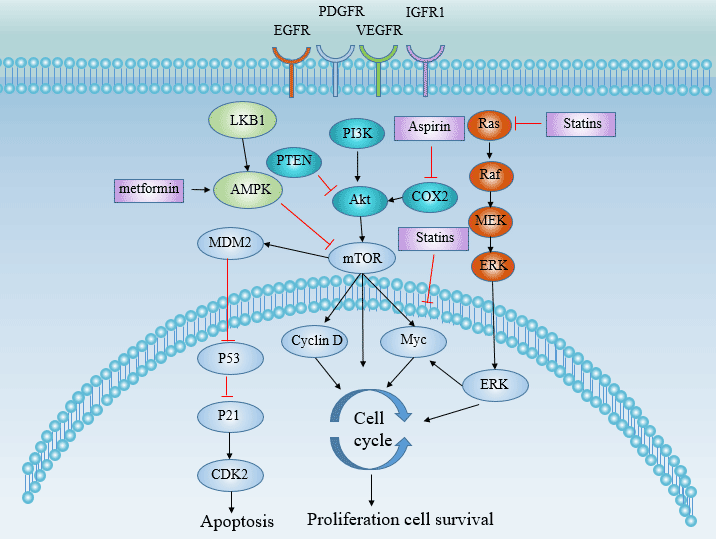
AQP5 AQP5 Antibody CSB-PA978373 Human ELISA,IHC
AQP6 AQP6 Antibody CSB-PA16139A0Rb Human ELISA,IHC
AQP6 AQP6 Antibody,FITC conjugated CSB-PA16139C0Rb Human ELISA
AQP6 AQP6 Antibody,HRP conjugated CSB-PA16139B0Rb Human ELISA
AQP7 AQP7 Antibody CSB-PA276755 Human ELISA,IHC
AQP7 AQP7 Antibody CSB-PA390632 Human ELISA,IHC
AQP8 AQP8 Antibody CSB-PA186529 Human ELISA,IHC
AQP8 AQP8 Antibody CSB-PA905788 Human ELISA,IHC
MIP MIP Antibody CSB-PA013834LA01HU Human ELISA,IHC,IF
MIP MIP Antibody,Biotin conjugated CSB-PA013834LD01HU Human ELISA
MIP MIP Antibody,HRP conjugated CSB-PA013834LB01HU Human ELISA
MIP MIP Antibody CSB-PA000910 Human,Mouse,Rat ELISA,WB,IHC


Aquaporin related ELISA kits for your research

Target Product Name Code Expression System
AQP1 Human Aquaporin 1,AQP-1 ELISA Kit CSB-E08248h serum, plasma, cell culture supernates, cell lysates 3.9 pg/mL
AQP2 Human Aquaporin 2,AQP-2 ELISA Kit CSB-E08242h serum, plasma, urine, tissue homogenates 0.078 ng/mL
AQP3 Human Aquaporin 3,AQP-3 ELISA Kit CSB-E08251h serum, plasma, tissue homogenates 0.078 ng/mL
AQP4 Human Aquaporin 4,AQP-4 ELISA Kit CSB-E08254h serum, plasma, tissue homogenates 0.039 ng/mL
AQP5 Human Aquaporin 5,AQP-5 ELISA Kit CSB-E08257h serum, plasma, tissue homogenates 0.039 ng/mL
AVP Human antidiuretic hormone/vasopressin/arginine vasopressin,ADH/VP/AVP ELISA Kit CSB-E09080h serum, plasma, tissue homogenates 0.312 pg/mL

A Switch that Controls the Entry and Exit of Ions


Transmembrane protein series two: Ion Channels

What are Ion Channels?

Transmembrane proteins are widely present in organisms and play important physiological functions. Its function differs significantly depending on the type of difference. It constitutes a variety of ion channels, which inputs nutrients and some inorganic electrolytes into cells, and excretes toxic or useless metabolites from cells.

Ion channels are commonly called channel proteins. Some stimuli bind to and interact with the channel proteins on the cell membrane, changing the conformation of ion channles. Conformational alteration of ion channels regulates the opening and closing of ion channels, modulating the ability of corresponding substances to enter and exit cells, thereby regulating various key physiological processes of the living system. Sometimes ion channle gating is controlled by the membrane potential. Almost all cellular processes rely on ion channels.

Ion channel function

  • Substance absorption
  • Osmotic pressure regulation
  • Electrical impulse formation: The various sensory mechanisms of animals, such as vision, taste, smell and temperature perception, are related to changes in potential caused by ion channels.
  • Signal transmission: Certain ions (Ca2+, K+, etc.) in the cytoplasm rapidly increase in concentration to form signal stimulation, further affecting downstream signaling pathways. Ion channels are the primary channel for adjusting ion concentration.

Ion channels and diseases

Ion channel genetic variation and dysfunction are associated with the development and progression of many diseases. The ion channel disease, which is well studied, mainly involves the fields of potassium, sodium, calcium and chloride channels.

Ion channels have a certain relationship with tumors. Ion channels also play a conservative role in development.

To learn more about ion channels, there may be information you want to know in this article: A Switch that Controls the Entry and Exit of Ions.

Ion channels and their correlation with nerves, cardiovasculars, immune diseases, and cancer make ion channels an important target for drug design. Different target drugs can be designed according to different types of channels.

These ion channel related products produced by CUSABIO provide convenience for your research. At present, we have produced more than 1200 ion channel related products: 1106 antibody products, 70 protein products, 37 ELISA kits.

Hot Products

Recombinant human Calmodulin
(CSB-EP004445HU)

Recombinant Human Chitinase domain-containing protein 1(CHID1)
(CSB-MP883614HU)

Recombinant Human Potassium channel subfamily K member 3(KCNK3)
(CSB-CF012071HU)

Recombinant Human Voltage-dependent calcium channel subunit alpha-2/delta-1(CACNA2D1),partial
(CSB-EP004407HU1)

Ion channels related proteins for your research

Target Product Name Code Expression System
KCNJ10 Recombinant Human ATP-sensitive inward rectifier potassium channel 10(KCNJ10) ,partial CSB-EP012048HU E.coli
KCNJ1 Recombinant Human ATP-sensitive inward rectifier potassium channel 1(KCNJ1),partial CSB-YP012047HU Yeast
KCNJ1 Recombinant Human ATP-sensitive inward rectifier potassium channel 1(KCNJ1),partial CSB-EP012047HU E.coli
KCND2 Recombinant Human Potassium voltage-gated channel subfamily D member 2(KCND2),partial CSB-YP012023HU Yeast
KCND1 Recombinant Human Potassium voltage-gated channel subfamily D member 1(KCND1),partial CSB-YP012022HU Yeast
KCND1 Recombinant Human Potassium voltage-gated channel subfamily D member 1 protein(KCND1),partial CSB-EP012022HU E.coli
KCNAB2 Recombinant Human Voltage-gated potassium channel subunit beta-2(KCNAB2) CSB-EP012014HU E.coli
KCNA1 Recombinant Human Potassium voltage-gated channel subfamily A member 1 protein(KCNA1),partial CSB-EP012005HU E.coli
HTR3E Recombinant Human 5-hydroxytryptamine receptor 3E(HTR3E),partial CSB-CF010894HU in vitro E.coli expression system
HTR3D Recombinant Human 5-hydroxytryptamine receptor 3D(HTR3D),partial CSB-CF747864HU in vitro E.coli expression system
GRIN2A Recombinant Human Glutamate receptor ionotropic, NMDA 2A(GRIN2A),partial CSB-EP618634HU1 E.coli
GRIN1 Recombinant Human Glutamate [NMDA] receptor subunit zeta-1(GRIN1),partial CSB-EP009911HU E.coli
GRIA3 Recombinant Human Glutamate receptor 3(GRIA3),partial CSB-YP009900HU Yeast
GRIA3 Recombinant Human Glutamate receptor 3(GRIA3),partial CSB-EP009900HU E.coli
GABRA4 Recombinant Human Gamma-aminobutyric acid receptor subunit alpha-4(GABRA4),partial CSB-EP009143HU E.coli
FXYD3 Recombinant Human FXYD domain-containing ion transport regulator 3(FXYD3),Partial CSB-EP622789HU E.coli
FXYD2 Recombinant Human Sodium/potassium-transporting ATPase subunit gamma(FXYD2) CSB-EP009090HU E.coli
CYBB RecombinantHumanCytochromeb-245heavychain(CYBB),partial CSB-YP006325HU1 Yeast
EGF Recombinant Human Pro-epidermal growth factor(EGF) ,partial CSB-RP062444h E.coli
CYBB Recombinant Human Cytochrome b-245 heavy chain(CYBB),partial CSB-EP006325HU1a2 E.coli
CTNNB1 Recombinant Human Catenin beta-1(CTNNB1) CSB-YP006169HU Yeast
CTNNB1 Recombinant Human Catenin beta-1(CTNNB1) CSB-EP006169HU E.coli
CNGA4 Recombinant Human Cyclic nucleotide-gated cation channel alpha-4(CNGA4) CSB-CF808540HU in vitro E.coli expression system
CLIC4 Recombinant Human Chloride intracellular channel protein 4(CLIC4) CSB-EP005549HU E.coli
CAV3 Recombinant Human Caveolin-3(CAV3) CSB-EP004573HU E.coli
CALM1 Recombinant Human Calmodulin(CALM1) CSB-EP004445HU E.coli
ATP6V1F Recombinant Human V-type proton ATPase subunit F(ATP6V1F) CSB-RP015744h E.coli
ATP6V1G1 Recombinant Human V-type proton ATPase subunit G 1(ATP6V1G1) CSB-RP001344h E.coli
ATP6V0D1 Recombinant Human V-type proton ATPase subunit d 1(ATP6V0D1) CSB-EP002390HU E.coli
ANXA5 Recombinant Human Annexin A5(ANXA5) CSB-YP001846HU Yeast
ANXA2 Recombinant Human Annexin A2(ANXA2) CSB-EP001840HUb2 E.coli
ANXA2 Recombinant Human Annexin A2(ANXA2) CSB-EP001840HUa0 E.coli
ANXA2 Recombinant Human Annexin A2(ANXA2) CSB-EP001840HU E.coli
ANXA2 Recombinant Human Annexin A2(ANXA2) CSB-EP001840HUe0 E.coli
YWHAQ Recombinant Human 14-3-3 protein theta(YWHAQ) CSB-EP026290HU E.coli
YWHAH Recombinant Human 14-3-3 protein eta(YWHAH),partial CSB-RP021744h E.coli
UCP1 Recombinant Human Mitochondrial brown fat uncoupling protein 1(UCP1) CSB-YP025554HU Yeast
UCP1 Recombinant Human Mitochondrial brown fat uncoupling protein 1(UCP1) CSB-EP025554HU E.coli
TRPA1 Recombinant Human Transient receptor potential cation channel subfamily A member 1(TRPA1),partial CSB-EP025074HU E.coli
SLC8A1 Recombinant Human Sodium/calcium exchanger 1 (SLC8A1),partial CSB-YP021723HU Yeast
RYR3 Recombinant Human Ryanodine receptor 3(RYR3),partial CSB-EP020621HU E.coli
RYR1 Recombinant Human Ryanodine receptor 1(RYR1),partial CSB-EP020619HU E.coli
PRKCSH Recombinant Human Glucosidase 2 subunit beta(PRKCSH),partial CSB-RP029254h E.coli
NCS1 Recombinant Human Neuronal calcium sensor 1(NCS1) CSB-EP008983HU E.coli
KCNMA1 Recombinant Human Calcium-activated potassium channel subunit alpha-1(KCNMA1),partial CSB-EP614255HU E.coli
KCNK3 Recombinant Human Potassium channel subfamily K member 3(KCNK3) CSB-CF012071HU in vitro E.coli expression system
KCNK2 Recombinant Human Potassium channel subfamily K member 2(KCNK2),partial CSB-YP012070HU Yeast
KCNJ10 Recombinant Human ATP-sensitive inward rectifier potassium channel 10(KCNJ10) CSB-CF012048HU in vitro E.coli expression system

Ion channels related antibodies for your research

Target Product Name Code Expression System Tested Applications
ANO2 ANO2 Antibody CSB-PA001813GA01HU Human, Mouse, Rat ELISA, WB, IF
ANXA2 ANXA2 Antibody CSB-PA001840GA01HU Human, Mouse, Rat ELISA, WB, IHC
ANXA7 ANXA7 Antibody CSB-PA001848GA01HU Human, Mouse, Rat ELISA, WB, IHC
AP2M1 AP2M1 Antibody CSB-PA00627A0Rb Human, Mouse, Rat ELISA, WB, IHC
ARRB1 ARRB1 Antibody CSB-PA002134GA01HU Human, Mouse, Rat ELISA, WB, IHC, IF
ATP1B1 ATP1B1 Antibody CSB-PA002326LA01HU Human, Rat, Mouse ELISA, WB, IHC, IF
CACNA1B CACNA1B Antibody CSB-PA004398GA01HU Human, Mouse, Rat ELISA, WB, IHC
CAMK2D CAMK2D Antibody CSB-PA004467GA01HU Human, Mouse, Rat ELISA, WB, IHC, IF
CASQ2 CASQ2 Antibody CSB-PA004557GA01HU Human, Mouse, Rat ELISA, WB, IHC
CAV1 CAV1 Antibody CSB-PA004571GA01HU Human, Mouse, Rat ELISA, WB, IHC
CHP1 CHP1 Antibody CSB-PA860773LA01HU Human, Mouse, Rat ELISA, WB, IHC, IF
CHRNA3 CHRNA3 Antibody CSB-PA005389GA01HU Human, Mouse, Rat ELISA, WB, IHC
CHRNB1 CHRNB1 Antibody CSB-PA005395GA01HU Human, Mouse, Rat ELISA, WB, IHC
CIB1 CIB1 Antibody CSB-PA005426GA01HU Human, Mouse, Rat ELISA, WB, IHC
CLCNKA CLCNKA Antibody CSB-PA005487GA01HU Human, Mouse, Rat ELISA, WB, IHC
CLIC1 CLIC1 Antibody CSB-PA005545GA01HU Human, Mouse, Rat ELISA, IHC, IF
CTNNB1 CTNNB1 Antibody CSB-PA006169GA01HU Human, Mouse, Rat, Zebrafish ELISA, WB, IHC
CYBB CYBB Antibody CSB-PA006325KA01HU Human, Mouse, Rat ELISA, WB, IHC
DIAPH1 DIAPH1 Antibody CSB-PA006890GA01HU Human, Mouse, Rat ELISA, WB, IF
FXYD2 FXYD2 Antibody CSB-PA009090GA01HU Human, Mouse, Rat ELISA, WB, IHC
FXYD6 FXYD6 Antibody CSB-PA009094GA01HU Human, Mouse, Rat ELISA, WB, IHC
GABRG2 GABRG2 Antibody CSB-PA009152GA01HU Human, Mouse, Rat ELISA, WB, IF
GRIA3 GRIA3 Antibody CSB-PA009900KA01HU Human, Mouse, Rat ELISA, WB, IHC
GRID1 GRID1 Antibody CSB-PA009902GA01HU Human, Mouse, Rat ELISA, WB, IHC
GRIN2A GRIN2A Antibody CSB-PA14129A0Rb Human, Mouse, Rat ELISA, WB, IHC, IF
HTR3A HTR3A Antibody CSB-PA010890GA01HU Human, Mouse, Rat ELISA, WB, IHC
KCNA1 KCNA1 Antibody CSB-PA012005LA01HU Human, Mouse, Rat ELISA, WB, IF
KCNAB1 KCNAB1 Antibody CSB-PA012013GA01HU Human, Mouse, Rat ELISA, WB, IHC, IF
KCNIP1 KCNIP1 Antibody CSB-PA012043GA01HU Human, Mouse, Rat ELISA, WB, IHC
KCNJ11 KCNJ11 Antibody CSB-PA012049GA01HU Human, Mouse, Rat ELISA, WB, IF
KCNS2 KCNS2 Antibody CSB-PA012096GA01HU Human, Mouse, Rat ELISA, WB, IHC
NCS1 NCS1 Antibody CSB-PA008983ESR1HU Human, Mouse, Rat ELISA, WB, IHC
P2RX4 P2RX4 Antibody CSB-PA017322GA01HU Human, Mouse, Rat ELISA, WB, IHC
P2RX4 P2RX4 Antibody CSB-PA859513LA01HU Human, Mouse, Rat ELISA, WB, IHC
PACSIN3 PACSIN3 Antibody CSB-PA017375GA01HU Human, Mouse, Rat ELISA, WB, IHC
PANX2 PANX2 Antibody CSB-PA839405LA01HU Human, Mouse, Rat ELISA, WB, IHC, IF
POMP POMP Antibody CSB-PA018365GA01HU Human, Mouse, Rat ELISA, WB, IF
PRKCSH PRKCSH Antibody CSB-PA018709GA01HU Human, Mouse, Rat ELISA, WB, IHC
SCNN1G SCNN1G Antibody CSB-PA020851GA01HU Human, Mouse, Rat ELISA, WB, IHC
SGK3 SGK3 Antibody CSB-PA021191GA01HU Human, Mouse, Rat ELISA, WB, IHC
SLC39A7 SLC39A7 Antibody CSB-PA021636GA01HU Human, Mouse, Rat ELISA, WB, IHC
STIM1 STIM1 Antibody CSB-PA022829GA01HU Human, Mouse, Rat, Zebrafish ELISA, WB, IHC
VDAC1 VDAC1 Antibody CSB-PA025821GA01HU Human, Mouse, Rat ELISA, WB, IHC
VDAC3 VDAC3 Antibody CSB-PA025827GA01HU Human, Mouse, Rat ELISA, WB, IHC
YES1 YES1 Antibody CSB-PA026253GA01HU Human, Mouse, Rat ELISA, WB, IHC
YWHAE YWHAE Antibody CSB-PA026287GA01HU Human, Mouse, Rat ELISA, WB, IHC, IF
YWHAQ YWHAQ Antibody CSB-PA026290GA01HU Human, Mouse, Rat ELISA, WB, IF

Ion channels related ELISA kits for your research

Target Product Name Code Sample Type Sensitivity
S100A10 Human Protein S100-A10(S100A10) ELISA kit CSB-EL020623HU serum, plasma, tissue homogenates 0.078 ng/mL
S100A1 Human Soluble protein-100,S-100 ELISA Kit CSB-E08067h serum, plasma, tissue homogenates 0.039 ng/mL
P2RX7 Human P2X purinoceptor 7(P2RX7) ELISA kit CSB-EL017325HU serum, plasma, tissue homogenates, cell lysates 6.25 pg/mL
GRIN1 Human Glutamate [NMDA] receptor subunit zeta-1(GRIN1) ELISA kit CSB-EL009911HU serum, plasma, tissue homogenates, cell lysates 31.25 pg/mL
EGF Human Epidermal growth factor,EGF ELISA Kit CSB-E08027h serum, plasma, cell culture supernates, tissue homogenates_x005f_x000D_ 0.39 pg/mL
CTNNB1 Human Catenin beta-1 (CTNNB1/CTNNB/OK/SW-cl.35/PRO2286) ELISA kit CSB-E08963h serum, plasma, cell culture supernates, tissue homogenates, cell lysates 3.9 pg/mL
CLDN4 Human Claudin 4 (CLDN4)ELISA Kit CSB-E17961h serum, plasma, tissue homogenates, cell lysates 1.56 pg/mL
CFTR Human Cystic fibrosis transmembrane conductance regulator(CFTR) ELISA kit CSB-EL005292HU serum, plasma, tissue homogenates, cell lysates 7 pg/mL
CDH5 Human Vascular Endothelial-Cadherin,VE-cad ELISA Kit CSB-E09372h serum, plasma, tissue homogenates 0.98 ng/mL
CAV1 Human Caveolin-1,Cav-1 ELISA KIT CSB-E09682h serum, plasma, tissue homogenates 7.8 pg/mL
ATP6V0A2 Human V-type proton ATPase 116 kDa subunit a isoform 2(ATP6V0A2) ELISA kit CSB-EL002386HU serum, plasma, tissue homogenates 7.81 pg/mL
ATP5MC1 Human ATP synthase lipid-binding protein, mitochondrial(ATP5G1) ELISA kit CSB-EL002359HU serum, plasma, tissue homogenates, cell lysates 5.86 pg/mL
ANXA5 Human Annexin Ⅴ,ANX-Ⅴ ELISA Kit CSB-E04882h serum, plasma, cell culture supernates, cell lysates 0.078 ng/mL
ANXA2 Human Annexin Ⅱ(ANX-Ⅱ) ELISA Kit CSB-E12156h serum, urine, saliva, cell lysates 0.098 ng/mL
ANXA1 Human Annexin Ⅰ(ANX-Ⅰ) ELISA Kit CSB-E12155h serum, plasma, tissue homogenates 0.078 ng/mL
SGK1 Human Serine/threonine-protein kinase Sgk1(SGK1) ELISA kit CSB-EL021189HU serum, plasma, tissue homogenates, cell lysates 5.86 pg/mL
STX1A Human Syntaxin-1A(STX1A) ELISA kit CSB-EL022895HU serum, tissue homogenates 3.9 pg/mL
TRPV1 Human transient receptor potential cation channel subfamily V member 1 (TrpV1)ELISA Kit CSB-E14198h serum, plasma, tissue homogenates, cell lysates 0.078 ng/mL
TRPV4 Human transient receptor potential cation channel subfamily V member 4 (TrpV4)ELISA Kit CSB-E14199h serum, plasma, tissue homogenates, cell lysates 3.9 pg/mL
UCP1 Human Mitochondrial brown fat uncoupling protein 1(UCP1) ELISA kit CSB-EL025554HU serum, plasma, tissue homogenates 7.81 pg/mL
YWHAE Human 14-3-3 protein epsilon(YWHAE) ELISA kit CSB-EL026287HU serum, plasma, tissue homogenates, cell lysates 19.5 pg/mL
YWHAH Human 14-3-3 protein eta(YWHAH) ELISA kit CSB-EL026289HU serum, plasma, tissue homogenates 0.156 ng/mL
YWHAQ Human 14-3-3 protein theta(YWHAQ) ELISA kit CSB-EL026290HU serum, plasma, tissue homogenates, cerebrospinal fluid (CSF) 0.11 ng/mL
YWHAZ Human 14-3-3 protein zeta/delta(YWHAZ) ELISA kit CSB-EL026293HU serum, plasma, tissue homogenates 0.156 ng/mL

A transport machine - ATP-binding cassette

ATP-binding cassette (ABC) transporters are the largest known transmembrane protein superfamily, widely distributed in eukaryotes and prokaryotes. ABC proteins bind and hydrolyze ATP and use energy to drive various molecules across the plasma membrane as well as the endoplasmic reticulum (ER), peroxisome and mitochondrial inner membrane [1][2]. It is involved in the transport of specific molecules on the lipid membrane and the resistance in all organisms.

The Structure of ATP-binding cassette

Figure 1 The Structure of ATP-binding cassette, The picture is from wikipedia

1. The Structure of ABC

Three basic organization forms of ABC transporters

Figure 2 Three basic organization forms of ABC transporters

ABC protein has a typical modular structure, consisting of four protein domains or subunits: two hydrophobic transmembrane domains (TMD) are considered to constitute transmembrane transport pathways or channels (usually composed of six transmembrane alpha helix); two hydrophilic nucleotide binding domains (NBDs) (ATP binding domains, known as nucleotide binding folding (NBF)) provide energy for active transport [2]. NBF contains three conserved domains: Walker A and B domains, and C motif [3]. The C domain is unique to the ABC transporter.

In addition to the complete transporters, there are also half transporters that contain one of each domain.

Figure 3 The structure and transport process of ABC

Figure 3 The structure and transport process of ABC

2. The Substrate

The ATP-binding cassette (ABC) transporter superfamily transfers a variety of substrates, including sugars, amino acids, metal ions, peptides and proteins, as well as a large number of hydrophobic compounds and metabolites across the extracellular and intracellular membranes.

3. ABC Superfamily

ABC transporters are one of the largest known protein superfamily: there are 49 ABC transporters in humans and 80 in gram-negative E. coli bacteria.

The ABC gene in the human genome is divided into 7 subfamilies (ABCA~ABCG) based on amino acid sequence similarity and phylogeny. The following table is a list of human ABC genes.

Table 1 List of human ABC genes and function

Subfamily Member Function
ABCA ABCA1 Cholesterol efflux onto HDL
ABCA2 Drug resistance
ABCA3 Phosphatidyl choline efflux
ABCA4 N-retinylidiene-PE efflux
ABCA5ABCA6; ABCA7;ABCA8ABCA9; ABCA10; ABCA11; ABCA12ABCA13. /
ABCB ABCB1 Multidrug resistance
ABCB3 Peptide transport
ABCB4 PC transport
ABCB5 Iron transport
ABCB6 Fe/S cluster transport
ABCB11 Bile salt transport
ABCB2; ABCB7ABCB8ABCB9ABCB10. /
ABCC ABCC1 Drug resistance
ABCC2 Organic anion efflux
ABCC3 Drug resistance
ABCC4 Nucleoside transport
ABCC5 Nucleoside transport
CFTR (ABCC7) Chloride ion channel
ABCC8 Sulfonylurea receptor
ABCC9 Potassium channel regulation
ABCC6ABCC10ABCC11ABCC12. /
ABCD ABCD1 VLCFA transport regulation
ABCD2ABCD3ABCD4. /
ABCE ABCE1 Elongation factor complex
ABCF ABCF1ABCF2ABCF3. /
ABCG ABCG1 Cholesterol transport
ABCG2 Toxin efflux, drug resistance
ABCG4 Cholesterol transport
ABCG5 Sterol transport
ABCG8 Sterol transport

4. The Function of ABC

The ATP-binding cassette (ABC) superfamily gene plays an important role in transporting compounds between the intestinal tract, the blood-brain barrier, and the placenta. It also plays an important role in the body’s core function of resisting foreign bodies and detoxification [4]. Different subtypes have different functions and can act as drug efflux transporters (ABCB1, ABCC subfamily and ABCG2) or sterol transporters (ABCA1, ABCA7, ABCG1, ABCG5 and ABCG8).

4.1 T Eukaryotic

In eukaryotes, most ABC genes move compounds from the cytoplasm to the extracellular or extracellular compartment (endoplasmic reticulum, mitochondria, peroxisomes). Its importance in eukaryotic systems has been fully demonstrated, characterized by its association with genetic diseases and its role in multidrug resistance to cancer.

4.2 Prokaryote

In bacteria, the ABC gene is mainly involved in the import of essential compounds that cannot pass through cells through diffusion (eg, sugars, vitamins, metal ions, etc.). These genes play a role in nutrient absorption, toxin secretion and antibacterial drugs. The role of ABC transporters is mainly in the pathogenicity and toxicity of bacteria. In pathogenic bacteria, these functions are usually to evade or resist the host’s defense. Therefore, many cell surfaces or secreted factors can be useful targets for antibacterial therapy or vaccine development.

4.3 Plant

ATP-binding cassette (ABC) transporter in plant is an important membrane protein used to transport a variety of compounds, including heavy metals, antibiotics, phytohormones and secondary metabolites [5]. The number of ABC family members in the plant genome has more than doubled compared to animals and insects.

In plants, ATP-binding cassette transporters have important physiological functions such as cell detoxification, cuticle formation, stomatal regulation, seed germination, and resistance to pathogenic bacteria.

In rice, several ATP-binding cassette transporters of the ABCG family are essential for cuticle formation [6].

In tomato, the ATP-binding cassette transporter is involved in the transport of tomato fruit auxin. As a member of the subfamily ABCB, SlABCB4 plays an important role in the transport of auxin during tomato fruit development.

In Arabidopsis, some members of the ABC transporter ABCB subfamily also are involved in auxin transport [7].

ABC transporters also play an important role in the detoxification of pesticides.

The ATP-binding cassette (ABC) transporter [8] was identified as an important detoxifying enzyme in Plutella xylostella.

Epis et al[9] demonstrated the role of ABC transporters in insecticide defense.

5. ABC and Disease

The ATP-binding cassette (ABC) superfamily genes encode membrane proteins that transport different substrates on the cell membrane. The genetic variation of the ATP-binding cassette (ABC) transporter superfamily gene is the cause or contributing factor of various Mendelian diseases and complex genetic diseases in humans. The heterozygous variation in ABC gene mutations is related to the susceptibility of specific complex diseases.

Currently, there are 17 ABC genes related to Mendelian inheritance. These include adrenoleukodystrophy, cystic fibrosis, retinal degeneration, hypercholesterolemia and cholestasis, neurological diseases, anemia and drug reactions. Many of the ABC genes that cause disease lead to many different clinical phenotypes. For example, cystic fibrosis associated with specific CFTR genotypes includes pancreatic sufficiency and pancreatic insufficiency.

5.1 ABCA1

ABCA1 mediates the transport of intracellular free cholesterol and phospholipids through the cell membrane to poor apoA1 to form HDL [10]. It plays a key role in reverse cholesterol transport and cellular lipid efflux, is the initial rate-limiting step of reverted cholesterol transport (RCT) in macrophages [11], and is the most critical determinant of plasma high density lipoprotein (HDL) levels in hepatic cells.

Mutations in the ABCA1 gene may induce Tangier disease [12] and familial hypolipoproteinemia [13] and may also result in loss of cellular cholesterol homeostasis in prostate cancer. In the general population, some common single-nucleotide polymorphisms of ABCA1 also affect blood lipid levels, atherosclerosis generation and the severity of coronary heart disease [14].

In recent years, a large amount of evidence indicates that abnormal cholesterol metabolism may play an important role in the development of Alzheimer’s disease AD (ABCA2/ABCA7), and apoE gene is also closely related to the metabolism of cholesterol in the brain [15].

Regulatory Factors of ABC1

The expression of the ABCA1 gene is mainly regulated by the liver X receptor (LXR)/retinoid X receptor (RXR).In addition, interferon- γ (IFN- γ ) inhibits liver X receptor α (LXR α ) through the JAK/STAT signaling pathway, which can down-regulate ABCA1 expression and intracellular cholesterol outflow [16].

5.2 ABCA4

Mutations in the ABCA4 gene lead to retinitis pigmentosa, recessive pyramidal malnutrition, and Stargardt disease [17]. All of these diseases are characterized by retinal degeneration, the severity of which is roughly related to the predicted severity of ABCA4 mutation. Patients with heterozygous mutation of ABCA4 gene are more likely to suffer from late-onset macular degeneration – age-related macular degeneration (AMD) [18]. Women with heterozygous mutations of liver transporters that lead to cholestasis may have a higher risk of cholestasis during pregnancy [19].

5.3 ABCG

Members of the ABCG subfamily, ABCG5 and G8 are involved in the development of glutathione, a genetic disorder of lipid metabolism. ABCG1 mainly mediates the transport of intracellular free cholesterol to mature HDL. It works synergistically with ABCA1 to promote extracellular lipids out of the cell and complete reverse cholesterol transport in the body [20]. ABCG1 deficiency can also participate in foam cell formation, endothelial cell dysfunction and inflammatory response, and thus affects the formation and development of atherosclerosis.

Table 2 Diseases and phenotyes caused by ABC genes

Gene Mendelian disorder Complex disease Animal model
ABCA1 Tangier disease, FHDLD HDL levels Mouse, chicken
ABCA3 Surfactant deficiency / /
ABCA4 Stargardt/FFM, RP, CRD AMD Mouse
ABCA12 Lamellar ichthyosis / /
ABCB1 Ivermectin sensitivitya Digoxin uptake Mouse, dog
ABCB2 Immune deficiency / Mouse
ABCB3 Immune deficiency / Mouse
ABCB4 PFIC-3 ICP /
ABCB7 XLSA/A / /
ABCB11 PFIC-2 / /
ABCC2 Dubin-Johnson Syndrome / Rat, sheep, monkey
ABCC6 Pseudoxanthoma elasticum / Mouse
ABCC7 Cystic Fibrosis, CBAVD Pancreatitis, bronchiectasis Mouse
ABCC8 FPHHI Mouse
ABCC9 DCVT
ABCD1 ALD Mouse
ABCG5 Sitosterolemia Mouse
ABCG8 Sitosterolemia Mouse

This table derived from literature “Evolution of the ATP-binding cassette (ABC) transporter superfamily in vertebrates” [21]

6. ABC and Drug Resistance

Currently, chemotherapy is one of the approaches to cancer treatment, and multidrug resistance (MDR) is a major obstacle to successful chemotherapy [22]. MDR refers to the phenomenon that tumor cells are cross-resistant to a variety of structurally unrelated chemotherapeutic drugs. Clinically, drug resistance is caused by multiple factors. The efflux of cytotoxic drugs mediated by ATP-binding cassette transporters is the “classical MDR” pathway [23]. ABC transporters pump drugs out of cells and reduce the concentration of intracellular chemotherapy drugs. Overexpression of ABC transporters in tumor cells is the main mechanism of tumor MDR.

6.1 ATP Binding Cassette (ABC) Multidrug Transporter

ATP binding cassette transporters (ABC transporters) can transport chemotherapy drugs to the extracellular environment and reduce intracellular drug concentration. There are four main types ATP binding cassette multidrug transporter: P-glycoprotein (P-gp, MDR1, ABCB1), Multidrug resistance-associated protein (MDR-related protein /MRP, ABCC), lung resistance-related protein (LRP) and breast cancer resistance protein (BCRP, ABCG2) [24].

Breast cancer resistance protein (BCRP, ABCG2) belongs to the ABCG gene family. The gene is located on chromosome 4 and is a recently discovered ABC drug efflux transporter. In structure, BCRP is “half transporter” [25].

6.2 Liver Cancer MDR

Acquired MDR of hepatocellular carcinoma may be related to the expression of MRP1, MRP3 and MRP5 genes, and MRP2 is the main target of endogenous drug resistance, BCRP is a new drug efflux pump related to tumor MDR. Liver cancer MDR mediated by ATP-binding cassette transporter severely limits chemotherapy efficacy and prognosis.

6.3 Pancreatic Cancer

Pancreatic cancer is resistant to a variety of chemotherapeutic drugs, and resistance is associated with the ATP-binding cassette (ABC) transport vector superfamily [26].

7. How to overcome ABC drug efflux transporter-mediated drug resistance?

For MDR induced by ATP-binding cassette transporters, inhibition of ATP-binding cassette transporter-mediated drug efflux is the easiest and direct route.

7.1 ABC Inhibitors

Inhibitors of ABC transporters include competitive and non-competitive inhibitors. Currently, there are 3 generations of chemical reversals, most of which are competitive inhibitors of MDR1.

First-generation drugs: (including verapamil, tamoxifen, cyclosporine A, quinine).Calcium channel blocker verapamil inhibits MDR1 synthesis and its activity at mRNA level while competing for MDR1 binding sites, thereby reversing drug resistance [27].

Second-generation drugs: (including biricodar, elacridar and valspodar) failed to show overall efficacy improvements in multiple randomized clinical trials due to poor efficacy and increased toxicity.

Third-generation drugs: have high transporter affinity and low pharmacokinetics, including Tariquidar, Zosuquidar and Laniquidar.

Strategies for circumvention of MDR also include small interfering RNA (siRNA) [28] and microRNA (miRNA) to down-regulate the expression of ABC transporters.

Plant Anti-Tumor Drugs: Plant anti-tumor drugs provide new ideas for the development of MDR reversal agents due to their small side effects, natural sources and low cost.

It has been found that artemisinin, quercetin, magnolia officinalis phenol, emodin, zhejiang fritillaria alkaloid, osmanthus cnidii, ginsenoside, total saponin of panax notoginseng, root of mahonia mahogany, ganoderma lucidum and other traditional Chinese medicines or their effective components can reverse the drug resistance of tumor cells by down-regulating the expression of MDR1.

Psoralen, tetramethylpyrazine, tetrandrine, and paeonol are calcium antagonists, which can inhibit the function of pumping drugs out of cells by binding to P-gp, increase the concentration of intracellular chemotherapeutic drugs, and then reverse Resistance.

Studies have found that diosgenin can reverse the resistance of leukemia cells by inhibiting NF-κB down-regulation of MDR1 [29].

7.2 Other

In addition, immunotherapy reversion, gene reversion, somatostatin and its analogues reversion and Chinese herbal reversion were also used to reverse MDR.

The increased oxidative stress and the activated NF-κB transcription factor may be involved in the inhibition of ABCG1 expression by high glucose.

References

[1] Momburg F, Roelse J, Howard J C, et al. Selectivity of MHC-encoded peptide transporters from human, mouse and rat [J]. Nature (London), 1994, 367(6464): 648-651.
[2] Higgins C F. ABC transporters: from microorganisms to man [J]. Annu Rev Cell Biol, 1991, 8(1): 67-113.
[3] Hyde S C, Emsley P, Hartshorn M J, et al. Structural model of ATP-binding proteing associated with cystic fibrosis, multidrug resistance and bacterial transport [J]. Nature, 1990, 346(6282): 362-365.
[4] Fletcher J I, Haber M, Henderson M J, et al. ABC transporters in cancer: more than just drug efflux pumps [J]. Nature Reviews Cancer, 2010, 10(2): 147.
[5] Yazaki K, Shitan N, Sugiyama A, et al. Cell and molecular biology of ATP-binding cassette proteins in plants [J]. Int Rev Cell Mol Biol, 2009, 276: 263-299.
[6] Bessire M, Borel S, Fabre G, et al. A Member of the PLEIOTROPIC DRUG RESISTANCE Family of ATP Binding Cassette Transporters Is Required for the Formation of a Functional Cuticle in Arabidopsis [J]. Plant Cell, 2011, 23(5): 1958-1970.
[7] Cho M, Cho H T. The function of ABCB transporters in auxin transport [J]. Plant Signaling & Behavior, 2013, 8(2): e22990.
[8] He W, You M, Vasseur L, et al. Developmental and insecticide-resistant insights from the de novo assembled transcriptome of the diamondback moth, Plutella xylostella [J]. Genomics, 2012, 99(3): 169-177.
[9] Epis S D, Porretta V, Mastrantonio S, et al. Temporal dynamics of the ABC transporter response to insecticide treatment: insights from the malaria vector Anopheles stephensi [J]. Sci Rep, 2014, 4: 7435-7435.
[10] Oram J F, Heinecke J W. ATP-Binding Cassette Transporter A1: A Cell Cholesterol Exporter That Protects Against Cardiovascular Disease [J]. Physiological Reviews, 2005, 85(4): 1343.
[11] Jiang Z, Zhou R, Xu C, et al. Genetic variation of the ATP-binding cassette transporter A1 and susceptibility to coronary heart disease [J]. Molecular Genetics and Metabolism, 2011, 103(1): 0-88.
[12] Santamarina-Fojo S, Remaley A T, Neufeld E B, et al. Regulation and intracellular trafficking of the ABCA1 transporter [J]. Journal of Lipid Research, 2001, 42(9): 1339-1345.
[13] Brookswilson A, Marcil M, Clee S M, et al. Mutations in ABC1 in Tangier disease and familial high-density lipoprotein deficiency [J]. Nature Genetics, 1999, 22(4): 336-345.
[14] Cenarro A, Artieda M, Castillo S, et al. A common variant in the ABCA1 gene is associated with a lower risk for premature coronary heart disease in familial hypercholesterolaemia [J]. Journal of Medical Genetics, 2003, 40(3): 163-168.
[15] Lahiri D. Apolipoprotein e as a target for developing new therapeutics for Alzheimer’s disease based on studies from protein, RNA, and regulatory region of the gene [J]. Journal of Molecular Neuroscience, 2004, 23(3): 225-234.
[16] Hao X, Cao D, Hu Y, et al. IFN-γ down-regulates ABCA1 expression by inhibiting LXRα in a JAK/STAT signaling pathway-dependent manner [J]. Atherosclerosis, 2009, 203(2): 417-428.
[17] Amalia MartínezMir, Paloma E, Allikmets R, et al. Retinitis pigmentosa caused by a homozygous mutation in the Stargardt disease gene ABCR [J]. Nature Genetics, 1998, 18(1): 11.
[18] Allikmets R. Simple and Complex ABCR: Genetic Predisposition to Retinal Disease [J]. American Journal of Human Genetics, 2000, 67(4): 793-799.
[19] Pauli-Magnus C, Lang T, Meier Y, et al. Sequence analysis of bile salt export pump (ABCB11) and multidrug resistance p-glycoprotein 3 (ABCB4, MDR3) in patients with intrahepatic cholestasis of pregnancy [J]. Pharmacogenetics, 2004, 14(2): 91-102.
[20] Gelissen I C, Harris M, Rye K A, et al. ABCA1 and ABCG1 Synergize to Mediate Cholesterol Export to ApoA-I [J]. Arteriosclerosis Thrombosis & Vascular Biology, 2006, 7(3): 541-541.
[21] Dean M, Annilo T. EVOLUTION OF THE ATP-BINDING CASSETTE (ABC) TRANSPORTER SUPERFAMILY IN VERTEBRATES*[J]. Annu Rev Genomics Hum Genet, 2005, 6(1): 123-142.
[22] Binkhathlan Z, Lavasanifar A. P-glycoprotein Inhibition as a Therapeutic Approach for Overcoming Multidrug Resistance in Cancer: Current Status and Future Perspectives [J]. Curr Cancer Drug Targets, 2013, 13(3):-.
[23] Borst P, Elferink R O. Mammalian ABC transporters in health and disease [J]. Ann Rev Biochem, 2003, 71(1): 537.
[24] Igarashi A, Konno H, Tanaka T, et al. Liposomal photofrin enhances therapeutic efficacy of photodynamic therapy against the human gastric cancer [J]. Toxicology Letters, 2003, 145(2): 133-141.
[25] Chang G, Roth C B. Structure of MsbA from E. coli: a homolog of the multidrug resistance ATP binding cassette (ABC) transporters [J]. Science, 2001, 293(5536): 1793-800.
[26] Nieth C, Priebsch A, Stege A, et al. Modulation of the classical multidrug resistance (MDR) phenotype by RNA interference (RNAi) [J]. Febs Letters, 2003, 545(2): 144-150.
[27] Kohno M, Pouyssegur J. Targeting the ERK signaling pathway in cancer therapy [J]. Annals of Medicine, 2006, 38(3): 200-211.
[28] Abbasi M, Lavasanifar A, Uludag H. Recent attempts at RNAi-mediated P-glycoprotein downregulation for reversal of multidrug resistance in cancer [J]. Medicinal Research Reviews, 2013, 33(1): 33-53.
[29] Wang L, Meng Q, Wang C, et al. Dioscin restores the activity of the anticancer agent adriamycin in multidrug-resistant human leukemia K562/adriamycin cells by down-regulating MDR1 via a mechanism involving NF-κB signaling inhibition [J]. Journal of Natural Products, 2013, 76(5): 909-914.

Transmembrane protein series four: ATP-binding cassette transporters

What is ATP-binding cassette transporters?

Transmembrane proteins span the lipid bilayer. According to the number of transmembrane times, it can be divided into one transmembrane protein or multiple transmembrane proteins. Many naturally occurring transmembrane proteins act as channels for specific substances to pass through the biofilm. Some transmembrane proteins can receive or transmit cellular signals. Transmembrane proteins also play an important role in molecular transport.

ATP-binding cassette (ABC) transporters is a transmembrane protein involved in molecular transport.

ATP-binding cassette transporters are the largest known transmembrane protein superfamily. ABC proteins bind and hydrolyze ATP and use energy to drive various molecules across the plasma membrane as well as the endoplasmic reticulum (ER), peroxisome and mitochondrial inner membrane. It is involved in the transport of specific molecules on the lipid membrane and the resistance in all organisms.

ATP-binding cassette (ABC) transporters and disease

ABC gene mutations are associated with the development of certain diseases. These diseases include adrenal white matter dystrophy, cystic fibrosis, retinal degeneration, hypercholesterolemia and cholestasis, neurological diseases, anemia and drug reactions.

Some common single nucleotide polymorphisms in ABCA1 also affect blood lipid levels, atherogenesis, and the severity of coronary heart disease.

ATP-binding cassette (ABC) transporters and drug resistance

MDR refers to the phenomenon that tumor cells have cross-resistance to multiple chemotherapy drugs. This condition is mainly caused by the efflux of cytotoxic drugs mediated by ATP-binding cassette transporter. In the chemotherapy of liver cancer, pancreatic cancer and other tumors, ATP-binding cassette transporter-mediated MDR seriously restricts the efficacy and prognosis of chemotherapy.

ATP-binding cassette (ABC) transporters inhibitors

Evading MDR can inhibit MDR1 synthesis and its activity at the mRNA level, or compete for MDR1 binding sites. In addition, the reversal methods of MDR include immunotherapy reversal, gene reversal, reversal of somatostatin and its analogues, and reversal of Chinese herbal medicine. More information about ABC, you can have a look at this article: A transport machine — ATP-binding cassette.

In many studies of ABC-related diseases, drug resistance, you may need ABC – related products. CUSABIO has a special transmembrane protein expression system. We could produce 36000+ transmembrane proteins and could also provide marker proteins at mg level for NMR and X-ray studies.

At present, we can produce 133 ABC-related products: 124 antibody products, 5 protein products, 4 ELISA products.

Hot Products

Recombinant Human ABCB8, partial
(CSB-CF868325HU)

Recombinant Human ABCC1, partial
(CSB-EP001056HU)

Recombinant Human ABCD1
(CSB-CF001068HU)

Recombinant Human ABCG1, partial
(CSB-YP001080HU)


ATP-binding cassette (ABC) transporters related proteins for your research

Target Product Name Code Expression System
ABCB1 Recombinant Human Multidrug resistance protein 1(ABCB1),partial CSB-RP117374h(A6) E.coli
ABCB8 Recombinant Human ATP-binding cassette sub-family B member 8, mitochondrial(ABCB8),partial CSB-CF868325HU in vitro E.coli expression system
ABCC1 Recombinant Human Multidrug resistance-associated protein 1 (ABCC1),partial CSB-EP001056HU E.coli
ABCD1 Recombinant Human ATP-binding cassette sub-family D member 1(ABCD1) CSB-CF001068HU in vitro E.coli expression system
ABCG1 Recombinant Human ATP-binding cassette sub-family G member 1(ABCG1),partial CSB-YP001080HU Yeast

ATP-binding cassette (ABC) transporters related antibodies for your research

Target Product Name Code Species Reactivity Tested Applications
ABCB10 ABCB10 Antibody CSB-PA001047GA01HU Human, Mouse, Rat ELISA, WB
ABCA9 ABCA9 Antibody CSB-PA812864LA01HU Human ELISA, IF
ABCB1 ABCB1 Antibody CSB-PA11737A0Rb Human ELISA, WB, IHC, IF
ABCA3 ABCA3 Antibody CSB-PA859942ESR1HU Human ELISA, IHC
ABCA5 ABCA5 Antibody CSB-PA845163LA01HU Human ELISA, IHC
ABCA2 ABCA2 Antibody CSB-PA887172LA01HU Human ELISA, IHC
ABCA12 ABCA12 Antibody CSB-PA774804LA01HU Human ELISA, IF
ABCA2 ABCA2 Antibody CSB-PA001038GA01HU Human, Mouse, Rat ELISA, WB
ABCB4 ABCB4 Antibody CSB-PA001050LA01HU Human ELISA, IHC, IF
ABCB5 ABCB5 Antibody CSB-PA643574LA01HU Human ELISA, IHC, IF
ABCB6 ABCB6 Antibody CSB-PA001052GA01HU Human, Mouse, Rat ELISA, WB
ABCB7 ABCB7 Antibody CSB-PA001053GA01HU Human, Mouse, Rat ELISA, WB
ABCB7 ABCB7 Antibody CSB-PA001053LA01HU Human ELISA, IHC
ABCB8 ABCB8 Antibody CSB-PA868325ESR1HU Human, Mouse ELISA, WB, IHC
ABCB8 ABCB8 Antibody CSB-PA868325ESR2HU Human ELISA, IHC
ABCB9 ABCB9 Antibody CSB-PA001055GA01HU Human, Mouse, Rat ELISA, WB
ABCB9 ABCB9 Antibody CSB-PA889064LA01HU Human, Mouse ELISA, WB, IF
ABCC11 ABCC11 Antibody CSB-PA846641ESR1HU Human ELISA, IHC
ABCC2 ABCC2 Antibody CSB-PA001061GA01HU Human ELISA, WB
ABCC2 ABCC2 Antibody CSB-PA852918LA01HU Human ELISA, IHC, IF
ABCC3 ABCC3 Antibody CSB-PA001062LA01HU Human ELISA, IF
ABCC4 ABCC4 Antibody CSB-PA001063KA01HU Human, Mouse, Rat ELISA, WB, IHC
ABCC4 ABCC4 Antibody CSB-PA001063LA01HU Human ELISA, IF
ABCC5 ABCC5 Antibody CSB-PA001064LA01HU Human ELISA, IHC, IF
ABCC6 ABCC6 Antibody CSB-PA001065LA01HU Human ELISA, IF
ABCC8 ABCC8 Antibody CSB-PA22439A0Rb Human ELISA, IHC, IF
ABCC9 ABCC9 Antibody CSB-PA001067LA01HU Human ELISA, IHC, IF
ABCD1 ABCD1 Antibody CSB-PA001068GA01HU Human, Mouse, Rat ELISA, WB, IHC
ABCD1 ABCD1 Antibody CSB-PA001068LA01HU Human ELISA, IHC
ABCD2 ABCD2 Antibody CSB-PA866202LA01HU Human ELISA, IHC
ABCD4 ABCD4 Antibody CSB-PA001075LA01HU Human ELISA, IHC
ABCE1 ABCE1 Antibody CSB-PA001076GA01HU Human, Mouse, Rat ELISA, WB, IHC
ABCE1 ABCE1 Antibody CSB-PA001076ESR2HU Human ELISA, WB, IHC
ABCF1 ABCF1 Antibody CSB-PA001077GA01HU Human, Mouse, Rat ELISA, WB
ABCF2 ABCF2 Antibody CSB-PA001078GA01HU Human, Mouse, Rat ELISA, WB, IHC
ABCF2 ABCF2 Antibody CSB-PA871400ESR2HU Human ELISA, WB, IHC
ABCF3 ABCF3 Antibody CSB-PA889115LA01HU Human ELISA, IHC
ABCG1 ABCG1 Antibody CSB-PA001080GA01HU Human, Mouse, Rat ELISA, WB, IHC
ABCG1 ABCG1 Antibody CSB-PA001080LA01HU Human ELISA, IHC
ABCG2 ABCG2 Antibody CSB-PA001081GA01HU Human, Mouse, Rat ELISA, WB
ABCG2 ABCG2 Antibody CSB-PA891568LA01HU Human ELISA, IF
ABCG4 ABCG4 Antibody CSB-PA001082GA01HU Human, Mouse, Rat ELISA, WB, IHC
ABCG5 ABCG5 Antibody CSB-PA863956LA01HU Human ELISA, WB, IHC, IF
ABCG8 ABCG8 Antibody CSB-PA875651LA01HU Human, Mouse ELISA, WB, IHC, IF
TAP1 TAP1 Antibody CSB-PA023120GA01HU Human, Mouse, Rat ELISA, WB, IHC, IF
TAP1 TAP1 Antibody CSB-PA023120ESR1HU Human ELISA, IHC
TAP2 TAP2 Antibody CSB-PA22539A0Rb Human ELISA, IHC

ATP-binding cassette (ABC) transporters related ELISA kits for your research

Target Product Name Code Sample Type Sensitivity
ABCB1 Human permeability glycoprotein,P-gp/ABCB1 ELISA Kit CSB-E11709h serum, plasma, ascitic fluid, tissue homogenates 0.78 ng/mL
ABCC5 Human Soluble Mesothelin Related Peptide(SMRP)ELISA Kit CSB-E13725h serum, plasma, tissue homogenates, cell lysates 0.078 pmol/mL
ABCG2 Human ATP-binding cassette transporter G2,ABCG2 ELISA Kit CSB-E11251h serum, plasma, cell lysates, cell culture supernates 0.039 ng/mL
CFTR Human Cystic fibrosis transmembrane conductance regulator(CFTR) ELISA kit CSB-EL005292HU serum, plasma, tissue homogenates, cell lysates 7 pg/mL

A Transport Machine -- ATP-binding Cassette

ATP-binding cassette (ABC) transporters are the largest known transmembrane protein superfamily, widely distributed in eukaryotes and prokaryotes. ABC proteins bind and hydrolyze ATP and use energy to drive various molecules across the plasma membrane as well as the endoplasmic reticulum (ER), peroxisome and mitochondrial inner membrane [1][2]. It is involved in the transport of specific molecules on the lipid membrane and the resistance in all organisms.

The Structure of ATP-binding cassette
Figure 1 The Structure of ATP-binding cassette, The picture is from wikipedia.

1. The Structure of ABC

Three basic organization forms of ABC transporters

Figure 2 Three basic organization forms of ABC transporters

ABC protein has a typical modular structure, consisting of four protein domains or subunits: two hydrophobic transmembrane domains (TMD) are considered to constitute transmembrane transport pathways or channels (usually composed of six transmembrane alpha helix); two hydrophilic nucleotide binding domains (NBDs) (ATP binding domains, known as nucleotide binding folding (NBF)) provide energy for active transport [2]. NBF contains three conserved domains: Walker A and B domains, and C motif [3]. The C domain is unique to the ABC transporter.

In addition to the complete transporters, there are also half transporters that contain one of each domain.

Figure 3 The structure and transport process of ABC

Figure 3 The structure and transport process of ABC

2. The Substrate

The ATP-binding cassette (ABC) transporter superfamily transfers a variety of substrates, including sugars, amino acids, metal ions, peptides and proteins, as well as a large number of hydrophobic compounds and metabolites across the extracellular and intracellular membranes.

3. ABC Superfamily

ABC transporters are one of the largest known protein superfamily: there are 49 ABC transporters in humans and 80 in gram-negative E. coli bacteria.

The ABC gene in the human genome is divided into 7 subfamilies (ABCA~ABCG) based on amino acid sequence similarity and phylogeny. The following table is a list of human ABC genes.

Table 1 List of human ABC genes and function

Subfamily Member Function
ABCA ABCA1 Cholesterol efflux onto HDL
ABCA2 Drug resistance
ABCA3 Phosphatidyl choline efflux
ABCA4 N-retinylidiene-PE efflux
ABCA5ABCA6ABCA7;ABCA8ABCA9ABCA10ABCA11ABCA12; ABCA13. /
ABCB ABCB1 Multidrug resistance
ABCB3 Peptide transport
ABCB4 PC transport
ABCB5 Iron transport
ABCB6 Fe/S cluster transport
ABCB11 Bile salt transport
ABCB2ABCB7ABCB8ABCB9; ABCB10. /
ABCC ABCC1 Drug resistance
ABCC2 Organic anion efflux
ABCC3 Drug resistance
ABCC4 Nucleoside transport
ABCC5 Nucleoside transport
CFTR (ABCC7) Chloride ion channel
ABCC8 Sulfonylurea receptor
ABCC9 Potassium channel regulation
ABCC6ABCC10ABCC11ABCC12. /
ABCD ABCD1 VLCFA transport regulation
ABCD2ABCD3ABCD4. /
ABCE ABCE1 Elongation factor complex
ABCF ABCF1ABCF2ABCF3. /
ABCG ABCG1 Cholesterol transport
ABCG2 Toxin efflux, drug resistance
ABCG4 Cholesterol transport
ABCG5 Sterol transport
ABCG8 Sterol transport

4. The Function of ABC

The ATP-binding cassette (ABC) superfamily gene plays an important role in transporting compounds between the intestinal tract, the blood-brain barrier, and the placenta. It also plays an important role in the body’s core function of resisting foreign bodies and detoxification [4]. Different subtypes have different functions and can act as drug efflux transporters (ABCB1, ABCC subfamily and ABCG2) or sterol transporters (ABCA1, ABCA7, ABCG1, ABCG5 and ABCG8).

4.1 T Eukaryotic

In eukaryotes, most ABC genes move compounds from the cytoplasm to the extracellular or extracellular compartment (endoplasmic reticulum, mitochondria, peroxisomes). Its importance in eukaryotic systems has been fully demonstrated, characterized by its association with genetic diseases and its role in multidrug resistance to cancer.

4.2 Prokaryote

In bacteria, the ABC gene is mainly involved in the import of essential compounds that cannot pass through cells through diffusion (eg, sugars, vitamins, metal ions, etc.). These genes play a role in nutrient absorption, toxin secretion and antibacterial drugs. The role of ABC transporters is mainly in the pathogenicity and toxicity of bacteria. In pathogenic bacteria, these functions are usually to evade or resist the host’s defense. Therefore, many cell surfaces or secreted factors can be useful targets for antibacterial therapy or vaccine development.

4.3 Plant

ATP-binding cassette (ABC) transporter in plant is an important membrane protein used to transport a variety of compounds, including heavy metals, antibiotics, phytohormones and secondary metabolites [5]. The number of ABC family members in the plant genome has more than doubled compared to animals and insects.

In plants, ATP-binding cassette transporters have important physiological functions such as cell detoxification, cuticle formation, stomatal regulation, seed germination, and resistance to pathogenic bacteria.

In rice, several ATP-binding cassette transporters of the ABCG family are essential for cuticle formation [6].

In tomato, the ATP-binding cassette transporter is involved in the transport of tomato fruit auxin. As a member of the subfamily ABCB, SlABCB4 plays an important role in the transport of auxin during tomato fruit development.

In Arabidopsis, some members of the ABC transporter ABCB subfamily also are involved in auxin transport [7].

ABC transporters also play an important role in the detoxification of pesticides.

The ATP-binding cassette (ABC) transporter [8] was identified as an important detoxifying enzyme in Plutella xylostella.

Epis et al[9] demonstrated the role of ABC transporters in insecticide defense.

5. ABC and Disease

The ATP-binding cassette (ABC) superfamily genes encode membrane proteins that transport different substrates on the cell membrane. The genetic variation of the ATP-binding cassette (ABC) transporter superfamily gene is the cause or contributing factor of various Mendelian diseases and complex genetic diseases in humans. The heterozygous variation in ABC gene mutations is related to the susceptibility of specific complex diseases.

Currently, there are 17 ABC genes related to Mendelian inheritance. These include adrenoleukodystrophy, cystic fibrosis, retinal degeneration, hypercholesterolemia and cholestasis, neurological diseases, anemia and drug reactions. Many of the ABC genes that cause disease lead to many different clinical phenotypes. For example, cystic fibrosis associated with specific CFTR genotypes includes pancreatic sufficiency and pancreatic insufficiency.

5.1 ABCA1

ABCA1 mediates the transport of intracellular free cholesterol and phospholipids through the cell membrane to poor apoA1 to form HDL [10]. It plays a key role in reverse cholesterol transport and cellular lipid efflux, is the initial rate-limiting step of reverted cholesterol transport (RCT) in macrophages [11], and is the most critical determinant of plasma high density lipoprotein (HDL) levels in hepatic cells.

Mutations in the ABCA1 gene may induce Tangier disease [12] and familial hypolipoproteinemia [13] and may also result in loss of cellular cholesterol homeostasis in prostate cancer. In the general population, some common single-nucleotide polymorphisms of ABCA1 also affect blood lipid levels, atherosclerosis generation and the severity of coronary heart disease [14].

In recent years, a large amount of evidence indicates that abnormal cholesterol metabolism may play an important role in the development of Alzheimer’s disease AD (ABCA2/ABCA7), and apoE gene is also closely related to the metabolism of cholesterol in the brain [15].

Regulatory Factors of ABC1

The expression of the ABCA1 gene is mainly regulated by the liver X receptor (LXR)/retinoid X receptor (RXR).In addition, interferon- γ (IFN- γ ) inhibits liver X receptor α (LXR α ) through the JAK/STAT signaling pathway, which can down-regulate ABCA1 expression and intracellular cholesterol outflow [16].

5.2 ABCA4

Mutations in the ABCA4 gene lead to retinitis pigmentosa, recessive pyramidal malnutrition, and Stargardt disease [17]. All of these diseases are characterized by retinal degeneration, the severity of which is roughly related to the predicted severity of ABCA4 mutation. Patients with heterozygous mutation of ABCA4 gene are more likely to suffer from late-onset macular degeneration – age-related macular degeneration (AMD) [18]. Women with heterozygous mutations of liver transporters that lead to cholestasis may have a higher risk of cholestasis during pregnancy [19].

5.3 ABCG

Members of the ABCG subfamily, ABCG5 and G8 are involved in the development of glutathione, a genetic disorder of lipid metabolism. ABCG1 mainly mediates the transport of intracellular free cholesterol to mature HDL. It works synergistically with ABCA1 to promote extracellular lipids out of the cell and complete reverse cholesterol transport in the body [20]. ABCG1 deficiency can also participate in foam cell formation, endothelial cell dysfunction and inflammatory response, and thus affects the formation and development of atherosclerosis.

Table 2 Diseases and phenotyes caused by ABC genes

Gene Mendelian disorder Complex disease Animal model
ABCA1 Tangier disease, FHDLD HDL levels Mouse, chicken
ABCA3 Surfactant deficiency / /
ABCA4 Stargardt/FFM, RP, CRD AMD Mouse
ABCA12 Lamellar ichthyosis / /
ABCB1 Ivermectin sensitivitya Digoxin uptake Mouse, dog
ABCB2 Immune deficiency / Mouse
ABCB3 Immune deficiency / Mouse
ABCB4 PFIC-3 ICP /
ABCB7 XLSA/A / /
ABCB11 PFIC-2 / /
ABCC2 Dubin-Johnson Syndrome / Rat, sheep, monkey
ABCC6 Pseudoxanthoma elasticum / Mouse
ABCC7 Cystic Fibrosis, CBAVD Pancreatitis, bronchiectasis Mouse
ABCC8 FPHHI Mouse
ABCC9 DCVT
ABCD1 ALD Mouse
ABCG5 Sitosterolemia Mouse
ABCG8 Sitosterolemia Mouse

This table derived from literature “Evolution of the ATP-binding cassette (ABC) transporter superfamily in vertebrates” [21]

6. ABC and Drug Resistance

Currently, chemotherapy is one of the approaches to cancer treatment, and multidrug resistance (MDR) is a major obstacle to successful chemotherapy [22]. MDR refers to the phenomenon that tumor cells are cross-resistant to a variety of structurally unrelated chemotherapeutic drugs. Clinically, drug resistance is caused by multiple factors. The efflux of cytotoxic drugs mediated by ATP-binding cassette transporters is the “classical MDR” pathway [23]. ABC transporters pump drugs out of cells and reduce the concentration of intracellular chemotherapy drugs. Overexpression of ABC transporters in tumor cells is the main mechanism of tumor MDR.

6.1 ATP Binding Cassette (ABC) Multidrug Transporter

ATP binding cassette transporters (ABC transporters) can transport chemotherapy drugs to the extracellular environment and reduce intracellular drug concentration. There are four main types ATP binding cassette multidrug transporter: P-glycoprotein (P-gp, MDR1, ABCB1), Multidrug resistance-associated protein (MDR-related protein /MRP, ABCC), lung resistance-related protein (LRP) and breast cancer resistance protein (BCRP, ABCG2) [24].

Breast cancer resistance protein (BCRP, ABCG2) belongs to the ABCG gene family. The gene is located on chromosome 4 and is a recently discovered ABC drug efflux transporter. In structure, BCRP is “half transporter” [25].

6.2 Liver Cancer MDR

Acquired MDR of hepatocellular carcinoma may be related to the expression of MRP1, MRP3 and MRP5 genes, and MRP2 is the main target of endogenous drug resistance, BCRP is a new drug efflux pump related to tumor MDR. Liver cancer MDR mediated by ATP-binding cassette transporter severely limits chemotherapy efficacy and prognosis.

6.3 Pancreatic Cancer

Pancreatic cancer is resistant to a variety of chemotherapeutic drugs, and resistance is associated with the ATP-binding cassette (ABC) transport vector superfamily [26].

7. How to overcome ABC drug efflux transporter-mediated drug resistance?

For MDR induced by ATP-binding cassette transporters, inhibition of ATP-binding cassette transporter-mediated drug efflux is the easiest and direct route.

7.1 ABC Inhibitors

Inhibitors of ABC transporters include competitive and non-competitive inhibitors. Currently, there are 3 generations of chemical reversals, most of which are competitive inhibitors of MDR1.

First-generation drugs: (including verapamil, tamoxifen, cyclosporine A, quinine).Calcium channel blocker verapamil inhibits MDR1 synthesis and its activity at mRNA level while competing for MDR1 binding sites, thereby reversing drug resistance [27].

Second-generation drugs: (including biricodar, elacridar and valspodar) failed to show overall efficacy improvements in multiple randomized clinical trials due to poor efficacy and increased toxicity.

Third-generation drugs: have high transporter affinity and low pharmacokinetics, including Tariquidar, Zosuquidar and Laniquidar.

Strategies for circumvention of MDR also include small interfering RNA (siRNA) [28] and microRNA (miRNA) to down-regulate the expression of ABC transporters.

Plant Anti-Tumor Drugs: Plant anti-tumor drugs provide new ideas for the development of MDR reversal agents due to their small side effects, natural sources and low cost.

It has been found that artemisinin, quercetin, magnolia officinalis phenol, emodin, zhejiang fritillaria alkaloid, osmanthus cnidii, ginsenoside, total saponin of panax notoginseng, root of mahonia mahogany, ganoderma lucidum and other traditional Chinese medicines or their effective components can reverse the drug resistance of tumor cells by down-regulating the expression of MDR1.

Psoralen, tetramethylpyrazine, tetrandrine, and paeonol are calcium antagonists, which can inhibit the function of pumping drugs out of cells by binding to P-gp, increase the concentration of intracellular chemotherapeutic drugs, and then reverse Resistance.

Studies have found that diosgenin can reverse the resistance of leukemia cells by inhibiting NF-κB down-regulation of MDR1 [29].

7.2 Other

In addition, immunotherapy reversion, gene reversion, somatostatin and its analogues reversion and Chinese herbal reversion were also used to reverse MDR.

The increased oxidative stress and the activated NF-κB transcription factor may be involved in the inhibition of ABCG1 expression by high glucose.

References

[1] Momburg F, Roelse J, Howard J C, et al. Selectivity of MHC-encoded peptide transporters from human, mouse and rat [J]. Nature (London), 1994, 367(6464): 648-651.
[2] Higgins C F. ABC transporters: from microorganisms to man [J]. Annu Rev Cell Biol, 1991, 8(1): 67-113.
[3] Hyde S C, Emsley P, Hartshorn M J, et al. Structural model of ATP-binding proteing associated with cystic fibrosis, multidrug resistance and bacterial transport [J]. Nature, 1990, 346(6282): 362-365.
[4] Fletcher J I, Haber M, Henderson M J, et al. ABC transporters in cancer: more than just drug efflux pumps [J]. Nature Reviews Cancer, 2010, 10(2): 147.
[5] Yazaki K, Shitan N, Sugiyama A, et al. Cell and molecular biology of ATP-binding cassette proteins in plants [J]. Int Rev Cell Mol Biol, 2009, 276: 263-299.
[6] Bessire M, Borel S, Fabre G, et al. A Member of the PLEIOTROPIC DRUG RESISTANCE Family of ATP Binding Cassette Transporters Is Required for the Formation of a Functional Cuticle in Arabidopsis [J]. Plant Cell, 2011, 23(5): 1958-1970.
[7] Cho M, Cho H T. The function of ABCB transporters in auxin transport [J]. Plant Signaling & Behavior, 2013, 8(2): e22990.
[8] He W, You M, Vasseur L, et al. Developmental and insecticide-resistant insights from the de novo assembled transcriptome of the diamondback moth, Plutella xylostella [J]. Genomics, 2012, 99(3): 169-177.
[9] Epis S D, Porretta V, Mastrantonio S, et al. Temporal dynamics of the ABC transporter response to insecticide treatment: insights from the malaria vector Anopheles stephensi [J]. Sci Rep, 2014, 4: 7435-7435.
[10] Oram J F, Heinecke J W. ATP-Binding Cassette Transporter A1: A Cell Cholesterol Exporter That Protects Against Cardiovascular Disease [J]. Physiological Reviews, 2005, 85(4): 1343.
[11] Jiang Z, Zhou R, Xu C, et al. Genetic variation of the ATP-binding cassette transporter A1 and susceptibility to coronary heart disease [J]. Molecular Genetics and Metabolism, 2011, 103(1): 0-88.
[12] Santamarina-Fojo S, Remaley A T, Neufeld E B, et al. Regulation and intracellular trafficking of the ABCA1 transporter [J]. Journal of Lipid Research, 2001, 42(9): 1339-1345.
[13] Brookswilson A, Marcil M, Clee S M, et al. Mutations in ABC1 in Tangier disease and familial high-density lipoprotein deficiency [J]. Nature Genetics, 1999, 22(4): 336-345.
[14] Cenarro A, Artieda M, Castillo S, et al. A common variant in the ABCA1 gene is associated with a lower risk for premature coronary heart disease in familial hypercholesterolaemia [J]. Journal of Medical Genetics, 2003, 40(3): 163-168.
[15] Lahiri D. Apolipoprotein e as a target for developing new therapeutics for Alzheimer’s disease based on studies from protein, RNA, and regulatory region of the gene [J]. Journal of Molecular Neuroscience, 2004, 23(3): 225-234.
[16] Hao X, Cao D, Hu Y, et al. IFN-γ down-regulates ABCA1 expression by inhibiting LXRα in a JAK/STAT signaling pathway-dependent manner [J]. Atherosclerosis, 2009, 203(2): 417-428.
[17] Amalia MartínezMir, Paloma E, Allikmets R, et al. Retinitis pigmentosa caused by a homozygous mutation in the Stargardt disease gene ABCR [J]. Nature Genetics, 1998, 18(1): 11.
[18] Allikmets R. Simple and Complex ABCR: Genetic Predisposition to Retinal Disease [J]. American Journal of Human Genetics, 2000, 67(4): 793-799.
[19] Pauli-Magnus C, Lang T, Meier Y, et al. Sequence analysis of bile salt export pump (ABCB11) and multidrug resistance p-glycoprotein 3 (ABCB4, MDR3) in patients with intrahepatic cholestasis of pregnancy [J]. Pharmacogenetics, 2004, 14(2): 91-102.
[20] Gelissen I C, Harris M, Rye K A, et al. ABCA1 and ABCG1 Synergize to Mediate Cholesterol Export to ApoA-I [J]. Arteriosclerosis Thrombosis & Vascular Biology, 2006, 7(3): 541-541.
[21] Dean M, Annilo T. EVOLUTION OF THE ATP-BINDING CASSETTE (ABC) TRANSPORTER SUPERFAMILY IN VERTEBRATES*[J]. Annu Rev Genomics Hum Genet, 2005, 6(1): 123-142.
[22] Binkhathlan Z, Lavasanifar A. P-glycoprotein Inhibition as a Therapeutic Approach for Overcoming Multidrug Resistance in Cancer: Current Status and Future Perspectives [J]. Curr Cancer Drug Targets, 2013, 13(3):-.
[23] Borst P, Elferink R O. Mammalian ABC transporters in health and disease [J]. Ann Rev Biochem, 2003, 71(1): 537.
[24] Igarashi A, Konno H, Tanaka T, et al. Liposomal photofrin enhances therapeutic efficacy of photodynamic therapy against the human gastric cancer [J]. Toxicology Letters, 2003, 145(2): 133-141.
[25] Chang G, Roth C B. Structure of MsbA from E. coli: a homolog of the multidrug resistance ATP binding cassette (ABC) transporters [J]. Science, 2001, 293(5536): 1793-800.
[26] Nieth C, Priebsch A, Stege A, et al. Modulation of the classical multidrug resistance (MDR) phenotype by RNA interference (RNAi) [J]. Febs Letters, 2003, 545(2): 144-150.
[27] Kohno M, Pouyssegur J. Targeting the ERK signaling pathway in cancer therapy [J]. Annals of Medicine, 2006, 38(3): 200-211.
[28] Abbasi M, Lavasanifar A, Uludag H. Recent attempts at RNAi-mediated P-glycoprotein downregulation for reversal of multidrug resistance in cancer [J]. Medicinal Research Reviews, 2013, 33(1): 33-53.
[29] Wang L, Meng Q, Wang C, et al. Dioscin restores the activity of the anticancer agent adriamycin in multidrug-resistant human leukemia K562/adriamycin cells by down-regulating MDR1 via a mechanism involving NF-κB signaling inhibition [J]. Journal of Natural Products, 2013, 76(5): 909-914.

Transmembrane protein series four: ATP-binding cassette transporters

What is ATP-binding cassette transporters?

Transmembrane proteins span the lipid bilayer. According to the number of transmembrane times, it can be divided into one transmembrane protein or multiple transmembrane proteins. Many naturally occurring transmembrane proteins act as channels for specific substances to pass through the biofilm. Some transmembrane proteins can receive or transmit cellular signals. Transmembrane proteins also play an important role in molecular transport.

ATP-binding cassette (ABC) transporters is a transmembrane protein involved in molecular transport.

ATP-binding cassette transporters are the largest known transmembrane protein superfamily. ABC proteins bind and hydrolyze ATP and use energy to drive various molecules across the plasma membrane as well as the endoplasmic reticulum (ER), peroxisome and mitochondrial inner membrane. It is involved in the transport of specific molecules on the lipid membrane and the resistance in all organisms.

ATP-binding cassette (ABC) transporters and disease

ABC gene mutations are associated with the development of certain diseases. These diseases include adrenal white matter dystrophy, cystic fibrosis, retinal degeneration, hypercholesterolemia and cholestasis, neurological diseases, anemia and drug reactions.

Some common single nucleotide polymorphisms in ABCA1 also affect blood lipid levels, atherogenesis, and the severity of coronary heart disease.

ATP-binding cassette (ABC) transporters and drug resistance

MDR refers to the phenomenon that tumor cells have cross-resistance to multiple chemotherapy drugs. This condition is mainly caused by the efflux of cytotoxic drugs mediated by ATP-binding cassette transporter. In the chemotherapy of liver cancer, pancreatic cancer and other tumors, ATP-binding cassette transporter-mediated MDR seriously restricts the efficacy and prognosis of chemotherapy.

ATP-binding cassette (ABC) transporters inhibitors

Evading MDR can inhibit MDR1 synthesis and its activity at the mRNA level, or compete for MDR1 binding sites. In addition, the reversal methods of MDR include immunotherapy reversal, gene reversal, reversal of somatostatin and its analogues, and reversal of Chinese herbal medicine. More information about ABC, you can have a look at this article: A transport machine — ATP-binding cassette.

In many studies of ABC-related diseases, drug resistance, you may need ABC – related products. CUSABIO has a special transmembrane protein expression system. We could produce 36000+ transmembrane proteins and could also provide marker proteins at mg level for NMR and X-ray studies.

At present, we can produce 133 ABC-related products: 124 antibody products, 5 protein products, 4 ELISA products.

Hot Products

Recombinant Human ABCB8, partial
(CSB-CF868325HU)

Recombinant Human ABCC1, partial
(CSB-EP001056HU)

Recombinant Human ABCD1
(CSB-CF001068HU)

Recombinant Human ABCG1, partial
(CSB-YP001080HU)

ATP-binding cassette (ABC) transporters related proteins for your research

Target Product Name Code Expression System
ABCB1 Recombinant Human Multidrug resistance protein 1(ABCB1),partial CSB-RP117374h(A6) E.coli
ABCB8 Recombinant Human ATP-binding cassette sub-family B member 8, mitochondrial(ABCB8),partial CSB-CF868325HU in vitro E.coli expression system
ABCC1 Recombinant Human Multidrug resistance-associated protein 1 (ABCC1),partial CSB-EP001056HU E.coli
ABCD1 Recombinant Human ATP-binding cassette sub-family D member 1(ABCD1) CSB-CF001068HU in vitro E.coli expression system
ABCG1 Recombinant Human ATP-binding cassette sub-family G member 1(ABCG1),partial

ATP-binding cassette (ABC) transporters related antibodies for your research

Target Product Name Code Species Reactivity Tested Applications
ABCB10 ABCB10 Antibody CSB-PA001047GA01HU Human, Mouse, Rat ELISA, WB
ABCA9 ABCA9 Antibody CSB-PA812864LA01HU Human ELISA, IF
ABCB1 ABCB1 Antibody CSB-PA11737A0Rb Human ELISA, WB, IHC, IF
ABCA3 ABCA3 Antibody CSB-PA859942ESR1HU Human ELISA, IHC
ABCA5 ABCA5 Antibody CSB-PA845163LA01HU Human ELISA, IHC
ABCA2 ABCA2 Antibody CSB-PA887172LA01HU Human ELISA, IHC
ABCA12 ABCA12 Antibody CSB-PA774804LA01HU Human ELISA, IF
ABCA2 ABCA2 Antibody CSB-PA001038GA01HU Human, Mouse, Rat ELISA, WB
ABCB4 ABCB4 Antibody CSB-PA001050LA01HU Human ELISA, IHC, IF
ABCB5 ABCB5 Antibody CSB-PA643574LA01HU Human ELISA, IHC, IF
ABCB6 ABCB6 Antibody CSB-PA001052GA01HU Human, Mouse, Rat ELISA, WB
ABCB7 ABCB7 Antibody CSB-PA001053GA01HU Human, Mouse, Rat ELISA, WB
ABCB7 ABCB7 Antibody CSB-PA001053LA01HU Human ELISA, IHC
ABCB8 ABCB8 Antibody CSB-PA868325ESR1HU Human, Mouse ELISA, WB, IHC
ABCB8 ABCB8 Antibody CSB-PA868325ESR2HU Human ELISA, IHC
ABCB9 ABCB9 Antibody CSB-PA001055GA01HU Human, Mouse, Rat ELISA, WB
ABCB9 ABCB9 Antibody CSB-PA889064LA01HU Human, Mouse ELISA, WB, IF
ABCC11 ABCC11 Antibody CSB-PA846641ESR1HU Human ELISA, IHC
ABCC2 ABCC2 Antibody CSB-PA001061GA01HU Human ELISA, WB
ABCC2 ABCC2 Antibody CSB-PA852918LA01HU Human ELISA, IHC, IF
ABCC3 ABCC3 Antibody CSB-PA001062LA01HU Human ELISA, IF
ABCC4 ABCC4 Antibody CSB-PA001063KA01HU Human, Mouse, Rat ELISA, WB, IHC
ABCC4 ABCC4 Antibody CSB-PA001063LA01HU Human ELISA, IF
ABCC5 ABCC5 Antibody CSB-PA001064LA01HU Human ELISA, IHC, IF
ABCC6 ABCC6 Antibody CSB-PA001065LA01HU Human ELISA, IF
ABCC8 ABCC8 Antibody CSB-PA22439A0Rb Human ELISA, IHC, IF
ABCC9 ABCC9 Antibody CSB-PA001067LA01HU Human ELISA, IHC, IF
ABCD1 ABCD1 Antibody CSB-PA001068GA01HU Human, Mouse, Rat ELISA, WB, IHC
ABCD1 ABCD1 Antibody CSB-PA001068LA01HU Human ELISA, IHC
ABCD2 ABCD2 Antibody CSB-PA866202LA01HU Human ELISA, IHC
ABCD4 ABCD4 Antibody CSB-PA001075LA01HU Human ELISA, IHC
ABCE1 ABCE1 Antibody CSB-PA001076GA01HU Human, Mouse, Rat ELISA, WB, IHC
ABCE1 ABCE1 Antibody CSB-PA001076ESR2HU Human ELISA, WB, IHC
ABCF1 ABCF1 Antibody CSB-PA001077GA01HU Human, Mouse, Rat ELISA, WB
ABCF2 ABCF2 Antibody CSB-PA001078GA01HU Human, Mouse, Rat ELISA, WB, IHC
ABCF2 ABCF2 Antibody CSB-PA871400ESR2HU Human ELISA, WB, IHC
ABCF3 ABCF3 Antibody CSB-PA889115LA01HU Human ELISA, IHC
ABCG1 ABCG1 Antibody CSB-PA001080GA01HU Human, Mouse, Rat ELISA, WB, IHC
ABCG1 ABCG1 Antibody CSB-PA001080LA01HU Human ELISA, IHC
ABCG2 ABCG2 Antibody CSB-PA001081GA01HU Human, Mouse, Rat ELISA, WB
ABCG2 ABCG2 Antibody CSB-PA891568LA01HU Human ELISA, IF
ABCG4 ABCG4 Antibody CSB-PA001082GA01HU Human, Mouse, Rat ELISA, WB, IHC
ABCG5 ABCG5 Antibody CSB-PA863956LA01HU Human ELISA, WB, IHC, IF
ABCG8 ABCG8 Antibody CSB-PA875651LA01HU Human, Mouse ELISA, WB, IHC, IF
TAP1 TAP1 Antibody CSB-PA023120GA01HU Human, Mouse, Rat ELISA, WB, IHC, IF
TAP1 TAP1 Antibody CSB-PA023120ESR1HU Human ELISA, IHC
TAP2 TAP2 Antibody CSB-PA22539A0Rb Human ELISA, IHC

ATP-binding cassette (ABC) transporters related ELISA kits for your research

Target Product Name Code Sample Type Sensitivity
ABCB1 Human permeability glycoprotein,P-gp/ABCB1 ELISA Kit CSB-E11709h serum, plasma, ascitic fluid, tissue homogenates 0.78 ng/mL
ABCC5 Human Soluble Mesothelin Related Peptide(SMRP)ELISA Kit CSB-E13725h serum, plasma, tissue homogenates, cell lysates 0.078 pmol/mL
ABCG2 Human ATP-binding cassette transporter G2,ABCG2 ELISA Kit CSB-E11251h serum, plasma, cell lysates, cell culture supernates 0.039 ng/mL
CFTR Human Cystic fibrosis transmembrane conductance regulator(CFTR) ELISA kit CSB-EL005292HU serum, plasma, tissue homogenates, cell lysates 7 pg/mL

Human Leukocyte Antigen: Distinguish between Friend and Foe

MHC proteins are found in all higher vertebrates. The glycoprotein encoded by human MHC is called human leukocyte antigen (HLA), which specifically presents short peptides to T cells and haves a key role in the immune defense of the body [1]. HLA is a protein or antigen found on the surface of cells in the body. The HLA gene is a family of genes that encode HLA complexes. The HLA system and MHC help the body’s immune system distinguish between autogenous and foreign or non-autogenous substances.

1. The Classification of HLA

The HLA antigen was first discovered by JeanDausset in 1958. MHC map to the short arm of chromosome 6, spanning about 3600 kilobase DNA [2], contains more than 200 genes [3]. It is considered to be the most polymorphic genetic system in humans, providing the immune system with the ability to resist multiple antigens. These genes are all located on chromosome 6. According to the differences in antigen structure, function and tissue distribution, HLA/MHC genes are generally divided into three classes: class I, class II and class III.

HLA is mainly divided into three groups: class I, class II and class III

Figure 1 The Classification of HLA

1.1 HLA/MHC Class I

  • Composition: There are 3 major MHC class I genes and 3 minor MHC class I genes in HLA.
    Major MHC class I: HLA-A, HLA-B, HLA-C.
    Secondary genes: HLA-E, HLA-F and HLA-G.
  • Structure: HLA class I proteins consist of a transmembrane heavy chain and three extracellular domains (a1, a2, a3) and b2-microglobulin light chains.
  • Function: HLA class I proteins present expired or defective intracellular proteins and peptides of invasive viruses to T cell receptors (TCR) on CD8+ T cells, resulting in an immune response.
  • Tissue distribution: Almost all nucleated cell surface expression.

1.2 HLA/MHC Class II

Table 1 The composition of HLA/MHC Class II

Composition Structure
HLA-DP α – chain encoded by HLA-DPA1 locus
β – chain is encoded by HLA-DPB1 locus
HLA-DQ α – chain is encoded by HLA-DQA1 locus
β – chain is encoded by HLA-DQB1 locus
HLA-DR α – chain is encoded by HLA-DRA site
4β-chains are encoded by HLA-DRB1DRB3DRB4, and DRB5 sites.
HLA-DM α – chain is encoded by HLA-DMA.
β – chain is encoded by HLA-DMB.
HLA-DO α – chain is encoded by HLA-DOA.
β – chain is encoded by HLA-DOB.
  • Structure: Each HLA class II protein consists of alpha – and beta – chains. Each chain has two extracellular domains.
  • Function: Class II proteins and binding peptides interact with CD4+ T cells (usually helper cells) and their receptors.
  • Tissue distribution: HLA class II protein expression was limited to immune-active cells, B lymphocytes, antigen presenting cells (APC) (monocytes, macrophages and dendritic cells) and activated T lymphocytes. In addition, the expression of other cells is also up-regulated in inflammatory environments

The structure of HLA class I and HLA class II

Figure 2 The structure of HLA class I and HLA class II

2. Antigen Processing Pathway

Cells contain two different antigen-processing pathways that provide peptides to T cells.

  • HLA class I molecules load peptides produced by the proteasome-degrading cytoplasmic protein on the endoplasmic reticulum. These HLA I/peptide complexes are surface expressed and presented to CD8+ T cells.

HLA class I antigen-processing and presentation pathway

Figure 3 HLA class I antigen-processing and presentation pathway
  • The HLA class II molecule exits the endoplasmic reticulum together with the invariant chain (II) which bound to its peptide binding groove. During endocytosis, the invariant chain is degraded by proteases, and the class II related invariant chain peptide (CLIP) remains in the binding groove. HLA-DM mediates the exchange of CLIP and antigenic peptides. HLA-class II peptide complexes are then transported to the cell surface and presented to CD4+ T cells.

HLA class II antigen-processing and presentation pathway

Figure 4 HLA class II antigen-processing and presentation pathway

Therefore, HLA class I usually detects the intracellular environment, while HLA class II detects antigens present in the extracellular environment [5].

3. Population Distribution

The distribution of HLA antigens in different ethnic groups has a unique pattern. Take HLA-B27 as an example, which is a gene associated with the prevalence of AS. The areas with higher B27 positive rates are mainly American Columbia, the Indian population along the coast of Canada and Canadian Haida. The positive rate of B27 in other regions is as follows: It was 6%-8% in European and American Caucasians; less than 1% in Japan and Africa; in Chinese population is 4% ~ 8%.

4. Polymorphism of HLA

MHC gene is the most polymorphic gene in human genome (total HLA allele 13023; HLA I allele 9749 and HLA II allele 3274) [6]. The specific information is shown in table 2 and table 3.

There are two main hypotheses about the polymorphism of HLA:

  • The first view is that polymorphism is maintained by heterozygote dominance. Since HLA gene expression is codominant, HLA heterozygous expression of functional HLA protein types is absolutely more than homozygous expression.
  • The second hypothesis is that HLA polymorphism is the result of frequency-dependent selection during evolution between pathogens and vertebrate immune systems.

The polymorphism of MHC, on the one hand, is an obstacle to finding a match, and on the other hand, it enable the immune system to recognize any invading pathogens.

Table 2 The number of alleles of HLA Class I

HLA Class I
Gene A B C E F G
Alleles 4,638 5,590 4,374 27 31 61
Proteins 3,172 3,923 2,920 8 6 19
Nulls 224 169 171 1 0 3

Table 3 The number of alleles of HLA Class II

HLA Class I
Gene Alleles Proteins Nulls
DRA 7 2 0
DRB 2,639 1,908 84
DQA1 100 36 4
DQB1 1316 878 35
DPA1 73 32 0
DPA2 5 2 0
DPB1 1097 728 34
DPB2 6 3 0
DMA 7 4 0
DMB 13 7 0
DOA 12 3 1
DOB 13 5 0

Data from IMGT – HLA database: https://www.ebi.ac.uk/ipd/imgt/hla/stats.html

5. HLA Test

Various HLA tests mainly include HLA typing, HLA antibody screening and identification.

5.1 HLA Typing

HLA genes play an important role in transplant rejection as well as infective and autoimmune diseases [7]. For these reasons, accurate HLA typing is important both in clinical and research. HLA typing has been performed using a variety of techniques, such as serology, cellular and molecular analysis [8].With the birth of DNA sequencing and polymerase chain reaction (PCR), specific oligonucleotide probe hybridization, sequence specific primer amplification, sequence typing (SBT) and other molecular typing techniques have been developed.

HLA antibody test: an HLA antibody test is performed on the recipient of a transplant to determine whether antibodies against the donor tissue or organ exist to determine whether they can be successfully transplanted to another individual.

5.2 The Application of HLA

The clinical application of HLA is mainly the matching of donor and recipient in organ transplantation [9]. Human leukocyte antigen (HLA) typing can be used to match patients and donors of bone marrow or cord blood transplantation.

Transplant

  • Bone marrow transplantation
    In bone marrow transplantation, the donor and the recipient must have the same or matching HLA gene, so that the transplantation can be successful and the donor tissue cannot be attacked or rejected by the recipient’s immune system.
  • Kidney transplant
    In addition to the classical HLA-A, HLA-B and HLA-DR antigens, the role of HLA-C and HLA-DQ antigens in graft survival or sensitization is now well documented [10] [11].
  • Hematopoietic stem cell transplantation
    HLA molecular allelic typing is often performed to provide HLA class I and class II allele matching.
  • HLA and clinical blood transfusion
    HLA is closely related to blood transfusion, mainly due to the immune response of HLA. Due to the high immunogenicity of HLA antigen, HLA antibody can be produced by immunizing the body through pregnancy, blood transfusion, transplantation and other ways. Heterologous HLA immunization causes various problems in transfusion therapy, such as platelet transfusion refractoriness (RTR), febrile non-hemolytic transfusion (FNHTR), transfusion-related acute lung injury (TRA-LI), Transfusion-associated graft-versus-host disease (TA-GVHD). In order to avoid these situations, HLA detection very necessary HLA detection is required for this situation.
    HLA genotyping plays an important role in determining the immune compatibility between donor and recipient, and is also part of the diagnosis of some autoimmune diseases.

Disease Diagnosis

HLA-B27 is a human leukocyte antigen and belongs to the MHC class I gene. In clinical work, B27 positive can help us diagnose ankylosing spondylitis (AS). However, the diagnosis of AS needs to be judged by combining clinical symptoms, physical examination, imaging examination and laboratory examination. HLA-B27 alone cannot diagnose and exclude AS. Nevertheless, B27 still has an important auxiliary value for the diagnosis of AS.

Currently, there are four commonly used methods for B27 detection: flow cytometry, PCR-SSP, ELISA and microcytotoxic assay. Flow cytometry is currently the most ideal method to detect B27, and it is also recommended internationally to detect B27.

6. HLA-Associated Diseases

Many HLA genes are linked to human diseases, including some autoimmune diseases and cancer. But the underlying mechanism has not been fully explained.

Human diseases associated with the HLA gene:

  • HLA-B* 2702, HLA-B* 2705 are associated with ankylosing spondylitis (AS).
  • HLA-DRB1 alleles (DRB1 * 04:01, DRB1 * 04:04 DRB1 * 04:05, DRB1 * 01:01) are associated with rheumatoid arthritis (RA) [1].
  • Celiac disease are associated with HLA-DQB1*02.
  • HLA-A1, B8, and DR17 haploids are often associated with autoimmune diseases;
  • Type I diabetes is associated with HLA-DR3 [1].
  • Psoriasis are associated with HLA-C.
  • Multiple sclerosis are associated with HLA-DR2.
  • Rheumatoid arthritis are associated with HLA-DR4.

7. HLA and HIV/AIDS

Many studies have shown that HLA alleles are associated with various aspects of HIV disease. Existing evidence indicates that HLA is the most important site for human HIV differential control [12]. Kaslow et al. evaluated the role of HLA I alleles in HIV infection and found that HLA B27 and B57 were closely related to the slow progress of AIDS [13].

HLA homozygous individuals are more likely to become infected with HIV than HLA heterozygous individuals. The reason is that individuals heterozygous at HLA sites will be able to provide T cells with a wider pool of antigenic peptides than homozygous individuals, thus exerting greater pressure on pathogens.

8. HLA and Pregnancy

Problems that can occur in pregnant women: recurrent miscarriage, pre-eclampsia, or hemolytic disease in the newborn. These conditions are considered to be an immune rejection.

Some mechanisms have been proposed to explain the immune privilege state of the diaphragm. Different assumptions can be grouped into five main points:

  • The mechanical barrier effect of the trophoblast.
  • Inhibition of the maternal immune system during pregnancy.
  • Deletion of HLA class I molecules in trophoblasts.
  • Cytokine changes.
  • Local immunosuppression mediated by Fas/FasL system.

There is such an interesting phenomenon in fetal tissues: low HLA – C expression, lack of highly polymorphic HLA – A molecule.

However, the tissues of fetal origin in the placenta expressed low polymorphism of HLA class I B molecules, HLA – E, HLA – F and HLA – G, which made people more and more interested in the immunological effects of these three proteins during pregnancy [14].

9. HLA and Drug Sensitivity

Allergic drug reactions usually occur in low molecular weight drugs. There are several explanations for the mechanism of drug allergy associated with HLA.

  • Drugs and their metabolites are too small to produce their own immunogenicity, but it can be used as a hapten to modify certain autologous proteins in the host, leading to immune recognition of the hapten produced: self – peptide complex as a new antigen [15]. An example of drug haptens is penicillin, which has chemical activity and stable covalent binding with proteins or polypeptides to produce immunogenic self-proteins [16].
  • Drugs can bridge TCR and HLA molecules without directly binding to peptide antigens. Drug complexes can directly activate the immune response of T cells without the need for specific peptide ligands [17]. For example, sulfamethoxazole can bind non-covalently to antigen-presenting structures (such as TCR or HLA) and directly cause irritation of immune responses.

10. HLA and Social Behavior

There is a certain correlation between the human leukocyte antigen and mate selection [18]. Individuals prefer to have a partner that is relatively different from their own HLA genotype. Choosing a different HLA partner can increase the heterozygosity of the offspring on HLA, potentially increasing the resistance of the offspring to the pathogen.

11. HLA and Longevity

Human life span may be directly related to the optimal function of the immune system. Studies conducted in mice have shown that HLA, which is known to control multiple immune functions, is associated with the lifespan of strains. But a conflicting results have been found in a number of cross-sectional studies comparing the frequency of HLA antigens in young and old people.

References

[1] Holoshitz J. The quest for better understanding of HLA-disease association: scenes from a road less travelled by [J]. Discovery Medicine, 2013, 16(87): 93-101.
[2] Beck S, Trowsdale J. THE HUMAN MAJOR HISTOCOMPATIBILITY COMPLEX: Lessons from the DNA Sequence [J]. Annu Rev Genomics Hum Genet, 2000, 1(1): 117-137.
[3] Jr C A J, Travers P, Walport M, et al. The major histocompatibility complex and its functions – Immunobiology – NCBI Bookshelf [J]. Garland Science, 2001.
[4] Shankarkumar U, Ghosh K, Mohanty D. The human leucocyte antigen (HLA) system [J]. Journal of the Association of Physicians of India, 2002, 50(50): 916.
[5] Wagner C S, Grotzke J E, Cresswell P. Intracellular events regulating cross-presentation [J]. Frontiers in Immunology, 2012, 3: 138.
[6] Robinson J, Halliwell J A, Hayhurst J D, et al. The IPD and IMGT/HLA database: allele variant databases [J]. Nucleic Acids Research, 2015, 43(D1): D423-D431.
[7] Matzaraki V, Kumar V, Wijmenga C, et al. The MHC locus and genetic susceptibility to autoimmune and infectious diseases [J]. Genome Biology, 2017, 18(1): 76.
[8] Lind C, Ferriola D, Mackiewicz K, et al. Next-generation sequencing: the solution for high-resolution, unambiguous human leukocyte antigen typing [J]. Human Immunology, 2010, 71(10): 0-1042.
[9] Bhadran Bose D W J, Campbell S B. Transplantation Antigens and Histocompatibility Matching [J]. Intech, 2013.
[10] Tran T H , Döhler, Bernd, Heinold A , et al. Deleterious Impact of Mismatching for Human Leukocyte Antigen-C in Presensitized Recipients of Kidney Transplants [J]. Transplantation, 2011, 92(4): 419.
[11] Tambur A R, Leventhal J R, Friedewald J J, et al. The Complexity of Human Leukocyte Antigen (HLA)-DQ Antibodies and Its Effect on Virtual Crossmatching [J]. Transplantation, 2016, 90(10): 1117-1124.
[12] Martin M P, Carrington M. Immunogenetics of HIV disease [J]. Immunological Reviews, 2013, 254(1): 245-264.
[13] Kaslow R A, Carrington M, Apple R, et al. Influence of combinations of human major histocompatibility complex genes on the course of HIV-1 infection [J]. Nature Medicine, 1996, 2(4): 405.
[14] Dahl M, Hviid T V F. Human leucocyte antigen class Ib molecules in pregnancy success and early pregnancy loss [J]. Human Reproduction Update, 2012, 18(1): 92.
[15] Yawalkar N, Pichler W J. Pathogenesis of Drug-Induced Exanthema [J]. International Archives of Allergy and Immunology, 2001, 124(1-3): 336-338.
[16] Warrington R. Drug allergy: causes and desensitization [J]. Human Vaccines & Immunotherapeutics, 2012, 8(10): 1513.
[17] Pichler W J, Beeler A, Keller M, et al. Pharmacological interaction of drugs with immune receptors: the p-i concept [J]. Allergology International, 2006, 55(1): 17-25.
[18] Havlicek J, Roberts S C. MHC-correlated mate choice in humans: A review [J]. Psychoneuroendocrinology,2009, 34(4): 497-512.

Transmembrane protein series five: Human leukocyte antigen (HLA)

What is Human leukocyte antigen?

Transmembrane proteins cross the phospholipid bilayer of the membrane, which determines the strong hydrophobicity of the transmembrane region. The transmembrane region of a transmembrane protein is completely or partially inserted into the membrane. The diversity of transmembrane proteins determines the diversity of their functional characteristics. It has the ability of material transport and signal transduction. In addition, transmembrane proteins also play an important role in the immune response.

Human leukocyte antigen is a kind of transmembrane protein, which is a protein or antigen present on the cell surface of the body. It plays a key role in the body’s immune defense. The HLA system and MHC help the body’s immune system distinguish between autologous substances and foreign or non-autologous substances.

Distribution of Human leukocyte antigen (HLA) in the population

HLA antigen has different distribution in different nationalities. Take HLA-B*27 as an example. In the US Columbia, Canadian coastal Indians and Canadian haida people, its positive rate is as high as 50%; among caucasians in Europe and the United States, the B27 positive rate was 6 to 8 percent; while the positive rate of B27 in Japanese and African blacks was less than 1%.

Human leukocyte antigen (HLA) application

The clinical application of HLA is mainly the matching of donor and recipient in organ transplantation and blood transfusion.

In organ transplantation, the donor and the recipient must have the same or matching HLA gene, so that the donor tissue is not attacked or rejected by the recipient’s immune system.

In clinical transfusion, allogeneic HLA immunity causes various problems in transfusion therapy, such as platelet transfusion refractoriness, fever-induced non-hemolytic transfusion reaction, transfusion-related acute lung injury, transfusion-related graft-versus host disease, etc. Therefore, HLA detection is necessary.

Human leukocyte antigen (HLA) and disease

The HLA gene is associated with autoimmune diseases and cancer.

Many studies have shown that HLA is associated with HIV disease. In addition, HLA is strongly associated with recurrent miscarriage during pregnancy, pre-eclampsia, or hemolytic disease in the newborn. HLA is also one of the causes of drug sensitivity.

In your experiment, you may need HLA products to assist your research. CUSABIO can help you. We have established a special transmembrane protein expression system. HLA related products are listed as follows:

Hot Products

Recombinant Human HLA class II histocompatibility antigen, DQ alpha 1 chain (HLA-DQA1),partial
(CSB-RP148394h)

Recombinant Human HLA class I histocompatibility antigen, alpha chain E (HLA-E), partial
(CSB-EP320269HU)

Recombinant Human HLA class I histocompatibility antigen, alpha chain G (HLA-G)
(CSB-EP010509HUa2)

Recombinant Human HLA class I histocompatibility antigen, alpha chain G (HLA-G)
(CSB-EP010509HU)

Human leukocyte antigen (HLA) related proteins for your research

Target Product Name Code Expression System
HLA-E Recombinant Human HLA class I histocompatibility antigen, alpha chain E(HLA-E),partial CSB-BP320269HU Baculovirus
HLA-E Recombinant Human HLA class I histocompatibility antigen, alpha chain E(HLA-E) ,partial CSB-EP320269HU E.coli
HLA-DRA Recombinant Human HLA class II histocompatibility antigen, DR alpha chain(HLA-DRA) CSB-YP360793HU Yeast
HLA-DRA Recombinant Human HLA class II histocompatibility antigen, DR alpha chain(HLA-DRA),partial CSB-RP179394h E.coli
HLA-DRA Recombinant Human HLA class II histocompatibility antigen, DR alpha chain(HLA-DRA) CSB-EP360793HU E.coli
HLA-DQB1 Recombinant Human HLA class II histocompatibility antigen,DQ beta 1 chain(HLA-DQB1),partial CSB-EP355782HU E.coli
HLA-DQA1 Recombinant Human HLA class II histocompatibility antigen, DQ alpha 1 chain(HLA-DQA1),partial CSB-RP148394h E.coli
HLA-A Recombinant Human HLA class I histocompatibility antigen,A-1 alpha chain(HLA-A),partial CSB-EP328989HU E.coli

Human leukocyte antigen (HLA) related antibodies for your research

Target Product Name Code Species Reactivity Tested Applications
HLA-E HLA-E Antibody, HRP conjugated CSB-PA320269LB01HU Human ELISA
HLA-E HLA-E Antibody CSB-PA320269LA01HU Human ELISA
HLA-DRB3 HLA-DRB3 Antibody, FITC conjugated CSB-PA302920LC01HU Human ELISA
HLA-DRB3 HLA-DRB3 Antibody, Biotin conjugated CSB-PA302920LD01HU Human ELISA
HLA-DRB3 HLA-DRB3 Antibody, HRP conjugated CSB-PA302920LB01HU Human ELISA
HLA-DRB3 HLA-DRB3 Antibody CSB-PA302920LA01HU Human, Mouse ELISA, WB, IF
CD38 CD38 Antibody CSB-PA004929ESR1HU Human ELISA, IHC
CD37 CD37 Antibody, Biotin conjugated CSB-PA004928LD01HU Human ELISA
CD37 CD37 Antibody, FITC conjugated CSB-PA004928LC01HU Human ELISA
CD37 CD37 Antibody, HRP conjugated CSB-PA004928LB01HU Human ELISA
CD37 CD37 Antibody CSB-PA004928LA01HU Human, Mouse ELISA, WB, IHC, IF
HLA-DRB4 HLA-DRB4 Antibody, Biotin conjugated CSB-PA03909D0Rb Human ELISA
HLA-DRB4 HLA-DRB4 Antibody, FITC conjugated CSB-PA03909C0Rb Human ELISA
HLA-DRB4 HLA-DRB4 Antibody, HRP conjugated CSB-PA03909B0Rb Human ELISA
HLA-DRB4 HLA-DRB4 Antibody CSB-PA03909A0Rb Human ELISA, WB, IHC
HLA-DRA HLA-DRA Antibody, Biotin conjugated CSB-PA17939D0Rb Human ELISA
HLA-DRA HLA-DRA Antibody, FITC conjugated CSB-PA17939C0Rb Human ELISA
HLA-DRA HLA-DRA Antibody, HRP conjugated CSB-PA17939B0Rb Human ELISA
HLA-DRA HLA-DRA Antibody CSB-PA17939A0Rb Human ELISA, IHC
HLA-DQB1 HLA-DQB1 Antibody, Biotin conjugated CSB-PA14849D0Rb Human ELISA
HLA-DQB1 HLA-DQB1 Antibody, FITC conjugated CSB-PA14849C0Rb Human ELISA
HLA-DQB1 HLA-DQB1 Antibody, HRP conjugated CSB-PA14849B0Rb Human ELISA
HLA-DQB1 HLA-DQB1 Antibody CSB-PA14849A0Rb Human, Mouse ELISA, WB, IHC
HLA-DQA1 HLA-DQA1 Antibody, Biotin conjugated CSB-PA14839D0Rb Human ELISA, IHC
HLA-DQA1 HLA-DQA1 Antibody, FITC conjugated CSB-PA14839C0Rb Human ELISA
HLA-DQA1 HLA-DQA1 Antibody, HRP conjugated CSB-PA14839B0Rb Human ELISA
HLA-DQA1 HLA-DQA1 Antibody CSB-PA14839A0Rb Human ELISA, IHC
HLA-E HLA-E Antibody, FITC conjugated CSB-PA320269LC01HU Human ELISA
HLA-E HLA-E Antibody, Biotin conjugated CSB-PA320269LD01HU Human ELISA
HLA-F HLA-F Antibody CSB-PA335552LA01HU Human ELISA, IF
HLA-F HLA-F Antibody, HRP conjugated CSB-PA335552LB01HU Human ELISA
HLA-F HLA-F Antibody, FITC conjugated CSB-PA335552LC01HU Human ELISA
HLA-F HLA-F Antibody, Biotin conjugated CSB-PA335552LD01HU Human ELISA

Human leukocyte antigen (HLA) related ELISA kits for your research

Target Product Name Code Sample Type Sensitivity
HLA-B Human leucocyte antigen B27,HLA-B27 ELISA Kit CSB-E08607h serum, urine, cerebrospinal fluid (CSF) 0.078 ng/mL
HLA-DRA Human HLA class II histocompatibility antigen, DR alpha chain (HLA-DRA) ELISA kit CSB-EL010497HU serum, plasma, tissue homogenates 4.68 pg/mL
HLA-E Human major histocompatibility complex, class I, E (HLA-E) ELISA kit CSB-EL010507HU serum, plasma, cell culture supernates, tissue homogenates 7.81 pg/mL

Related Products of CUSABIO Chemokine Receptor

What are Chemokine receptors?

Chemokine receptors are G protein-coupled receptors and consists of approximately 350 amino acids. The chemokine receptors are all seven-transmembrane (7TM) receptors with seven helical membrane-spanning regions that are found predominantly on the surface of leukocytes, making it one of the rhodopsin-like receptors. Recently, the chemokine receptors have come to attract more attention than cytokines themselves, partly because of their remarkable characteristics, and partly because a deficiency of cytokine receptors has now been directly linked to certain debilitating immunodeficiency states.

What are the function of chemokine receptors?

Chemokine receptor consists of approximately 350 amino acids and a class of GTP-protein-coupled transmembrane receptors (GPCRs) that mediate the function of chemokines and are normally expressed on cell membranes such as immune cells and endothelial cells.

The structure of chemokine receptors

As mentioned before, Chemokine receptors are cytokine receptors expressed on the cell surface as 7-transmembrane proteins that interact with a type of cytokine called a chemokine. The seven transmembrane regions divide the protein into several sections, including extracellular free N-terminals, three extracellular loops, three intracellular loops, and a C-terminus containing serine and threonine residues that act as phosphorylation sites during receptor regulation.

If you want to obtain more information about chemokine receptors, you can click here to read the article entilted Chemokine Receptors – One Kind of Powerful Seven Transmembrane Spanning G Protein-coupled Receptor.

The features of CUSABIO chemokine receptors proteins

At present, we can provide a series of chemokine receptors related products, involving proteins, antibodies and ELISA kits. Among the three types of products, CUSABIO chemokine receptors proteins are more popular than others with the following features:

  • Full-length transmembrane protein
  • Seven-times transmembrane
  • High Purity (>85%)
  • Active Verification

Hot Products

Recombinant Human Atypical chemokine receptor 2(ACKR2) (CSB-CF004618HU)

Recombinant Human C-C chemokine receptor type 2(CCR2) (CSB-CF004841HU)

Chemokine receptors related proteins for your research

Target Product Name Code Expression System
ACKR1 Recombinant Human Atypical chemokine receptor 1(ACKR1) CSB-CF624105HU in vitro E.coli expression system
ACKR2 Recombinant Human Atypical chemokine receptor 2(ACKR2) CSB-CF004618HU in vitro E.coli expression system
ACKR3 Recombinant Human Atypical chemokine receptor 3(ACKR3) CSB-CF006257HU in vitro E.coli expression system
ACKR4 Recombinant Human Atypical chemokine receptor 4(ACKR4) CSB-CF865095HU in vitro E.coli expression system
ADRB2 Recombinant Human Beta-2 adrenergic receptor(ADRB2) CSB-CF001392HU in vitro E.coli expression system
Agtr2 Recombinant Rat Type-2 angiotensin II receptor(Agtr2) ,partial CSB-EP001466RA E.coli
C5AR2 Recombinant Human C5a anaphylatoxin chemotactic receptor 2(C5AR2) CSB-CF868390HU in vitro E.coli expression system
CCR1 Recombinant Human C-C chemokine receptor type 1(CCR1) CSB-CF004839HU in vitro E.coli expression system
CCR10 Recombinant Human C-C chemokine receptor type 10(CCR10) CSB-CF004840HU in vitro E.coli expression system
CCR2 Recombinant Human C-C chemokine receptor type 2(CCR2) CSB-CF004841HU in vitro E.coli expression system
CCR3 Recombinant Human C-C chemokine receptor type 3(CCR3) CSB-CF004842HU in vitro E.coli expression system
CCR4 Recombinant Human C-C chemokine receptor type 4(CCR4) CSB-CF004842HU in vitro E.coli expression system
CCR5 Recombinant Human C-C chemokine receptor type 5(CCR5) CSB-CF004844HU in vitro E.coli expression system
CCR6 Recombinant Human C-C chemokine receptor type 6(CCR6) CSB-CF004845HU in vitro E.coli expression system
CCR7 Recombinant Human C-C chemokine receptor type 7(CCR7) CSB-CF004846HU in vitro E.coli expression system
CCR8 Recombinant Human C-C chemokine receptor type 8(CCR8) CSB-CF004847HU in vitro E.coli expression system
CCR9 Recombinant Human C-C chemokine receptor type 9(CCR9) CSB-CF004848HU in vitro E.coli expression system
CCRL2 Recombinant Human C-C chemokine receptor-like 2(CCRL2) CSB-CF004852HU in vitro E.coli expression system
CREB1 Recombinant Human Cyclic AMP-responsive element-binding protein 1(CREB1) CSB-EP005947HU E.coli
CRHR1 Recombinant Human Corticotropin-releasing factor receptor 1(CRHR1),partial CSB-EP005965HU E.coli
CX3CR1 Recombinant Human CX3C chemokine receptor 1(CX3CR1) CSB-CF006236HU in vitro E.coli expression system
CXCR1 Recombinant Human C-X-C chemokine receptor type 1(CXCR1) CSB-CF006236HU in vitro E.coli expression system
CXCR2 Recombinant Human C-X-C chemokine receptor type 2(CXCR2) CSB-CF011673HU in vitro E.coli expression system
CXCR3 Recombinant Human C-X-C chemokine receptor type 3(CXCR3) CSB-CF006253HU in vitro E.coli expression system
CXCR4 Recombinant Human C-X-C chemokine receptor type 4(CXCR4) CSB-CF006254HU in vitro E.coli expression system
CXCR5 Recombinant Human C-X-C chemokine receptor type 5(CXCR5) CSB-CF006255HU in vitro E.coli expression system
CXCR6 Recombinant Human C-X-C chemokine receptor type 6(CXCR6) CSB-CF006256HU in vitro E.coli expression system
DGKE Recombinant Human Diacylglycerol kinase epsilon(DGKE) CSB-CF006835HU in vitro E.coli expression system
FPR1 Recombinant Human fMet-Leu-Phe receptor(FPR1),partial CSB-EP008854HU1d1 E.coli
Fpr2 Recombinant Mouse Formyl peptide receptor 2(Fpr2),partial CSB-EP008855MO1 E.coli
FSHR Recombinant Human Follicle-stimulating hormone receptor(FSHR),partial CSB-EP009021HU E.coli
Gcgr Recombinant Mouse Glucagon receptor(Gcgr),partial CSB-EP009316MO E.coli
GLP1R Recombinant Human Glucagon-like peptide 1 receptor(GLP1R) CSB-CF009514HU(A4) in vitro E.coli expression system
GNAI3 Recombinant Human Guanine nucleotide-binding protein G(k) subunit alpha(GNAI3) CSB-EP009591HU E.coli
GNAZ Recombinant Human Guanine nucleotide-binding protein G(z) subunit alpha(GNAZ) CSB-EP009601HU E.coli
GPR157 Recombinant Human G-protein coupled receptor 157(GPR157) CSB-CF713185HU in vitro E.coli expression system
HCRTR1 Recombinant Human Orexin receptor type 1(HCRTR1) CSB-YP010231HU1 Yeast
HTR1F Recombinant Human 5-hydroxytryptamine receptor 1F(HTR1F) CSB-CF010886HU in vitro E.coli expression system
HTR2B Recombinant Human 5-hydroxytryptamine receptor 2B(HTR2B) CSB-CF010888HU in vitro E.coli expression system
HTR7 Recombinant Human 5-hydroxytryptamine receptor 7(HTR7) CSB-CF010899HU in vitro E.coli expression system
LGR5 Recombinant Human Leucine-rich repeat-containing G-protein coupled receptor 5(LGR5),partial CSB-EP012906HU E.coli
NPFFR2 Recombinant Human Neuropeptide FF receptor 2(NPFFR2) CSB-YP015983HU Yeast
OR1A1 Recombinant Human Olfactory receptor 1A1(OR1A1) CSB-CF865201HU in vitro E.coli expression system
P2RY12 Recombinant Human P2Y purinoceptor 12(P2RY12) CSB-CF861997HU in vitro E.coli expression system
XCR1 Recombinant Human Chemokine XC receptor 1(XCR1) CSB-CF026188HU in vitro E.coli expression system

Chemokine receptors related antibodies for your research

Target Product Name Code Species Reactivity Tested Applications
ACKR1 ACKR2 Antibody CSB-PA001619 Human ELISA, WB, IHC, IF
CCR1 CCR1 Antibody CSB-PA004839LA01HU Human ELISA, IHC, IF
CCR10 CCR10 Antibody CSB-PA004840LA01HU Human ELISA, WB, IHC, IF
CCR2 CCR2 Antibody CSB-PA004841GA01HU Human ELISA, WB
CCR2 CCR2 Antibody CSB-PA004841LA01HU Human ELISA, IF
CCR3 CCR3 Antibody CSB-PA004842LA01HU Human, Mouse ELISA, WB, IHC, IF
CCR5 CCR5 Antibody CSB-PA004844GA01HU Human, Mouse, Rat ELISA, WB
CCR6 CCR6 Antibody CSB-PA004845EA01HU Human ELISA, WB
CCR8 CCR8 Antibody CSB-PA091770 Human ELISA, WB, IHC
CCR8 CCR8 Antibody CSB-PA172806 Human ELISA, WB, IHC
CCR9 CCR9 Antibody CSB-PA035070 Human, Mouse ELISA, IHC
CCR9 CCR9 Antibody CSB-PA293047 Human, Mouse ELISA, IHC
CCRL2 CCRL2 Antibody CSB-PA004852LA01HU Human ELISA, WB, IHC
CX3CR1 CX3CR1 Antibody CSB-PA006236GA01HU Human, Mouse, Rat ELISA, WB
CX3CR1 CX3CR1 Antibody CSB-PA006236LA01HU Human ELISA, IHC, IF
CXCR2 Phospho-CXCR2 (S347) Antibody CSB-PA009566 Human, Mouse ELISA, WB, IHC, IF
CXCR2 CXCR2 Antibody CSB-PA011673GA01HU Human ELISA, WB, IF
CXCR2 CXCR2 Antibody CSB-PA011673HA01HU Human ELISA, IHC, IF
CXCR3 CXCR3 Antibody CSB-PA006253LA01HU Human ELISA, WB, IHC, IF
CXCR4 Phospho-CXCR4 (S339) Antibody CSB-PA060127 Human, Mouse, Rat, Monkey ELISA, WB, IHC, IF
CXCR4 CXCR4 Antibody CSB-PA006254GA01HU Human, Mouse, Rat ELISA, WB
CXCR4 CXCR4 Antibody CSB-PA006254YA01HU Human, Mouse ELISA, WB, IHC, IF
CXCR5 CXCR5 Antibody CSB-PA006255LA01HU Human ELISA, IHC, IF
CXCR6 CXCR6 Antibody CSB-PA224134 Human, Mouse ELISA, WB, IHC
CXCR6 CXCR6 Antibody CSB-PA188638 Human, Mouse ELISA, WB, IHC

Chemokine receptors related ELISA kits for your research

Target Product Name Code Sample Type Sensitivity
CCR2 Human CC-Chemokine Receptor 2,CCR2 ELISA Kit CSB-E14200h serum, plasma, tissue homogenates, cerebrospinal fluid (CSF) 5.86 pg/mL
CCR2 Mouse C-C chemokine receptor type 2(CCR2) ELISA kit CSB-EL004841MO serum, plasma, tissue homogenates 3.9 pg/mL
CX3CR1 Human CX3C-chemokine receptor 1,CX3CR1 ELISA Kit CSB-E09942h serum, plasma, tissue homogenates 7.8 pg/mL

Chemokine Receptors – One Kind of Powerful Seven Transmembrane Spanning G Protein-coupled Receptor

Chemokine Receptors – One Kind of Powerful Seven Transmembrane Spanning G Protein-coupled Receptor

Chemokine receptors are cytokine receptors expressed on the cell surface as 7-transmembrane proteins that interact with a type of cytokine called a chemokine. Recently, the chemokine receptors have come to attract more attention than cytokines themselves, partly because of their remarkable characteristics, and partly because a deficiency of cytokine receptors has now been directly linked to certain debilitating immunodeficiency states.

Some time ago, a guy from CUSABIO has written an article named “The Overview of Chemokine”. The article involves the definition, function, family and signaling pathway of chemokines. Here, we focus on chemokine receptor, including function, structure, family, signaling pathway and related diseases and so on.

1. What is the function of chemokine receptors?

Chemokine receptors are a class of GTP-protein-coupled transmembrane receptors (GPCRs) that mediate the function of chemokines and are normally expressed on cell membranes such as immune cells and endothelial cells.

2. What is the structure of chemokine receptors?

Chemokine receptor are G protein-coupled receptors and consists of approximately 350 amino acids. The chemokine receptors are all seven-transmembrane (7TM) receptors with seven helical membrane-spanning regions that are found predominantly on the surface of leukocytes, making it one of the rhodopsin-like receptors. The seven transmembrane regions divide the protein into several sections, including extracellular free N-terminals, three extracellular loops, three intracellular loops, and a C-terminus containing serine and threonine residues that act as phosphorylation sites during receptor regulation. As the figure 1 shows:

Schematic presentation of the chemokine receptor structure

Figure 1. Schematic presentation of the chemokine receptor structure

The N terminus and three ECLs are exposed outside the cell, whereas the C terminus and three intracellular loops face the cytoplasm [1]. The first two extracellular loops of chemokine receptors are linked together by disulfide bonding between two conserved cysteine residues. The N-terminal end of a chemokine receptor binds to chemokine(s) and is important for ligand specificity. G-proteins couple to the C-terminal end, which is important for receptor signaling following ligand binding. The intracellular second loop has a characteristic aspartate-arginine-tyrosine box (DRY box) amino acid sequence.

Although chemokine receptors share high amino acid identity in their primary sequences, they typically bind a limited number of ligands. Chemokine receptors are redundant in their function as more than one chemokine is able to bind to a single receptor.

3. What are the members of chemokine receptor family?

According to the classification of chemokines, receptors that bind to CC-like chemokines are called CC receptors (CCR), and receptors that bind to CXC-like chemokines are called CXC-like receptors (CXCR). C and CX3C receptors (CR, CX3CR). Approximately 20 signaling chemokine receptors have been reported as well as 3 non-signaling scavenger receptors that dampen the immune response by binding, internalizing, and, in the case of D6, degrading chemokines [2] [3] [4] (Table 1).

Table 1. Chemokine receptors and their ligands

Chemokine Receptors Ligands
CCR1 CCL3CCL5CCL7CCL13CCL14CCL15CCL16CCL23
CCR2 CCL2CCL7CCL8CCL13CCL16
CCR3 CCL5CCL7CCL8CCL11CCL13CCL15CCL16CCL24CCL26CCL28
CCR4 CCL17CCL22
CCR5 CCL3CCL4CCL5CCL8CCL11CCL14CCL16
CCR6 CCL20
CCR7 CCL19CCL21
CCR8 CCL1
CCR9 CCL25
CCR10 CCL27CCL28
CXCR1 CXCL6, CXCL7, CXCL8
CXCR2 CXCL1CXCL2CXCL3CXCL6, CXCL7, CXCL8
CXCR3-A CXCL9CXCL10CXCL11
CXCR3-B CXCL4, CXCL9, CXCL10CXCL11
CXCR4 CXCL12
CXCR5 CXCL13
CXCR6 CXCL6
CXCR7 CXCL12
XCR1 XCL1XCL2
CX3CR1 CX3CL1
CCX-CKR CXCL9CCL21CCL25
D6 CCL2, CCL3L1CCL4CCL5CCL7CCL8CCL11CCL13CCL14CCL17CCL22
DARC/Duffy CCL2CCL7CCL8CCL11CCL13CCL14CCL16CCL17CXCL1CXCL5CXCL6,

CXCL7, CXCL8CXCL9CXCL11CXCL13

*The content of Table 1 is derived from reference No. 1.

4. Chemokine receptor signaling

Chemokine receptors are also called G protein–coupled receptors (GPCRs). Intracellular signaling by chemokine receptors is dependent on G-protein which exists as a (αβγ) heterotrimer [5]. They are composed of three distinct subunits.

The Gα subunit interacts directly with the chemokine receptor intracellular loops and with the Gβ subunit, which in turn forms a tight complex with the Gγ subunit. The Gα subunit contains a GTPase domain involved in binding and hydrolysis of GTP. When the molecule GDP is bound to the G-protein subunit, the G-protein is in an inactive state.

When a chemokine ligand binds a chemokine receptor, it induces the chemokine receptor into a conformation that activates the heterotrimeric G protein inside the cell, causing the exchange of GDP for another molecule called GTP. Gα-GTP then dissociates from the receptor and from the Gβγ heterodimer, and the subunit called Gα activates an enzyme known as Phospholipase C (PLC) that is associated with the cell membrane.

PLC cleaves Phosphatidylinositol (4,5)-bisphosphate (PIP2) to form two second messenger molecules called inositol triphosphate (IP3) and diacylglycerol (DAG); DAG activates another enzyme called protein kinase C (PKC), and IP3 triggers the release of calcium from intracellular stores. These events promote many signaling cascades, effecting a cellular response [6] [7]. You can click here to view the signaling pathway.

5. Chemokine receptor and diseases

Many tumor cells over-express functional chemokine receptors, undetectable on their normal counterparts. These receptors respond to chemokine signals by promoting cell survival, proliferation, adhesion, or migration, but also direct metastasis formation on tissues or organs where the corresponding ligands are secreted [8] [9]. Here, we focus on the infectious diseases (such as HIV) and cancer.

5.1 Chemokine receptor and HIV

HIV, an abbreviation of Human Immunodeficiency Virus, is a lentivirus that belongs to the class of retroviruses. Since its discovery in 1983 as the etiological agent of AIDS, the disease has spread in successive waves in various regions around the world. Two major subtypes of HIV, HIV-1 and HIV-2, have been characterized. HIV-1 is the predominant HIV type throughout the world [10]. Click the article entitled “One World, One Hope——What Can We Do about AIDS?”, and you can view more information about HIV, including its history, classification, structure and testing.

In recent years, accumulating evidence has uncovered that chemokine receptors are the human cofactors required along with CD4 for fusion and infection by HIV [11]. This discovery has opened new directions in AIDS research on mechanisms of viral entry, tropism, and pathogenesis [12].

HIV entry into cells is a multistep process. First, the viral envelope protein gp120 binds to CD4 receptor on the host cell surface, which induces conformational changes in the gp120 subunit setting free the co-receptor binding site on gp120. After binding of the glycoprotein with the co-receptor, HIV-1 gp41 ‘unfolds’ by a hinge mechanism followed by insertion of the fusion peptide into the cell membrane, anchoring the virus to the cellular membrane. Then, gp41 folds into a ‘hairpin structure’ and brings the cell membrane and the viral membrane into close proximity whereafter fusion can take place. Following membrane fusion, the viral contents are expelled into the cell [13]. As the figure 2 shows:

Schematic presentation of the HIV-1 entry process

Figure 2. Schematic presentation of the HIV-1 entry process

The chemokine receptors CXCR4 and CCR5 are the main co-receptors used by the T-cell-tropic (CXCR4-using, X4) and macrophage tropic (CCR5-using, R5) HIV-1 strains, respectively, for entering their CD4+ target cells, although it has been postulated that other receptors, including the non-signaling chemokine receptor D6, may also play a role.

Based on these discoveries, several low-molecular weight CCR5 and CXCR4 antagonistic compounds have been described with potent antiviral activity. The best CXCR4 antagonists described are the bicyclam derivatives, which consistently block X4 but also R5/X4 viral replication in PBMCs. We believe that chemokine receptor antagonists will become important new antiviral drugs to combat AIDS. Both CXCR4 and CCR5 chemokine receptor inhibitors will be needed in combination and even in combinations of antiviral drugs that also target other aspects of the HIV replication cycle to obtain optimum antiviral therapeutic effects.

5.2 Chemokine receptor and Cancer

As you know, chemokines, a family of small chemotactic cytokines, play a central role in homeostasis and the maintenance of innate and acquired immunity by controlling leukocyte trafficking and recruitment [14]. The biological effects of chemokines are exerted through their interaction with chemokine receptors. Chemokines and their receptors have been implicated in the pathogenesis of many inflammatory and infectious diseases, but also in cancer [15] [16].

Expression of chemokines and their receptors play a dual role in tumorigenicity. On one hand, chemokines, secreted by either the cancer-initiating cells or the normal cells surrounding them, can limit tumor development by increasing leukocyte migration toward the site, and induce long-term anti-tumor immunity. On another hand, they may facilitate survival, proliferation, and metastatic potential of tumor cells [17] [18].

The most frequently over-expressed chemokine receptor in malignant cells is CXCR4 [19]. It presents in over 23 different types of human cancer, such as lung, brain, prostate, breast, pancreas, ovarian, colorectal, leukemia, and melanomas [20]. CXCR4 expression on malignant cells correlates with cell survival, tumor growth, angiogenesis, higher metastatic potential, and resistance to therapeutic agents. Its ligand, CXCL12, is secreted in large amounts by bone marrow, lymphnode, liver, and lung cells.

During the last years, a broad effort has been centered on targeting regulatory molecules from the host immune system that act as “immune checkpoints” with mAbs. With the monoclonal technology development, monoclonal antibodies against CXCR4, CCR2, and CCR4 have entered clinical trials for cancer therapy.

References

[1] Samantha J. Allen, Susan E. Crown, et al. Chemokine: Receptor Structure, Interactions, and Antagonism [J]. Annu. Rev. Immunol. 2007. 25:787–820.
[2] Graham GJ, McKimmie CS. Chemokine scavenging by D6: a movable feast [J]? Trends Immunol. 2006, 27:381–86.
[3] Weber M, Blair E, et al. The chemokine receptor D6 constitutively traffics to and from the cell surface to internalize and degrade chemokines [J]. Mol. Biol. Cell. 2004, 15:2492–508.
[4] Nibbs R, Graham G, et al. Chemokines on the move: control by the chemokine“interceptors” Duffy blood group antigen and D6 [J]. Semin. Immunol. 2003, 15:287–94.
[5] Lefkowitz RJ. Historical review: a brief history and personal retrospective of seventransmembrane receptors [J]. Trends Pharmacol. Sci. 2004, 25:413–22.
[6] Murdoch.Craig, Finn. Adam. Chemokine receptors and their role in inflammation and infectious diseases [J]. Blood. 2000, 95: 3032–3043.
[7] Annelien J.M. Zweemer, Jimita Toraskar, et al. Bias in chemokine receptor signaling [J]. Trends in Immunology. 2014, 35 (6):243-252.
[8] MukaidaN, BabaT. Chemokines in tumor development and progression [J]. Exp CellRes. 2012, 318(2):95–102.
[10] MariaVela, Mariana Aris, et al. Chemokine receptor-specific antibodies in cancer immunotherapy: achievements and challenges [J]. 2015, 6 (12): 1-15.
[11] Katrien Princen, Dominique Schols. HIV chemokine receptor inhibitors as novel anti-HIV drugs [J]. Cytokine & Growth Factor Reviews. 2005, 16: 659–677.
[12] Broder CC, Collman RG. Chemokine receptors and HIV [J]. J Leukoc Biol. 1997, 62(1):20-9.
[13] Doms RW. Beyond receptor expression: the influence of receptor conformation, density, and affinity in HIV-1 infection [J]. Virology. 2000, 276:229–37.
[14] RotA, von Andrian UH. Chemokines in innate and adaptive host defense: basic chemokinese grammar for immune cells [J]. Annu Rev Immunol. 2004, 22: 891–928.
[15] WhiteGE, IqbalAJ,et al.CC chemokine receptors and chronic inflammation therapeutic opportunities and pharmacological challenges [J]. Pharmacol Rev. 2013, 65(1):47–89.
[16] BarbieriF, BajettoA, et al. Role of chemokine network in the development and progression of ovarian cancer: a potential novel pharmacological target [J]. J Oncol. 2010, 42: 56-69.
[17] Balk will F. Chemokine biology in cancer [J]. Semin Immunol. 2003, 15(1):49–55.
[18] Viola A, Sarukhan A, et al. The pros and cons of chemokines in tumor immunology [J]. Trend sImmunol. 2012, 33(10):496–504.
[19] Burger JA, Kipps TJ. CXCR4: a key receptor in the crosstalk between tumor cells and their micro environment [J]. Blood. 2006, 107(5):1761–7.
[20] Balkwill F. The significance of cancer cell expression of the chemokine receptor CXCR4 [J]. Semin Cancer Biol. 2004, 14(3):171–9.

A Resume for Transmembrane Proteins

Transmembrane protein (TP), also known as intact protein, is a type of membrane protein exists in the whole biofilm, that is, transmembrane proteins span from one side of the membrane to another side. Transmembrane proteins play an important role in molecular transport, signal transduction, energy utilization and other basic physiological processes.

For example, many natural transmembrane proteins act as channels for specific substances to pass through the biofilm, and some transmembrane proteins receive or transmit cellular signals. The strong attachment of the transmembrane protein to the biofilm is due to the hydrophobic interaction of the membrane lipid with the hydrophobic region of the protein.

High-resolution solution structure of outer membrane protein A transmembrane domain

Figure 1 The structure of outer membrane protein A transmembrane domain

2. Classification of Transmembrane Proteins

2.1 Alpha Helix

Most of the transmembrane regions are alpha – helical. This protein is present in the inner membrane of bacterial cells or in the plasma membrane of eukaryotic cells and sometimes in the outer membrane of eukaryotic cells. It is estimated that 27% of all proteins in the body are alpha – helical membrane proteins.

2.2 β – Barrel Membrane Protein

So far, such proteins have been found only in the outer membrane of gram-negative bacteria, the cell wall of gram-positive bacteria, and the outer membrane of mitochondria and chromatin. All β – tubular transmembrane proteins have common evolutionary origins and similar folding mechanisms.

2.3 Other Classification

It can also be classified according to the position of its N-terminal and C-terminal domains.

Type I transmembrane proteins are anchored to the lipid membrane by stop-transport anchoring sequences, and their N-terminal domains target the endoplasmic reticulum cavity during synthesis.

Types II and III are anchored by signal anchoring sequences. The type II targets the ER cavity with its C-terminal domain. Type III targets the ER cavity with its N-terminal domain.

There are two subtypes of type IV: IV-a and IV-b. IV-a targets the cytoplasm with its N-terminal domain, while IV-b targets the cavity with its N-terminal domain.

In these four types, type I, II and III are single-transmembrane proteins, while type IV is multi-transmembrane proteins.

Schematic representation of transmembrane proteins

Figure 2 Schematic representation of transmembrane proteins
①1a single transmembrane α-helix.
②2a polytopic transmembrane α-helical protein.
③3a polytopic transmembrane β-sheet protein.
Note: This image is from wikipedia

3. Studies on Transmembrane Proteins

3.1 Studies on Transmembrane Proteins

Transmembrane proteins are located at the interface between cells and the outside world, mediating the signal transduction between cells and the outside world, and performing many important cellular biological functions.

For example, it is a receptor for various signaling molecules, hormones and other substrates; it is involved in the exchange of substances, energy and signal between the inside and outside of the cell membrane; it constitutes a channel for various ion transmembranes, which inputs nutrients and some inorganic electrolytes into cells, and discharges toxic or useless metabolites into cells; and it constitutes respiratory chains and transporters.

The function of transmembrane proteins: Transport, signal transduction, enzymatic activity, Cell-Cell recognition, etc.

Figure 3 The function of transmembrane proteins

Different transmembrane proteins also have different functional characteristics, and understanding the functions of these membrane proteins provides clues to further elucidate the functions of different membrane systems [1].

Interferon Induces Transmembrane Proteins

IFITM1 is a member of the interferon-induced transmembrane protein family, and its gene product is one of the leukocyte antigens involved in the transmission of anti-proliferation and homogenous anti-adhesion signal complexes on lymphocytes [2]. Studies have found that IFITM protein can help inhibit viral infection caused by viral bacteria in the cell, which is the most effective method for HIV transmission [3]. IFITM proteins, especially IFITM2 and IFITM3, block HIV cell-to-cell transmission. IFITM proteins often affect the lipid properties of cell membranes and impede the fusion of different viruses with host cells.

In recent years, studies on high expression of IFITM1 have reported in colon cancer, lung cancer, rectal cancer, gastric cancer and head and neck squamous cell carcinoma. Its role in the occurrence, proliferation and invasion of malignant tumors is attracting more and more attention. Studies on gene expression analysis of ovarian cancer showed that IFITM1 was the most promising new molecular marker [4].

Experiments show that [5] IFITM1 mRNA is overexpressed in both early and late stages of murine and human intestinal tumors. In humans, its mechanism is activated by Wnt/β-catenin signaling pathway, and IFITM1 is a candidate target gene. It can be used as a genetic marker for clinical diagnosis of colorectal cancer.

Transmembrane Protein Gp41

AIDS is an acquired immune deficiency syndrome (AIDS) caused by human immune deficiency virus (HIV) infection. Early diagnosis of AIDS allows patients to be treated early and prevents further spread of HIV. High purity specific HIV antigen is necessary for the development of HIV diagnostic kit. The transmembrane protein Gp41 is a key protein in the fusion process between the HIV-1 envelope and the target cell membrane. Gp41 is exposed to the surface of HIV and is the dominant epitope for inducing antibodies in the body. It is therefore the preferred target for HIV antibody detection and an epitope for protein vaccine development [6] [7].

Gp41, with a conserved sequence and no homology with human proteins, is considered to be an ideal target for HIV-1 fusion inhibitors. Currently, the developed peptides hiv-1 fusion inhibitor Fuzeon (enfuvirtide, T-20) targeting Gp41 has been approved by FDA, which is the first and the only anti-HIV-l fusion inhibitor applied in clinical practice.

Transmembrane 4 superfamily

The four-pass transmembrane protein is a protein that can be linked to intramembrane-transmembrane and extramembranous proteins, and plays the role of connecting biological signals inside and outside the membrane. CD151 is one of the important members of the four-transmembrane protein superfamily, which has been confirmed to play an important role in the invasion and metastasis of liver cancer, and the overexpression of CD151 can promote the invasion and metastasis of liver cancer [8].

Tetraspanins are evolutionarily highly conserved four-time transmembrane proteins that interact with surrounding molecules to form a broad molecular network of interactions, namely four transmembrane protein networks (TEMs). Tetraspanins are associated with the pathogenesis of malignant tumors, the immune system, and infectious diseases, so they have become potential targets for the treatment of these diseases. It is currently known that TEMs can provide access to or exit for human papillomavirus (HPV) and human immunodeficiency virus (HIV) [9] and other viruses [10] [11].

CD81, a member of the Tetraspanins family, is a four-transmembrane protein molecule with multiple biological activities. It plays a role in many different cell types, such as brain development, retinal pigment epithelial cell development and fertilization.

In addition, CD81 plays an important role in hepatitis c virus (HCV) infection, plasmodium falciparum parasitism and listeria monocyte proliferation. The results showed that [12] CD81 could provide a platform for the endocytosis triggered by HPV pseudovirus (PsV).

In Drosophila, a large transmembrane protein uninflatable (Uif) with EGF like repeats is shown to combat classical Notch signals [13].

3.2 Applied Research on Transmembrane Proteins

Membrane proteins play an important role in life sciences, but only a few of them have been studied thoroughly due to their difficulty in expression and activity in vitro. In order to maintain the hydrophobic structure and protein activity of membrane proteins, integrated membrane proteins with lipids and detergents have been studied. The integration of transmembrane proteins into vesicles for structural and functional studies is a hot topic in membrane protein research.

There are several advantages to properly integrating membrane proteins into vesicles:

  • Studies on transport and catalytic functions can be carried out without interference from other membrane components.
  • A large number of membrane proteins are integrated into the vesicles to form a crystal structure, which solves the problem that the hydrophobic membrane protein cannot form a crystal structure and a natural conformation in an aqueous solution.

In addition, scientists have demonstrated that it is now possible to accurately design complex multi-transmembrane proteins that can be expressed in cells [14]. This will make it possible for researchers to design transmembrane proteins with novel structures and functions. These proteins, like naturally occurring transmembrane proteins, can pass through the membrane multiple times and assemble into a stable multiprotein complex.

4. Transcription Factor

Transmembrane proteins are widely present in plants and play important physiological functions. In current genomic data, 20%-30% of gene products are predicted to be transmembrane proteins [15]. As a special class of proteins, transmembrane proteins have some special physical and chemical properties, which may make it difficult to study. Nevertheless, the expression of plant membrane proteins in different systems (e.g., yeast, insect cells, etc.) has been successfully used to study the transport activities of ions and solute transporters in cells.

Currently, there are five main expression systems: E. coli expression system, yeast expression system, insect cell expression system, mammalian cell expression system and cell-free protein expression system.

The cell-free protein expression system is particularly suitable for the expression of transmembrane proteins and toxic proteins. In recent years, proteins that are difficult to express by conventional intracellular means have been successfully expressed in cells in vitro [16] [17] [18].

The cell-free protein expression system is also known as the in vitro translation system. The cell-free protein synthesis system is a rapid and efficient method for synthesizing target proteins by supplementing various substrates and energy substances required for transcription and translation in the enzyme system of cell extracts. In recent years, the advantages of cell-free protein expression systems in the expression of complex proteins, toxic proteins and membrane proteins have gradually emerged, demonstrating their potential applications in the biopharmaceutical field.

Cell-free technology can easily and controllably add a variety of unnatural amino acids to achieve complex modification processes that are difficult to solve after conventional recombinant expression [19]. Cell-free protein expression systems have high application value and potential for drug delivery and vaccine development using viroid-like particle VLPs. A large number of membrane proteins have been successfully expressed in cells free.

CUSABIO’s cell-free expression platform can provide you with complete technical services. It can solve specific problems related to protein expression, such as low protein yield, expression of special proteins (such as membrane proteins, toxic proteins, etc.), protein complexes production, parallel synthesis of many different proteins.

There are some features of cell-free protein expression systems as follows:

  • Compared with the traditional protein expression system, many processes are omitted, such as transformation of plasmids, cell culture, collection, crushing and centrifugation etc., which greatly improves the working efficiency.
  • The reaction system is small and can simultaneously parallel synthesizing many different proteins.
  • Short reaction cycles, meeting the scientific requirements for high-throughput ligand screening and proteomics.
  • The open reaction system is more easy change the reaction conditions and is conducive to the regulation of gene transcription, protein synthesis and post-translational modifications, and avoids the formation of inclusion bodies.
  • Stable reaction system, can be coupled with other processes to form automated, procedural, and scale production and accelerate the purification, functional characterization and subsequent structural analysis of recombinant proteins.
  • No cell structures restriction, can be used to produce exogenous proteins that are toxic to the host as it seeks to avoid the lethal effect of protein expression on host cells.
  • Add unnatural amino acids or isotopically labeled amino acids to express specific proteins.
  • Significant improvements have been made in items that have difficulty to express in common cell lines due to multiple transmembrane or hydrophobic conditions.

Based on these advantages, CUSABIO can express 36000+ multiple transmembrane proteins, mainly including:

  • G-protein-coupled receptors (GPCR).
  • Aquaporin (AQP).
  • Ion channel.
  • ATP-Binding Cassette (ABC).
  • Human leucocyte antigen (HLA).
  • Others.

Product applications include: Antibody production; protein crystallization; immunoprecipitation; receptor-ligand interaction studies; mass spectrometry; NMR; protein array construction.

References
[1] Cui S, Huang F, Wang J, et al. A proteomic analysis of cold stress responses in rice seedlings [J]. Proteomics, 2005, 5(12): 3162-3172.
[2] Deblandre G A, Marinx O P, Evans S S, et al. Expression Cloning of an Interferon-inducible 17-kDa Membrane Protein Implicated in the Control of Cell Growth [J]. Journal of Biological Chemistry, 1995, 270(40): 23860-23866.
[3] Yu J, Li M, Wilkins J, et al. IFITM Proteins Restrict HIV-1 Infection by Antagonizing the Envelope Glycoprotein [J]. Cell Reports, 2015, 13(1): 145-156.
[4] Maria Rosa Bani. Gene expression correlating with response to paclitaxel in ovarian carcinoma xenografts [J]. Molecular Cancer Therapeutics, 2004, 3(2): 111-121.
[5] Andreu P, Colnot S, Godard C, et al. Identification of the IFITM family as a new molecular marker in human colorectal tumors [J]. Cancer Research, 2006, 66(4): 1949.
[6] Huang X, Xu J, Qiu C, et al. Mucosal priming with PEI/DNA complex and systemic boosting with recombinant TianTan vaccinia stimulate vigorous mucosal and systemic immune responses [J]. Vaccine, 2007, 25(14): 0-2629.
[7] Keenan P A, Keenan J M, Branson B M. Rapid HIV testing. Wait time reduced from days to minutes [J]. Postgraduate Medicine, 2005, 117(3): 47-52.
[8] Ai-Wu K, Guo-Ming S, Jian Z, et al. CD151 amplifies signaling by integrin α6β1 to PI3K and induces the epithelial-mesenchymal transition in HCC cells [J]. Gastroenterology, 2011, 140(5): 1629-1641.e15.
[9] Krementsov D N, Rassam P, Margeat E, et al. HIV-1 Assembly Differentially Alters Dynamics and Partitioning of Tetraspanins and Raft Components [J]. Traffic, 2010, 11(11): 1401-1414.
[10] Spriel A B V, Figdor C G. The role of tetraspanins in the pathogenesis of infectious diseases [J]. Microbes & Infection, 2010, 12(2): 106-112.
[11] Monk P N, Partridge L J. Tetraspanins – Gateways for Infection [J]. Infectious Disorders – Drug Targets, 2012, 12(1):-.
[12] Homsi Y, Schloetel J G, Scheffer K, et al. The Extracellular δ-Domain is Essential for the Formation of CD81 Tetraspanin Webs [J]. Biophysical Journal, 2014, 107(1): 100-113.
[13] Krogh A, Larsson B, Heijne G V, et al. Predicting transmembrane protein topology with a hidden markov model: application to complete genomes [J]. Journal of Molecular Biology, 2001, 305(3): 0-580.
[14] Peilong Lu, Duyoung Min, Frank DiMaio, et al. Accurate computational design of multipass transmembrane proteins. Science, 2018, 359(6379): 1042-1046.
[15] Xie G, Zhang H, Du G, et al. Uif, a Large Transmembrane Protein with EGF-Like Repeats, Can Antagonize Notch Signaling in Drosophila [J]. PLOS ONE, 2012, 7.
[16] Goerke A R, Swartz J R. Development of cell-free protein synthesis platforms for disulfide bonded proteins [J]. Biotechnology and bioengineering, 2008, 99(2): 351-367.
[17] Kanter G, Yang J, Voloshin A, et al. Cell-free production of scFv fusion proteins: An efficient approach for personalized lymphoma vaccines [J]. Blood, 2007, 109(8): 3393-3399.
[18] Yang J, Kanter G, Voloshin A, et al. Rapid expression of vaccine proteins for B-cell lymphoma in a cell-free system [J]. Biotechnology & Bioengineering, 2010, 89(5): 503-511.
[19] Jennings G T, Bachmann M F. The coming of age of virus-like particle vaccines [J]. Biological Chemistry, 2008, 389(5).

Neurological Research

Neuroscience, also known as a science of nervous system, is a branch of medicine dealing with disorders of the nervous system. Neurology deals with the diagnosis and treatment of all categories of conditions and disease involving the central and peripheral nervous systems (and their subdivisions, the autonomic and somatic nervous systems), including their coverings, blood vessels, and all effector tissue, such as muscle.

Do You Really Know Neurons and Glia Cells in Nerve System?

The nervous system is a system that plays a leading role in the regulation of physiological function. It is mainly composed of nerve tissue and is divided into two parts: the central nervous system and the peripheral nervous system. The central nervous system includes the brain and spinal cord, and the peripheral nervous system includes the brain and spinal nerves. Both the central nervous system and the peripheral nervous system are consist of two types of cells in nervous system, including neuron and neuroglia. One of them, neuron, also known as nerve cell, is a type of electrically excitable cell that receives, processes, and transmits information through electrical and chemical signals, and it is a specific feature of nervous system. Another one, neuroglia, also known as glial cell or glia, is non-neuronal cell in the central nervous system (brain and spinal cord) and the peripheral nervous system. Its main function is maintaining homeostasis, myelin formation, and providing support and protection for neurons[1]. In this article, we focus on the markers of them, we are prepared a table for you, which contains the hot research markers of neuron and neuroglia. We hope this form will facilitate your neurological research.

Classification

Both neurons and neuroglia have several different subtypes. Neurons exist in a number of different shapes and sizes and can be classified by their morphology and function. Neuroglia can be classified by their morphology.

1. The neuron classification

Neurons can be classified by function, locality, cell morphology and neurotransmitter, respectively. In this part, we illustrate the four different classification of neurons in details.

Function

Firstly, according to various function, the neurons are divided into three subtypes, named sensory neurons, motor neurons and interneurons. Among of them, sensory neurons, also known as afferent neurons, respond to one particular type of stimulus such as touch, sound and all other stimuli affecting the cells of the sensory organs or tissues, and converts it into an electrical signal via transduction, which is then sent to the central nervous. The second one, motor neurons, also known as efferent neurons, receive signals from the brain and spinal cord to control everything from muscle contractions to glandular output. As opposed to sensory neurons that arrive at the region. The third one, Interneurons, also called internuncial neuron, create neural circuits and connect neurons to other neurons within specific regions of the central nervous system. Interneurons can be further split into two subtypes through diversity structures: local interneurons and relay interneurons[2]. Local interneurons have short axons and form circuits with nearby neurons to analyze small pieces of information[3]. Relay interneurons have long axons and connect circuits of neurons in one region of the brain with those in other regions.

Locality

Secondly, the neurons can be sorted through distrinct locality, including central neurons and peripheral neurons. Moreover, central neurons can be further divided into several groups – spinal cord neurons, cortical neurons, hippocampal neurons, thalamic neurons, et al. – based on the different parts of brain.

Cell Morphology

Thirdly, most neurons can be anatomically characterized as unipolar, bipolar and multipolar in cell morphology. Morphologically similar neurons tend to be concentrated in a specific region of the nervous system and have similar functions. General speaking, a unipolar neuron is a type of neuron in which only one protoplasmic process extends from the cell body and is common in invertebrate nervous system. A bipolar neuron, also called bipolar cell, is a type of neuron which has two extensions, a dendrite and an axon, and is a prototype of neuron. Bipolar cells are specialized sensory neurons for the transmission of special senses, including hearing, seeing and smelling. A multipolar neuron, also known as multipolar neuron, is a type of neuron that possesses a single axon and many dendrites (and dendritic branches), allowing for the integration of a great deal of information from other neurons. These processes are projections from the nerve cell body. Multipolar neurons constitute the majority of neurons in the central nervous system.

Neurotransmitter

Finally, the most important one, neurons are classified by different neurotransmitters production. Early biologists believed that a neuron can only secrete a neurotransmitter, and neurons are classified into GABAergic neurons, glutamatergic neurons, cholinergic neurons, Dopaminergic neurons, and serotonergic neurons according to their secreted transmitters. Although it has been found that a variety of neurotransmitters can coexist in a single neuron, this classification remains useful. As shown in the Table 1, GABAergic neurons—gamma aminobutyric acid. GABA is one of two neuroinhibitors in the central nervous system (CNS), the other being Glycine. Glutamatergic neurons—glutamate. Glutamate is one of two primary excitatory amino acid neurotransmitter, the other being Aspartate. Cholinergic neurons—acetylcholine. Acetylcholine is released from presynaptic neurons into the synaptic cleft. Dopaminergic neurons—dopamine. Dopamine is a neurotransmitter that acts on D1 type (D1 and D5) Gs coupled receptors, which increase cAMP and PKA, and D2 type (D2, D3, and D4) receptors, which activate Gi-coupled receptors that decrease cAMP and PKA. Serotonergic neurons—serotonin. Serotonin (5-Hydroxytryptamine, 5-HT) can act as excitatory or inhibitory.

2. The Neuroglia Classification

Neuroglia, also called glial cells, are non-neuronal cells in the central nervous system (brain and spinal cord) and the peripheral nervous system. As the different structures, the neuroglia can be classified into three groups, astrocyte, microgalia and oligodendrocyte.

Microglia

Microglia is a type of neuronal support cell (neuroglia) in the central nervous system of invertebrates and vertebrates that functions primarily as an immune cell. As the name microglia indicates, these cells are the smallest of all the neuroglia. Microglia nuclei are typically oval-shaped, and projecting out from their cell bodies are slender elongated processes that enable the cells to move via chemotaxis (moved along a chemical gradient). For many years the function of microglia was unclear. However, today it is known that microglia mediates immune responses in the central nervous system via acting as macrophages, clearing cellular debris and dead neurons from nervous tissue through the process of phagocytosis (cell eating)[12]. The phagocytic role of microglia is displayed during early embryonic brain development in which the microglia ingests cellular debris of excess neurons that have undergone programmed cell death. Microglia are involved in multiple sclerosis, including Parkinson’s disease, Alzheimer’s disease, retinal degenerative diseases, HIV dementia, and many other conditions.

Astrocyte

Astrocyte are characteristic star-shaped glial cells in the brain and spinal cord. They contiguously tile the entire central nervous system (CNS) and exert many essential complex functions in the healthy CNS. Astrocytes respond to all forms of CNS insults through a process referred to as reactive astrogliosis, which has become a pathological hallmark of CNS structural lesions[4]. Previously in medical science, the neuronal network was considered the only important function of astrocytes, and they were looked upon as gap fillers. Recently, emerging numerous evidences reveal that astrocyte plays a number of active roles in the brain and includes maintenance of the blood–brain barrier[5], transmitter uptake and release[6], regulation of ion concentration in the extracellular space[7], modulation of synaptic transmission[8], nervous system repair[9], et al. Besides that, accumulating studies have demonstrated that astrocyte plays a key role in several neurologic diseases, including Astrocytomas[10], autism spectrum disorders (ASDs), schizophrenia[11], and so on.

Oligodendrocyte

Oligodendrocytes, highly specialized neural cells, are the myelinating cells of the central nervous system (CNS), and like Schwann cells in the peripheral nervous system (PNS), ensheathe axonal processes by their processes[13][14]. They are the end product of a cell lineage which has to undergo a complex and precisely timed program of proliferation, migration, differentiation, and myelination to finally produce the insulating sheath of axons. In addition to myelinating oligodendrocytes, oligodendrocyte progenitor cells persist in the adult brain and are capable of regenerating myelinating oligodendrocytes. The key issues are to determine the factors that regulate oligodendrocyte differentiation and myelination, which are relevant to demyelinating diseases and basic neurobiology, such as multiple sclerosis.

The Markers and Function of The Cells in Nerve System

In this part, we conclude the markers of the specific neurons and neuroglia and show them in the table 1.

Table 1a The Markers of Neurons

types Function Target
Cholinergic neurons A cholinergic neuron is a nerve cell which mainly utilizes the neurotransmitter acetylcholine (ACh) to send its messages and provides the primary source of acetylcholine to the cerebral cortex, and promote cortical activation during both wakefulness and rapid eye movement sleep. ACHE SLC5A7
CHAT SLC18A3
Dopaminergic neurons Dopaminergic neurons of the midbrain are the main source of dopamine (DA) in the mammalian central nervous system. Dopamine is connected to mood and behavior and modulates both pre and post synaptic neurotransmission. Their loss is associated with one of the most prominent human neurological disorders. DBH SLC6A2
SLC6A3 NR4A2
FOXA2 PRKN
KCNJ6 TH
LMX1B TOR1A
GABAergic neurons GABAergic neuron is one type of nerve cells, generates gamma aminobutyric acid (GABA) which is one of the two inhibitory neurotransmitters in the central nervous system (CNS), the other being Glycine. GABA is synthesized from glutamate neurotransmitters by the enzyme glutamate decarboxylase. ABAT GABBR1
PPP1R1B GABBR2
SLC6A1 GAD2
SLC6A13 GAD1
SLC32A1
Glutamatergic neurons Glutamatergic neurons produce glutamate, which is one of two primary excitatory amino acid neurotransmitter in the central nervous system (CNS), the other being Aspartate. Glutamate receptors are one of four categories, three of which are ligand-gated ion channels and one of which is a G-protein coupled receptor (often referred to as GPCR). Glutamate can cause excitotoxicity when blood flow to the brain is interrupted, resulting in brain damage. FOLH1 SLC17A8
GRIN1 HDLBP
GRIN2B GLS
SLC14A2 SLC1A1
SLC17A7 SLC1A6
SLC17A6 GOT1
Serotonergic neurons Serotonergic neurons produce serotonin, which can act as excitatory or inhibitory. Of the four 5-HT receptor classes, 3 are GPCR and 1 is ligand gated cation channel. FEV TPH2
SLC6A4

Table 1b The Markers of Neuroglia

types Function Target
Astrocyte Astrocyte, also known collectively as astroglia, star-shaped cell that is a type of neuroglia found in the nervous system in both invertebrates and vertebrates. They perform many functions, including biochemical support of endothelial cells that form the blood–brain barrier, provision of nutrients to the nervous tissue, maintenance of extracellular ion balance, and a role in the repair and scarring process of the brain and spinal cord following traumatic injuries. Astrocytes can be subdivided into fibrous and protoplasmic types. ALDH1L1 GFRA2
ATP1A3 GFRA3
TUBB3 GFRA4
ITGAM GFAP
CORO1A PINK1
SLC1A3 S100B
SLC1A2 SOX2
LGALS3 BIRC5
GAP43 TNFRSF19
GFRA1 VIM
SLC7A11
Microglia Microglia, known to react quickly in response to CNS injury or disease, proliferating and migrating into an injury site, are one of the three types of glial cells in the central nervous system (CNS). In contrast to neurons, astrocytes, and oligodendrocytes, they are not of ectodermal but rather of mesodermal origin. CD40LG CD68
PTPRC SLC2A5
AIF1
Oligodendrocyte Oligodendrocytes are neuroectodermally derived glial cells that have the major role of myelinating central axons. Their main functions are to provide support and insulation to axons in the central nervous system of some vertebrates, equivalent to the function performed by Schwann cells in the peripheral nervous system. Oligodendrocytes are found only in the central nervous system, which comprises the brain and spinal cord. ABCA2 RTN4R
CNTNAP2 OLIG1
CNP OLIG2
GALC OLIG3
MAG OMG
MBP CLDN11
RANGRF PDGFRA
RNF5 SOX10

References

[1] Jessen KR, Mirsky R. Glial cells in the enteric nervous system contain glial fibrillary acidic protein [J]. Nature. 1980, 286 (5774): 736–7.
[2] Carlson, Neil R. Physiology of Behavior (11th ed.) [M]. Pearson Higher Education. 2013, p. 28.
[3] Kandel, Eric; Schwartz, James; et al (2000). Principles of Neural Science (4th ed.) [M]. New York City, New York: McGraw Hill Companies. 2000, p. 25.
[4] Michael V. and Harry V. Vinters. Astrocytes: biology and pathology [J]. Acta Neuropathol. 2010 Jan; 119(1): 7–35.
[5] Figley CR, Stroman PW. The role(s) of astrocytes and astrocyte activity in neurometabolism, neurovascular coupling, and the production of functional neuroimaging signals [J]. The European Journal of Neuroscience. 2011, 33 (4): 577–88.
[6] Agulhon C, Fiacco T, et al. Hippocampal short- and long-term plasticity are not modulated by astrocyte Ca2+ signaling[J]. Science. 2010, 327 (5970): 1250–1257.
[7] Gabriel S, Njunting M, et al. Stimulus and Potassium-Induced Epileptiform Activity in the Human Dentate Gyrus from Patients with and without Hippocampal Sclerosis [J]. The Journal of Neuroscience. 2004, 24 (46): 10416–10430.
[8] Pascual O, Casper KB, et al. Astrocytic purinergic signaling coordinates synaptic networks [J]. Science. 2005, 310 (5745): 113–6.
[9] Anderson MA, Burda JE, et al. Astrocyte scar formation aids central nervous system axon regeneration [J]. Nature. 2016, 532 (7598): 195–200.
[10] Barker, AJ, Ullian, EM. New roles for astrocytes in developing synaptic circuits [J]. Communicative & integrative biology. 2008, 1 (2): 207–11.
[11] Sloan, SA, Barres, BA. Mechanisms of astrocyte development and their contributions to neurodevelopmental disorders [J]. Current Opinion in Neurobiology. 2014, 27C: 75–81.
[12] Nicolas G.Bazan, AnashehHalabi, et al. Basic Neurochemistry (Eighth Edition) [M]. 2012, Pages 610-620.
[13] B.M.Reuss. Encyclopedia of Neuroscience [M]. 2009, Pages 819-825.
[14] Monika Bradl, Hans Lassmann. Oligodendrocytes: biology and pathology [J]. Acta Neuropathol. 2010 Jan; 119(1): 37–53.

Neuronal Markers and Neurodegenerative Diseases

Neurons are highly specialized nerve cells that receive and transmit electrical or chemical signals to coordinate all of the necessary functions for life. Neurons are typically composed of a cell body, dendrites, an axon, and presynaptic terminals. The cell body contains a nucleus, Golgi body, endoplasmic reticulum, mitochondria, and other components. It is responsible for the synthesis of almost all neuronal proteins and membranes. Branch-like dendrites receive signals from other neurons and transmit signals to the cell body. The long tube-like axon, extending from the cell body, carries electrical impulses from the cell body to the axon terminals, which further passes the impulse to another neuron. The presynaptic terminals are structures on the end of an axon that can form a synapse with another neuron. Many axons are covered with a membraneous myelin sheath, which accelerates the transmission of electrical signals along the axon [1]. Oligodendrocytes make the sheath in the central nervous system (CNS) and Schwann cells in the peripheral nervous system (PNS) [1]. The presynaptic terminals possess vesicles full of neurotransmitters, which are released into the synaptic cleft when an action potential reaching the terminal, building chemical communication with the postsynaptic cell.

The structure of a neuron

Figure: The Structure of A Neuron (* Image courtesy of Wikipedia.)

There are three types of neurons and each has different roles. The sensory neurons receive nervous impulses and carry them from the sense organs to the spinal cord or brain; motor neurons carry impulses from the brain and spinal cord to muscles, organs, and glands of the body; interneurons are only found in the CNS and connect one neuron to another thus help to pass signals between two neurons.

Neurons are fragile and can be damaged by cutting, pressure, or stretching. Injuries of neurons can block the signals transmitted to and from the brain, causing the muscles in the damaged area to not work properly or to lose feeling. Neuronal dysfunction is an important factor in many neurological diseases, so identification, characterization, and analysis of these different cells and their functions are vital to neuroscience and neuropathology research. Identifying specialized neuronal markers on different neurons is therefore of importance.

Hereafter, we mainly introduce neuronal markers from four parts, including the definition, significance, classification, and their relationship with neurodegenerative diseases.

1. What Are Neuronal Markers?

Neuronal markers are proteins specifically expressed in neurons. They allow for the detection and identification of distinct neuron types by using different techniques. For example, certain subtypes of interneurons express calretinin, while pyramidal neurons or other subtypes of interneurons may not express calretinin. Different receptors, transcription factors, enzymes, and cytoskeletal proteins can specifically recognize different neuronal cells, which is of great significance for scientific research. Taken together, these characteristics can be used to describe and classify any neuron in the central nervous system (CNS), and can provide tangible information about the function and connectivity of specific cells.

2. The Significance of Neuronal Markers

Neurons are the basic units that constitute the structure and function of the nervous system. Using the specific markers of neurons in combination with the morphological characteristics of cells, researchers can identify the expression characteristics of specific genes and have an in-depth understanding of the types, functions and abnormal manifestations of neurons in some diseases. Neuronal markers are not only used in basic research, such as the development and differentiation of neurons, but also used in clinical diagnostic research of neurodegenerative diseases, psychosis, neurotumor and other diseases.

3. Lists of Neuronal Markers

Markers as cell surface signatures would allow the identification and isolation of many neuronal cell types, including sensory neurons, motor neurons, interneurons, oligodendrocytes, and Schwann cells. In the following table, we list some commonly used markers to distinguish neurons from other cells.

Cell Type Markers
Sensory Neuron Neuron Specific Enolase (NSE), βIII Tubulin, Tyrosine Hydroxylase (TH),

NeuroD2NeuroD6, Calretinin,

Microtubule-associated Protein 2 (MAP-2),

Neuronal Nuclei (NeuN), Doublecortin (DCX),

Choline Acetyltransferase (ChAT)Tau, NeuroD4, CALB1NEFLNEFMNEFHNeuroD1.

Motor Neuron Isl1Isl2Olig2En1, p75 Neurotrophin Receptor (p75NTR), ChAT,

Nkx6, Sim1, Chox10, Evx1, Evx2, Fibroblast growth factor-1/FGF1, HB9,

Lim3, REG2, SMI32, Zfh1.

Interneuron Calbindin, Calretinin(CR), Cholecystokinin(CCK), ChAT, Chx10,

DLX, EN1, ER81, Evx1, GABA, SPO, Pax2, Isl1, mGluR1, NMDAR2D, Lhx1, Lhx3, Lhx5, Lhx6,

SomatostatinVasoactive Intestinal Polypeptide (VIP)Parvalbumin (PV)Glutamic Acid Decarboxylase 65 (GAD65), Nkx2-2 (Nkx2.2), Mu Opioid Receptor (MOR), Substance P Receptor (SPR).

Cholinergic neurons ACHECHATSLC18A3
Dopaminergic neurons DAT1 / SLC6A3PITX3TH, SLC18A2
GABAergic neurons GAD1 / GAD67, GAD2GABBR1GABBR2
Glutamatergic neurons GLSGRIN1GRIN2B, SLC17A6, SLC17A7
Serotonergic neurons SLC6A4TPH1

4. Neuronal Markers and Neurodegenerative Diseases

As information messengers, neurons use chemical signals and electrical impulses to transmit information between different regions of the brain and between the brain and the rest of the nervous system. When neurons are damaged, the information transmitted by neurons is blocked, leading to some neurodegenerative disorders. Neuronal markers are valuable tools for checking the function of neuronal cells under normal conditions, as well as during disease and repair processes [2].

Amyotrophic Lateral Sclerosis (ALS), a fatal neurodegenerative disease resulting in a gradual loss of motor neuron function, is related to gene mutations in superoxide dismutase 1 (SOD1), TAR DNA-binding protein-43 (TDP-43), and the RNA-binding protein FUS [3-5]. Injuries to the upper motor neurons in the primary motor cortex and lower motor neurons in the brainstem and spinal cord are responsible for paralysis and other ALS symptoms [3]. Neuroinflammation, as a potential contributing factor in the pathogenesis and development of Alzheimer’s disease (AD), has received more and more attention. Extensive research has shown that amyloid-β (Aβ) plaques and hyper-phosphorylated tau-rich tangles are associated with the pathogenesis of AD [6]. Furthermore, activation of astrocytes and microglia, the immune cells of the brain also play a role in the formation and progression of neurofibrillary tangles (NFTs), resulting in neuronal dysfunction and loss [7] [8]. Lewy bodies are a pathological hallmark of Parkinson’s Disease (PD), characterized by the existence of α-synuclein protein inclusions, leading to loss of dopaminergic neurons [9].

References

[1] Susuki, K. Myelin: A Specialized Membrane for Cell Communication. Nature Education 2010, 3(9):59.
[2] Redwine JM and Evans CF. Markers of central nervous system glia and neurons in vivo during normal and pathological conditions [J]. Curr Top Microbiol Immunol 2002, 265: 119-40.
[3] Soldatov VO, Kukharsky MS, et al. Retinal Damage in Amyotrophic Lateral Sclerosis: Underlying Mechanisms [J]. Eye Brain. 2021;13:131- 146.
[4] van Rheenen W, Shatunov A, et al. Genome-wide association analyses identify new risk variants and the genetic architecture of amyotrophic lateral sclerosis [J]. Nat Genet. 2016;48(9):1043–1048.
[5] Pochet R. Genetics and ALS: cause for Optimism [J]. Cerebrum. 2017;2017.
[6] Ittner LM, Gotz J. Amyloid-beta and tau–a toxic pas de deux in Alzheimer’s disease [J]. Nat Rev Neurosci. 2011;12(2):65–72.
[7] Heppner FL, Ransohoff RM, et al. Immune attack: the role of inflammation in Alzheimer disease [J]. Nat Rev Neurosci. 2015;16(6):358–72.
[8] Teitsdottir, U.D., Jonsdottir, M.K., et al. Association of glial and neuronal degeneration markers with Alzheimer’s disease cerebrospinal fluid profile and cognitive functions [J]. Alz Res Therapy 12, 92 (2020).
[9] Xi-Xi Wang1, Ya Feng, et al. Prodromal Markers of Parkinson’s Disease in Patients With Essential Tremor [J]. Front. Neurol., 25 August 2020.

What You Should Know The Common Sense of Neurodegeneration

Neurodegeneration is a series of neurological diseases caused by the loss of nerve structure and function and will be worsen over time. Neurodegenerative diseases (NDD) are highly damaging brain lesions and have gradually become a worldwide health care problem, including Alzheimer’s disease (AD), Parkinson’s disease (PD), amyotrophic lateral sclerosis (ALS), and Huntington’s disease (HD). Unfortunately, for these diseases, effective treatment are still limited to the control of the disease, and there are no modified drugs that can prevent the disease from happening. Although these diseases have their own characteristics, in many cases, they have some of the same symptoms and neuropathological conditions. In this article, we introduce several common sence of the four neurological diseases, involving definition, incentives and current status of treatment.

1. Alzheimer’s disease

Alzheimer’s disease (AD) is a type of brain disorder that causes problems with memory, thinking and behavior.

Symptoms

AD causes a gradual decline in memory, thinking and reasoning skills. It is divided into three stages with progression of disease, including stage 1, stage 2 and stage 3. The symptoms are different in stages.

  • Stage 1, also known as early stage, which of symptoms include: misplacing items, forgetting the names of places and objects, repeating themselves regularly, such as asking the same question several times and becoming less flexible and more hesitant to try new things.
  • Stage 2, also known as middle stage, which of symptoms include: increasing confusion and disorientation, obsessive, repetitive or impulsive behavior, delusions, problems with speech or language, disturbed sleep and changeable mood.
  • Stage 3, also known as later stage, which of symptoms include: difficulty changing position or moving around without assistance, considerable weight loss – although some people eat too much and put on weight, gradual loss of speech and significant problems with short and long- term memory.

Incentives

AD is associated with genetic, lifestyle and environmental factors that affect the brain cells over time. From a genetic perspective, Alzheimer’s disease is heterogeneous and complex, with no single or simple genetic model. Risk genes for Alzheimer’s disease have significant effects on disease susceptibility and age of onset [1]. In general, carrying the Alzheimer’s gene gives you a higher chance of developing Alzheimer’s disease. So far, scientists have confirmed the correlation between multiple gene mutations and Alzheimer’s disease, such as amyloid protein precursor protein gene (APP), Presenilin-1(PS-1), Presenilin-2(PS-2), Apolipoprotein E (ApoE), etc[2]. Please click here to obtain more information about AD.

Current status of treatment

So far, although there are many drugs that can alleviate the development of symptoms, there is no thorough treatment. Prevention is better than cure, and effective prevention of controllable risk factors is quite effective.

2. Huntington’s disease

Huntington’s disease (HD), also known as Huntington’s chorea, is a progressive brain disorder caused by a defective gene on chromosome 4 – one of the 23 human chromosomes that carry a person’s entire genetic code. The defective gene codes the blueprint for a protein called huntingtin. For that reason, HD is a typical hereditary disease. In another word, you can’t “catch” it from another person. This disease causes uncontrolled movement, emotional problems, and loss of thinking ability.

Symptoms

HD causes movement, cognitive, and psychiatric disorders, with symptoms widely varying between individuals. Symptoms of HD usually develop between ages 30 and 50, but they can appear as early as age 2 or as late as 80, and they tend to develop in stages, including early stage, middle stage and late stage.

  • Early stage. In this stage, changes may be quite subtle and make it possible to keeping driving and working. Just slight changes occur to coordination, affecting balance or making you clumsy. Several fidgety movements that you can’t control. Your behavior will be slowing or stiffness. Besides that, sometimes you will trouble in thinking through problems. And more depression or irritability will emerge in your life.
  • Middle stage. Symptoms begin to interfere more with your day-to-day life along with the time. For example, you might start to drop things or to fall. Or you may have trouble speaking or swallowing. Staying organized may be difficult. And emotional changes may put pressure on relationships.
  • Late stage. In this stage, walking and speaking are not possible, most likely you will still be aware of loved ones around you. People with HD must depend on others for their care. Fidgety movements may become severe, or may subside.

Incentives

As mentioned earlier, HD, a genetic disease, results from a gene defect inherited in an autosomal dominant fashion from parents. In 1993, the gene caused HD was identified by researchers and named HTT. Everyone has the HTT gene, but in some families an abnormal copy of the gene gets passed from parent to child. If you have a parent with Huntington’s disease, you have a 50% chance of having the gene and developing the disease. The HTT gene provides instructions for making a protein called huntingtin. Although the function of this protein is unknown, it appears to play an important role in nerve cells (neurons) in the brain.

The HTT gene defect involves extra repeats of one specific chemical code in one small section of chromosome 4. The normal HTT gene includes 17 to 20 repetitions of this code among its total of more than 3,100 codes. The defect that causes HD includes 40 or more repeats. Genetic tests for HD measure the number of repeats present in an individual’s huntingtin protein gene. As shown in the Fig. 1.

The brief hereditary introduction of HD

Fig. 1. The brief hereditary introduction of HD

Current status of treatment

Since scientists have identified the defective gene that causes HD in 1993. A diagnostic genetic test is now available. The test can confirm that the defective gene for huntingtin protein is the cause of symptoms in people with suspected HD and can detect the defective gene in people who don’t yet have symptoms but are at risk because a parent has Huntington’s.

Experts strongly recommend professional genetic counseling both before and after genetic testing for Huntington’s disease [3].

Currently, there is no cure for Huntington’s disease and no way to slow the brain changes it causes. Treatments aim at managing the symptoms. Medication can help control fidgety movements. For example, monoamine depletors, one kind of drugs is to treat involuntary and writhing movements; antidepressants: antidepressants to treat depression; and antipsychotic drugs, one kind of drug is to reduce symptoms of mood disorders and psychosis, et al.

Furthermore, Self-care also is one way for treatment. For example:

  • Eat more than three meals a day for adequate nutrition.
  • Use utensils designed for your need.
  • Breakdown and organize your tasks.

Although scientists don’t yet understand the normal function of huntingtin protein or how a few dozen extra repeats in its genetic blueprint lead to the devastating symptoms of Huntington’s disease. Researchers are eager to solve these mysteries to find the answer to Huntington’s. These solutions also may offer important insights into a wide range of other brain disorders, including Alzheimer’s, Parkinson’s disease and amyotrophic lateral sclerosis (ALS).

3. Parkinson’s disease

Parkinson’s disease (PD), a progressive nervous system disorder, affects the nerve cells in the brain that produce dopamine. The symptoms generally start gradually, sometimes beginning with a barely noticeable tremor in just one hand.

Symptoms

The most recognizable symptoms in Parkinson’s disease are movement (“motor”) related. Non-motor symptoms, which include autonomic dysfunction, neuropsychiatric problems, and sensory and sleep difficulties, are also common. Parkinson’s disease signs and symptoms may vary from person to person. Early signs may be mild and go unnoticed. Symptoms often begin on one side of your body and usually get worse on the same side, even after symptoms begin to affect both sides.

Signs and symptoms may include:

  • Tremor. Trembling of hands, arms, legs, jaw and face. A tremor usually starts in a limb, often your hand or fingers. Your hand may tremor when it’s at rest.
  • Rigid muscles. Stiffness of the arms, legs and trunk.
  • Slowness of movement. Over time, Parkinson’s disease may slow your movement, making simple tasks difficult and time-consuming. Your steps may become shorter when you walk. It may be difficult to get out of a chair. You may drag your feet as you try to walk.
  • Poor balance and coordination. Your posture may become stopped, or you may have balance problems as a result of Parkinson’s disease.
  • Speech difficulty. You may speak fluently, quickly or hesitate before talking. Your speech may be more of a monotone rather than with the usual inflections.

Incentives

It is known that many of the symptoms of Parkinson’s disease are due to a loss of neurons that produce a chemical messenger in your brain called dopamine. However, the exact cause of this damage is still unknown. There are several factors which appear to play a role, including environment and genetics.

For the genetics, researchers have identified specific genetic mutations that can cause Parkinson’s disease, including SNCA, LRRK2, GBA, PRKN, PINK1, PARK7, VPS35, EIF4G1, DNAJC13 and CHCHD2 [4].

SNCA gene mutations are important in Parkinson’s disease because the protein that gene encodes, alpha-synuclein, is the main component of the Lewy bodies that accumulate in the brains of people with PD [5]. Mutations in some genes, including SNCA, LRRK2 and GBA, have been found to be risk factors for “sporadic” (non-familial) PD. A mutation in GBA presents the greatest genetic risk of developing Parkinson’s disease.

Several Parkinson-related genes are involved in the function of lysosomes, organelles that digest cellular waste products. It has been suggested that some cases of PD may be caused by lysosome dysfunctions that reduce the ability of cells to break down alpha-synuclein [6].

For the environmental factors, exposure to certain toxins or environmental factors may increase the risk of later Parkinson’s disease, but the risk is relatively small.

Moreover, certain medications, toxins and other diseases can produce symptoms, similar to Parkinson’s disease, and then it is known as secondary Parkinsonism, which may be reversible.

Besides, there are several risk factors for Parkinson’s disease, involving age, heredity and exposure to toxins.

Current status of treatment

Currently, because the cause of Parkinson’s is unknown, proven ways to prevent the disease also remain a mystery. Although Parkinson’s disease cannot be cured, medications can help control symptoms. In some later cases, surgery may be advised.

Medication, for example:

  • Dopamine precursor, one kind of drug which can pass through to the brain and readily get converted to dopamine, helps in managing Parkinson’s disease.
  • Levodopa, a Catechol-O-methyltransferase (COMT) inhibitors, inhibits the action of catechol-O-methyl transferase an enzyme which is involved in degrading neurotransmitters.
  • Dopamine agonists, one drug activates dopamine receptors and helps in managing the disease. MAO-B inhibitors, one type of drug, increase the amount of dopamine in the basal ganglia by inhibiting the activity an enzyme that breaks down dopamine.

Besides, self-care also very important for treatment, including getting educated about the disease and physical activities (improving strength, flexibility, posture, balance, aerobic capacity, coordination and agility).

4. Amyotrophic Lateral Sclerosis

Amyotrophic lateral sclerosis (ALS), also known as Lou Gehrig’s disease or motor neurone disease (MND), is progressive nervous system disease that affects nerve cells in the brain and the spinal cord and causes disability. There are two different types of ALS, sporadic and familial. Sporadic, the most common form of the disease in the U.S., accounts for 90 to 95% of all cases. Familial ALS means the disease is inherited and accounts for 5 to 10% of all cases in the U.S. In those families, there is a 50% chance each offspring will inherit the gene mutation and may develop the disease. Compared with other neurological disorder, ALS is very rare (fewer than 20,000 cases per year in US).

The brief introduction of ALS

Fig.2. The brief introduction of ALS

References

[1] Gets M, Reynolds C A, Fratiglioni L, et al. Role of Genes and Environments for Explaining Alzheimer Disease [J]. Arch Gen Psychiatry, 2006, 63(2):168-174.
[2] Bertram L, Lill C M, Tanzi R E. The genetics of Alzheimer disease: back to the future [J]. Neuron, 2010, 68(2):270-281.
[3] Caron NS, Dorsey ER, et al. Therapeutic approaches to Huntington disease: from the bench to the clinic [J]. Nat Rev Drug Discov. 2018, 17(10):729-750.
[4] Lorraine V Kalia, MD, Dr Anthony E Lang, MD, et al. Parkinson’s disease [J]. Lancet. 2015, 386 (9996): 896–912.
[5] Lesage S, Brice A. Parkinson’s disease: from monogenic forms to genetic susceptibility factors [J]. Human Molecular Genetics. 2009, 18 (R1): R48–59.
[6] Davie CA. A review of Parkinson’s disease [J]. British Medical Bulletin. 2008, 86 (1):109–27.
[7] Van Es MA, Hardiman O, et al. Amyotrophic lateral sclerosis [J]. Lancet. 2017, 390 (10107): 2084–2098.
[8] Corcia P, Couratier P, et al. Genetics of amyotrophic lateral sclerosis [J]. Revue Neurologique. 2017, 173 (5): 254–262.

The Memory thief – Alzheimer’s disease

A “trembling” Parkinson’s Disease

Ice Bucket Challenge – Pay your attention to amyotrophic lateral sclerosis

The Rare Disease-Huntington’s Disease

Infectious Disease

Infectious disease, also known as transmissible disease or communicable disease, is illness resulting from an infection. Infections are caused by infectious agents including viruses, viroids, prions, bacteria, nematodes such as parasitic roundworms and pinworms, arthropods such as ticks, mites, fleas, and lice, fungi such as ringworm, and other macroparasites such as tapeworms and other helminths.

The Encyclopedia of Common Virus

1. Influenza virus

What Is Influenza Virus?

Influenza virus, also known as Flu, is a respiratory tract infection disease mainly caused by influenza virus. Symptoms of flu involve a high fever, sore throat, chills, runny nose, muscle pains, headache, coughing, and feeling tired. These symptoms typically begin two days after exposure to the virus and most last less than a week. However, the cough, may last for more than two weeks. There are six features of influenza virus as follows: (I) Very common (more than 3 million cases per year in US); (II) Transmitted through airborne exposure; (III) May be preventable by vaccine; (IV) Diagnosis rarely requires lab test or imaging; (V) Treatment from medical professional advised; (VI) Can hast several days or weeks. Flu is caused by influenza virus of Class A, B and C. Flu spreads directly or indirectly through the air from sneezes or coughs.

The Types of Influenza Virus

Influenza virus, the representative species of Orthomyxoviridae, are RNA viruses. It includes human flu virus and animal influenza virus. Human flu virus can be divided into three types, including influenza virus A, influenza virus B and influenza virus C. Influenza type C infections generally cause a mild respiratory illness and are not thought to cause epidemics. The type A viruses are the most virulent human pathogens among the three influenza types and cause the severest disease. Influenza virus A are divided into subtypes based on two proteins on the surface of the virus: the hemagglutinin (H) and the neuraminidase (N). There are 18 different hemagglutinin subtypes and 11 different neuraminidase subtypes. Unlike type A flu viruses, type B flu is found only in humans. And is less common than influenza A. But both A and B cause seasonal epidemics of disease almost every winter in the United States. Influenza C viruses are also found in people. However, they are milder than either type A or B. People generally do not become very ill from the influenza type C viruses. Type C flu viruses do not cause epidemics. Recently, a fourth family of influenza viruses has been proposed – influenza D, which primarily affect cattle and are not known to infect or cause illness in people[1][2][3][4].

2. Hepatitis Viruses

What Is Hepatitis Viruses?

Viral hepatitis, as the name shown, is an inflammatory condition of the liver caused by a viral infection. But there are other possible causes of hepatitis, including autoimmune hepatitis and hepatitis that occurs as a secondary result of medications, drugs, toxins, and alcohol. Among of these causes, viral infection is the most universal, and it may present in acute (recent infection, relatively rapid onset) or chronic forms[5].

The Classification of Hepatitis Viruses

There are five different hepatitis virus in viral infections of the liver, including hepatitis A, B, C, D, and E. type A virus is responsible for each type of virally transmitted hepatitis. Hepatitis A is always an acute, short-term disease, while hepatitis B, C, and D are most likely to become ongoing and chronic. Hepatitis E is usually acute but can be particularly dangerous in pregnant women. As shown in Table 1, there are many differences between the five types virus.

Table 1. The Five Types of Hepatitis Viruses

HAV HBV HCV HDV HEV
Discovery(year) 1973 1965(HBsAg)
1970(HBV particle)
1989 1977(Delta antigen)
1986(HDV cloned)
1983(virus particle)
1990(HEV cloned)
classification Picornavirus Orthohepadnavirus Hepacivirus Deltavirus Hepevirus
Virus structure 28nm; nonenveloped nucleocapsid 42 nm; enveloped nucleocapsid 50 nm; enveloped nucleocapsid 40 nm; enveloped nucleocapsid 27–34 nm; nonenveloped nucleocapsid
Genome +ssRNA partially dsDNA +ssRNA -ssRNA +ssRNA
Mutation rate high (1/1,000–1/6,000 bases/year) low (1/100,000 bases/year) high (1/1,000 bases/year) high (1/300–1/3,000 bases/year) high (1/700 bases/year)
Genotype 3 major genotypes; 6 subtypes 8 genotypes (8% intergroup divergence) 6 major genotypes; more than 50 subtypes 8 genotypes 8 genotypes
Severity/Chronicity Mild; acute Occasionally severe; 5–10% chronic Subclinical; 70% chronic Exacerbates symptoms of HBV; chronic w/ HBV Normal patients, mild; pregnant women, severe; acute
Transmission enteral, fecal-oral parenteral parenteral parenteral enteral, fecal-oral; infrequent: parenteral
Vaccine 10 year protection 3 injections, lifetime protection None available None available Investigational (approved in China)
Treatment of persistent infection not applicable IFN-a-based therapy achieves seroconversion in a minority of patients; nucleoside analogs suppress but do not eradication of HBV pegylated IFN-a, ribavirin, direct acting antivirals; HCV clearance in 45%–80% of individuals depending on HCV genotype high doses of IFN-a-based therapy effective in only about 20% of patients; HDV relapses in most cases after cessation of treatment not applicable

*the content of the table 1 is derived from the Su-Hyung Park’s research[6] and wikipedia

The latest researches about Hepatitis viruses

In this part, we are listing some latest researches about Hepatitis viruses.

#1 in Tsachouridou O’s team, vaccination coverage for vaccine-preventable diseases was found to be insufficient for HIV positive adults in Northern Greece. Also, low educational level, lack of insurance coverage and economic distress have contributed to poor vaccine compliance, leading to poor protection of the HIV positive population and decreased immune coverage in the community. Please click here to view the article.

#2 the data of Edgren G’s research provide no evidence for transfusion transmission of agents causing liver disease after the implementation of screening for hepatitis B and C, and suggest that if such transmission does occur, it is rare. Please click here to view the article.

#3 Inoue J, Ninomiya M, et al. revealed that cellular membrane trafficking machineries ssed by the hepatitis viruses. Please click here to view the article.

3. Human Immunodeficiency Virus

What is Human Immunodeficiency Virus

The human immunodeficiency virus (HIV) is a type of virus called a Lentivirus (a subgroup of retrovirus) spread through certain body fluids that attacks the body’s immune system, especially the CD4 cells, often called T cells, and over time acquired immunodeficiency syndrome (AIDS) if not treated[7]. Unlike some other viruses, the human body can’t get rid of HIV completely, even with treatment. So once you get HIV, you have it for life. The HIV epidemic emerged in the early 1980s with HIV infection as a highly lethal disease among men who have sex with men and among frequent recipients of blood product transfusions[8]. General speaking, HIV is present to variable degrees in the blood and genital secretions of virtually all untreated individuals infected with HIV, regardless of whether or not they have symptoms. The spread of HIV can occur when these secretions come in contact with tissues such as those lining the vagina, anal area, mouth, eyes (the mucus membranes), or with a break in the skin, such as from a cut or puncture by a needle. The most common ways in which HIV is spreading throughout the world include sexual contact, sharing needles, and by mother-to-child transmission during pregnancy, labor (the delivery process), or breastfeeding.

The human immunodeficiency virus diagram

Fig.1 The structure of HIV

The Classification of Human Immunodeficiency Virus

HIV is a member of the genus Lentivirus, which belongs the family Retroviridae. Lentiviruses have many morphologies and biological properties in common. There are two types of HIV, which have been characterized, HIV-1 and HIV-2. As shown in the Table 2, HIV-1 is the virus that was initially discovered and termed both human T-lymphotropic virus 3 (HTLV-III) and lymphadenopathy associated virus (LAV). HIV-1 is more virulent and more infective than HIV-2, and is the cause of the majority of HIV infections globally. The lower infectivity of HIV-2, compared to HIV-1, implies that fewer of those exposed to HIV-2 will be infected per exposure. Due to its relatively poor capacity for transmission, HIV-2 is largely confined to West Africa[9][10].

Table 2. Comparison of HIV species

Species Virulence Infectivity Prevalence Inferred origin
HIV-1 High High Global Common chimpanzee
HIV-2 Lower Low West Africa Sooty mangabey

*The content of Table 2 is derived from Wikipedia

What Are The Stage of Human Immunodeficiency Virus?

HIV infection is generally a slowly progressive disease in which the virus is present throughout the body at all stages of the disease. When people get HIV and don’t receive treatment, they will typically progress through three stages of disease-acute HIV infection, chinical latency (HIV inactivity or dormancy) and acquired immunodeficiency syndrome (AIDS).

Stage 1. Acute HIV infection

The initial stage of infection (primary infection), which occurs within 2 to 4 weeks of acquiring the virus, often is characterized by a flu- or mono-like illness, which may last for a few weeks. This is natural response to infection in body. When people have acute HIV infection, they have a large amount of virus in their blood and are very contagious. But people with acute infection are often unaware that they’re infected because they may not feel sick right away or at all. If you think you have been exposed to HIV through sex or drug use and you have flu-like symptoms, seek medical care and ask for a test (a fourth-generation antibody/antigen test or a nucleic acid (NAT) test) to diagnose acute infection.

Stage 2: Clinical latency (HIV inactivity or dormancy)

This period is also called chronic asymptomatic infection (meaning a long duration of infection without symptoms) lasts an average of eight to 10 years without treatment, but some may progress through this phase faster. During this phase, IV is still active but reproduces at very low levels. People may not have any symptoms or get sick during this time. People who are taking medicine to treat HIV (ART) in the right way may be in this stage for several decades. It’s important to notice that people can still transmit HIV to others during this phase, but people who are on ART and stay virally suppressed (having a very low level of virus in their blood) are much less dangerous to transmit HIV than those who are not virally suppressed. At the end of this phase, a person’s viral load starts to go up and the CD4 cell count begins to go down. As this happens, the person may begin to have symptoms as the virus levels increase in the body, and the person moves into Stage 3

Stage 3. Symptomatic infection

The stage of symptomatic infection, in which the body’s immune (or defense) system has been suppressed and complications have developed, is called the acquired immunodeficiency syndrome (AIDS), also known as opportunistic illnesses. AIDS is the most severe phase of HIV infection. Without treatment, people with AIDS typically survive about 3 years. Common symptoms of AIDS include chills, fever, intellectual deterioration, swollen lymph glands, weakness, and weight loss. People are diagnosed with AIDS when their CD4 cell count drops below 200 cells/mm or if they develop certain opportunistic illnesses. People with AIDS can have a high viral load and be very infectious.

4. Rabies virus

What is Rabies virus?

Rabies virus is a neurotropic virus that causes rabies in humans and animals, belongs to the Lyssavirus genus of the Rhabdoviridae family, viruses with a nonsegmented, negative-stranded RNA genomes. The RNA genome of the virus encodes five genes whose order is highly conserved. These genes code for several key protein of virus, including nucleoprotein (N), phosphoprotein (P), matrix protein (M), glycoprotein (G) and the viral RNA polymerase (L)[11].

The rabies virus diagram

Fig.2 the structure of rabies virus

What you need to know about the common sense of rabies virus

In this part, we summarize the common sense of rabies from several aspects as following, including types, symptoms, transmission, incubation period and prevention.

1. The types of rabies

Based on different symptoms, rabies can be divided into two types, one is Furious/encephalitic rabies, which occurs in 80 percent of human cases. The person is more likely to experience hyperactivity and hydrophobia. Another is paralytic/dumb rabies, which dominant symptom is paralysis.

2. The general symptoms and incubation period of rabies

The first symptoms of rabies may be very similar to the flu including general weakness, fever, and may last for days. As the disease progress, later signs and symptoms may include: nausea, vomiting, agitation, anxiety, confusion, hyperactivity, difficulty swallowing, excessive salivation, fear of water (hydrophobia) because of the difficulty in swallowing, hallucinations, insomnia, and partial paralysis. General speaking, the time usually lasts from 3 to 12 weeks before symptoms appear, but it also can take as little as 5 days or more than 2 years.

3. Transmission

The rabies virus is usually transmitted by a bite through the saliva of animals and less commonly through contact with human saliva. Rabies virus, like many rhabdoviruses, has an extremely wide host range. In the United States, animals most likely to transmit rabies include bats, coyotes, foxes, raccoons and skunks. In rare cases, the virus has been transmitted to tissue and organ transplant recipients from an infected organ. Besides that, the virus can affect the body in one of two ways: It enters the peripheral nervous system (PNS) directly and migrates to the brain. It replicates within muscle tissue, where it is safe from the host’s immune system. From here, it enters the nervous system through the neuromuscular junctions. Note that, once inside the nervous system, the virus produces acute inflammation of the brain. Coma and death soon follow.

4. Prevention

If you want to keep away the rabies, you should follow some safety rules to reduce the chance of contracting rabies. (I) Vaccinate your pets. (II) Keep your pets confined. (III) Protect small pets from predators. (IV) Report stray animals to local authorities. (V) Don’t approach wild animals. (VI) Keep bats out of your home. (VII) Consider the rabies vaccine if you’re traveling. Keep mind, anyone who may have been exposed to the virus should seek medical help at once, without waiting for symptoms. By the time symptoms appear, rabies is usually fatal.

5. Epstein-Barr virus

What Is Epstein-Barr Virus

Epstein-Barr virus (EBV), also known as human herpesvirus 4 (HHV-4), is a member of the herpes virus family that causes mononucleosis, and it is one of the most common human viruses. You might know this disease better by its nickname, “kissing disease”, because kissing is one way you can spread it to someone else. EBV is found all over the world. Most people get infected with EBV at some point in their lives. EBV spreads most commonly through bodily fluids, primarily saliva. It is also associated with particular forms of cancer, such as Burkitt’s lymphoma, nasopharyngeal carcinoma, Hodgkin’s lymphoma, gastric cancer, and conditions associated with human immunodeficiency virus (HIV), such as hairy leukoplakia and central nervous system lymphomas[12].

The Common Sense of Epstein-Barr Virus

As well as the common sense of rabies virus, in this part, we summarize the common sense of rabies from several aspects as following, including types, symptoms, transmission, incubation period and prevention.

1. Subtypes of EBV

EBV can be divided into two major types, EBV type 1 and EBV type 2. These two subtypes have different EBNA-3 genes. As a result, the two subtypes differ in their transforming capabilities and reactivation ability. Type 1 is dominant throughout most of the world, but the two types are equally prevalent in Africa. One can distinguish EBV type 1 from EBV type 2 by cutting the viral genome with a restriction enzyme and comparing the resulting digestion patterns by gel electrophoresis[13].

2. Symptoms

Once you’re infected with EBV, symptoms can latent 4 to 6 weeks to show up. When they do, they’re often mild, especially in young children. Kids’ symptoms may be more like a cold or flu. Teens often have more obvious symptoms of mono. Symptoms of EBV infection can include fatigue, fever, lack of appetite, swollen lymph nodes in the neck, enlarged spleen, swollen liver, or rash.

3. Transmission

EBV is found in bodily fluids, especially saliva, so you can catch mono from kissing someone who’s infected. You can also get it from using objects, for instance, toothbrush or drinking glass that an infected person recently used. Besides that, EBV can also spread via blood and semen during sexual contact, blood transfusions, and organ transplantations. The first time you get infected with EBV (primary EBV infection) you can spread the virus for weeks and even before you have symptoms. Once the virus is in your body, it stays there in a latent state. If the virus activates again, you can potentially spread EBV to others no matter how much time has passed since the initial infection.

4. Prevention

Currently, there is not vaccine to protect you against the EBV virus. The best way to avoid catching it is to stay away from anyone who has mono, refuse to kiss, share drinks, food or personal items with others, who have EBV virus. In additional, if you get EBV infected by accident, these tips can be done to help you to relieve the symptoms, including drinking fluids to stay hydrated, getting plenty of rest, and taking over-the-counter medications for pain and fever.

Epstein-Barr Virus and Diseases

Epstein-Barr virus is famous as cause of mononucleosis. But accumulating evidence has implicated that it also can lead to other diseases, including infectious mononucleosis, Burkitt’s lymphoma, Hodgkin’s lymphoma, nasopharyngeal carcinoma, multiple sclerosis[14][15][16]. If you want to obtain the catalogue of related products of virus, please click here to download the PDF form.

References

[1] Vainionpää R, Hyypiä T. Biology of parainfluenza viruses [J]. Clin. Microbiol. Rev. 1994, 7 (2): 265–75.

[2] Hause BM, Collin EA, et al. Characterization of a novel influenza virus in cattle and swine: proposal for a new genus in the Orthomyxoviridae family [J]. MBio. 2014, 5 (2): e00031–14.

[3] Song H, Qi J, et al. An open receptor-binding cavity of hemagglutinin-esterase-fusion glycoprotein from newly-identified Influenza D Virus: Basis for its broad cell tropism [J]. PLoS Pathog. 2016, 12 (1): e1005411.

[4] Smith DB, Gaunt ER, et al. Detection of influenza C virus but not influenza D virus in Scottish respiratory samples [J]. J Clin Virol. 2016, 74: 50–53.

[5] Stanaway, Jeffrey D; Flaxman, Abraham D, et al. The global burden of viral hepatitis from 1990 to 2013: findings from the Global Burden of Disease Study 2013 [J]. The Lancet. 2016, 388 (10049): 1081–8.

[6] Su-Hyung Park1 and Barbara Rehermann. Immune Responses to HCV and Other Hepatitis Viruses [J]. Immunity.2014, 40(1):13-24.

[7] ouek DC, Roederer M, et al. Emerging Concepts in the Immunopathogenesis of AIDS [J]. Annual Review of Medicine. 2009, 60: 471–84.

[8] Hariri S,McKenna MT. Epidemiology of human immunodeficiency virus in the United States [J]. Clin Microbiol Rev. 2007, 20(3): 478-88.

[9] Gilbert PB, McKeague IW, E; et al. Comparison of HIV-1 and HIV-2 infectivity from a prospective cohort study in Senegal [J]. Statistics in Medicine. 2003, 22 (4): 573–593.

[10] Reeves JD, Doms RW. Human Immunodeficiency Virus Type 2 [J]. Journal of General Virology. 2002, 83 (Pt 6): 1253–65.

[11] inke S, Conzelmann KK. Replication strategies of rabies virus [J]. Virus Res. 2005, 111 (2): 120–131.

[12] Maeda E, Akahane M, et al. Spectrum of Epstein–Barr virus-related diseases: a pictorial review [J]. Jpn J Radiol. 2009, 27 (1): 4–19.

[13] Odumade, O. A.; Hogquist, Balfour. Progress and Problems in Understanding and Managing Primary Epstein–Barr Virus Infections [J]. American Society for Microbiology. 2011, 24 (1): 193–209.

[14] Weiss, LM; O’Malley, D. Benign lymphadenopathies [J]. Modern Pathology. 2013, 26 (Supplement 1): S88–S96.

[15] Dogan, S; Hedberg, ML; et al. Human papillomavirus and Epstein–Barr virus in nasopharyngeal carcinoma in a low-incidence population [J]. Head & neck. 2014, 36 (4): 511–6.

[16] Mechelli R, Manzari C, et al. Epstein–Barr virus genetic variants are associated with multiple sclerosis [J]. Neurology. 2015, 84 (13): 1362–8.

Please Take It Seriously-Influenza

1. History of Influenza

The Influenza was named flu, which was given by Gagliarde in 1733. The term “Influenza” first appeared in the UK in 1743 [1].

Since the 6th century, there has been a record of influenza epidemics in almost every century. The 1918 flu (the so-called Spanish flu) was by far the most devastating pandemic in the world, infecting more than half the world’s population and killing an estimated 20 to 50 million people. The flu swept through Eurasia and also invaded Africa and Oceania [2]. In addition, there were three influenza pandemics in the 20th century, It occurred from 1946 to 1947, from 1957 to 1958 (Asian flu), and from 1968 to 1969 (Hong Kong flu).The pandemics of 1957 and 1968 led to more than 4 million deaths and more than 1 million deaths (38,252), respectively.

2. Classification of Influenza Viruses

Influenza virus is an RNA virus of Orthomyxoviridae. The influenza virus is spherical, and the newly isolated strain is mostly filamentous, with a diameter between 80 and 120 nanometers, and a filamentous influenza virus with a length of up to 400 nanometers. Influenza viruses include human influenza viruses and animal influenza viruses. Human influenza viruses are divided into type A, B and C, which are the pathogens of influenza.

2.1 Influenza A Virus

Influenza A virus is an RNA virus with an eight-segment, single-stranded, negative-sense genome, belonging to the Orthomyxoviridae family. All subtypes of influenza A are known to exist in birds, particularly in waterfowl, and the virus can infect other animals such as pigs, horses, seals, whales and minks.

Structure

The influenza A virus genome consists of eight gene segments encoding 10 proteins: hemagglutinin (HA), neuraminidase (NA), matrix proteins M2 and M1, non-structural proteins NS1 and NS2, nucleocapsid and three A polymerase PB1 (polymerase basic 1), PB2 and PA (polymerase acid) protein [3]. According to the protein structure of hemagglutinin (HA) and neuraminidase (NA) on its surface, it can be divided into many subtypes. So far, influenza A virus has found 16 subtypes of hemagglutinin and 9 subtypes of neuraminidase.

Examples of Type A Virus

Avian Influenza: Avian influenza (AI) is an influenza A virus. Avian influenza viruses generally only infect birds. This interspecific transmission disorder is thought to be determined by a variety of viral genetic determinants, including viral HA and NA genes, as well as other internal genes such as nuclear proteins and PB2 genes. When the virus undergoes genetic reassortment during replication, resulting in structural changes, it is possible to acquire the ability to infect humans. The avian influenza virus subtypes that can directly infect humans are: H5N1, H7N1, H7N2, H7N3, H7N7, H9N2 and H7N9 subtypes.

Swine Influenza: Swine influenza (SI) is an acute, highly contagious, swine respiratory disease caused by the swine influenza virus of the Orthomyxoviridae, which is widely prevalent in pigs. The swine influenza virus is an influenza A virus. The main virus types are the classic swine H1N1, avian H1N1 and human H3N2 strains.

Both human influenza viruses and avian influenza viruses have established stable viral lineages in pigs, which may reflect the presence of both avian and human influenza virus receptors in pig epithelial cells [4]. For these reasons, pigs are considered to be possible intermediate hosts (mixed containers) for the production of pandemic influenza viruses by recombination [5]. The study [6] showed that the H1N1 virus genome is a mixture of avian influenza, swine flu and human influenza virus genes.

Variation of Influenza A Virus

Type A influenza virus often has antigenic variation, which is highly contagious and spreads rapidly, and can easily occur large-scale epidemics. Many influenza pandemics in history have been caused by influenza A viruses.

The variation of influenza A virus is reflected in the generation of new virus types through genetic recombination. The 1957 pandemic and the 1968 pandemic flu virus was produced by genetic recombination [7] [8] [9]. In contrast, the 1918 pandemic was thought to be due to a purely avian influenza virus that directly adapted to humans for effective transmission [10].

Therefore, the pandemic influenza virus is a zoonotic disease, and the avian influenza virus plays a key role in its occurrence.

In addition, influenza virus RNA does not have a corrective function during replication, and its frequency of mutation is higher than other viruses. This is also the main reason for the flu virus variation.

2.2 Influenza B Virus

No other natural hosts have been found other than infected people. Influenza B viruses often cause local outbreaks and do not cause a worldwide influenza pandemic. The nomenclature of influenza B and C viruses is the same as that of influenza A viruses, but there are no subtypes.

2.3 Influenza C Virus

It can infect pigs as well as people. Type C influenza virus mainly appears in the form of dispersal, mainly affecting infants, and generally does not cause pandemic.

Typical structural features and key components of influenza viruses: HA (Hemagglutinin), NA (Neuraminidase), and viral RNA

Figure 1 Typical structural features of influenza virus


3. Influenza Bacteria

Haemophilus Influenzae: Most Haemophilus influenzae are opportunistic infections, that is, they will survive in the host without causing any disease, but when certain factors (such as viral infection or decreased immunity) appear, it can cause the disease. Diseases naturally produced by Haemophilus influenzae occur only in humans. In infants and children, Haemophilus influenzae type B can cause bacteremia and acute bacterial meningitis.

4. The Cause of The Influenza

Pathological characteristics: clustered exfoliation of airway ciliated epithelial cells, metaplasia of epithelial cells, hyperemia of lamina propria cells, edema with mononuclear cell infiltration.

Influenza transmission: The flu is contagious and is mainly transmitted by airborne droplets. It can also be transmitted directly or indirectly through the mucous membranes of the mouth, nose, eyes, etc. Exposure to the patient’s respiratory secretions, body fluids, and contaminated items may also cause infection. Transmission through the respiratory tract through the aerosol remains to be confirmed.

Sources of infection: Influenza patients and latent infections are the main source of infection for the flu. It is contagious from the end of the incubation period to the acute phase of the onset.

4.1 Influenza Pathogenesis

On the one hand, when droplets of influenza virus particles are inhaled into the respiratory tract, the neuraminidase of the virus destroys the neuraminic acid, causing the mucin to hydrolyze and the glycoprotein receptor to be exposed. Hemagglutinin (HA) of influenza A and B viruses bind to the surface of epithelial cells containing sialic acid receptors to initiate infection.

On the other hand, influenza viruses enter cells by intracellular endocytosis. The M2 polypeptide ion channel on the viral envelope is activated by acidic pH, allowing nucleocapsid proteins to be released into the cytoplasm (uncoating). The nucleocapsid protein is transported to the host nucleus where the viral genome is transcribed and duplicated. The synthesized viral nucleoprotein enters the nucleus and binds to the viral RNA to form a nucleocapsid and is exported to the cytoplasm. After being completely processed and modified, the viral membrane protein is embedded in the cell membrane and releases progeny virus particles (buds) in a budding manner. Finally, the host protease hydrolyzes HA into HA1 and HA2, making the virus particles infectious.

The process of influenza virus infection, including uncoating, virus replication, protein synthesis and assembly

Figure 2 Influenza pathogenesis


5. Related Diseases Caused by Influenza Virus

Common symptoms and signs of influenza include: The flu is characterized by rapid onset, sore throat, cough, and salivation, accompanied by fever, chills, headache, weakness, nasal congestion, fatigue, and muscle and joint pain.

5.1 Clinical Typing

  • Simple influenza.
  • Pneumonia-type influenza: In essence, it is influenza virus pneumonia caused by influenza, which is more common in the elderly, children, and people with heart and lung disease.
  • Toxic influenza: manifested as high fever, shock, respiratory failure, central nervous system damage and disseminated intravascular coagulation (DIC) and other serious symptoms, high mortality.
  • Gastrointestinal influenza: fever, vomiting, abdominal pain and diarrhea are the prominent characteristics.

5.2 Complication

Suffering from influenza reduces the body’s resistance, and the body is susceptible to microbial and viral infections, causing complications.

Influenza and pneumonia

The most common complication of influenza is pneumonia, and influenza is the most serious form of pneumococcal pneumonia. Pneumococcal has an important synergistic lethal effect in influenza-related deaths. Other studies have confirmed that about 50% of patients who died of pneumonia in the 1918 influenza pandemic had pneumococcal bacteremia.

Other types of pneumonia: bacterial pneumonia, fungal pneumonia; other viral pneumonia (rhinovirus, coronavirus, respiratory syncytial virus, parainfluenza virus)

Influenza myositis

It often occurs after influenza in children. It is characterized by swelling of the calf or thigh, severe pain and tenderness in the muscles, and the symptoms are relieved automatically after one week. The disease is related to the spread of influenza A and B viruses, sporadic infections or parainfluenza virus infections.

Influenza meningitis

Haemophilus influenzae meningitis is a serious disease that jeopardizes children’s health. Haemophilus influenzae is one of the main pathogens of bacterial meningitis in children worldwide, and it has the characteristics of high incidence rate and high disability rate. The age of onset of H1 meningitis is younger, and the high-risk age group is from August to 4 years old, especially under 2 years old. This is related to the characteristics of humoral immunity in children during this period.

6. Susceptible Population

People at higher risk of developing flu complications include:

  • Pregnant women. Pregnancy was identified as a risk factor for severe H1N1 virus-associated disease [11]. These populations are more prone to respiratory complications (lung disease) and have higher rates of hospitalization and mortality compared to non-pregnant women [12]. This may be related to the immune response, as the immune function of pregnant and obese patients is impaired.
  • Patients with the following diseases or conditions. Chronic respiratory diseases, cardiovascular diseases (except hypertension), kidney disease, liver disease, blood system diseases, nervous system and neuromuscular diseases, metabolic and endocrine diseases, immune function inhibition.
  • Obese people. Body mass index (BMI) > 30(BMI = weight (kg) / height (m)). Epidemiological data have identified obesity as a risk factor for severe morbidity and increased mortality from influenza a (H1N1 pdm09) infection [13] [14].
  • Children younger than 5 years of age (ages younger than 2 years are more likely to have serious complications). During the influenza season, influenza virus infection in healthy children may present as mild influenza. The main symptoms are fever, cough, runny nose, nasal congestion, sore throat, headache, and a few cases of myalgia, vomiting and diarrhea.
  • Older people aged ≥65 years. Because the elderly often have respiratory system, cardiovascular system and other primary diseases, so the elderly infected with influenza virus more serious illness, rapid disease progression, pneumonia rate higher than young adults.

7. Diagnosis

The diagnosis of influenza disease is generally based on etiology, clinical presentation and laboratory examination.

7.1 Influenza Tests

  • Peripheral blood test. The total number of white blood cells is generally reduced and the number of lymphocytes is increased. If combined with bacterial infection, the white blood cell count and neutrophil increased.
  • Blood biochemical examination. In some cases, hypokalemia occurred, and in a few cases, creatine kinase, aspartate aminotransferase, alanine aminotransferase, lactate dehydrogenase, and creatinine were elevated.
  • Examination of pathogens. It mainly includes virus isolation, viral antigen, nucleic acid and antibody detection. Virus isolation is the primary method of laboratory testing; viral antigen and nucleic acid detection can be used for early diagnosis; antibody testing can be used for retrospective investigation, but it is of little significance for early diagnosis of cases.
  • Imaging examination. Some patients may present with signs of bronchial infection with increased bronchial texture. In severe cases, pulmonary invasive lesions or pleural effusion may occur, or even fused into a piece.

8. How to Prevent The Influenza?

Flu is highly capable of being transmitted from person to person, and it is more important to actively prevent and control than limited effective treatments.

8.1 Vaccination

Vaccination is considered the most effective way to prevent influenza. The centers for disease control and prevention (CDC) recommends everyone six months or older get a flu shot. The annual seasonal flu vaccine contains protection from three or four flu viruses that are expected to be the most common during the flu season that year. The best time to get a flu shot is before the start of the annual flu season.

Type of influenza vaccine. Whole virus inactivated flu vaccine, pyrolysis inactivated flu vaccine, cold adaptation live attenuated vaccine and subunit vaccines based on influenza surface glycoprotein molecules HA and NA (mainly producing specific antibodies against HA and NA).

Universal influenza vaccine. The rapid variability of influenza viruses severely limits the long-term protection of vaccines. Existing influenza vaccines do not induce cross-protection between different subtypes and are faced with the problem of timeliness and effectiveness.

Corti et al [15] published the results of the flu study on Science – a superantibody FI6 antibody that neutralizes all influenza A viruses. The antibody is isolated from human plasma cells and is demonstrated by animal experiments to protect against the entire influenza A virus. This result may be a turning point in the development of a universal influenza vaccine.

In February 2018, the National Institute of Allergy and Infectious Diseases launched the Universal Influenza Vaccine Strategic Plan. Regarding the study of universal influenza virus, the first concern is the highly conserved M2 and NP proteins in all antigens of influenza A virus, which is the main candidate antigen for the current universal influenza vaccine. The M2 protein is a transmembrane protein, and the NP protein is a major component of the ribonucleoprotein RNP. Currently, vaccines developed based on M2, M2e and NP include fusion protein subunit vaccine [16], DNA vaccine [17] [18], recombinant virus-like particle (V1F) vaccine [19] and peptide vaccine [20] [21]. These vaccines, based on M2, M2e and NP, can be induced to produce broad-spectrum cross-immune protection for different types or subtypes of viruses in animal models.

Influenza vaccine dose. Children between 12 and 35 months were given two doses, 0.25ml per dose, with an interval of one month. For children over 36 months and adults, 1 dose is given, 0.5ml per dose. Both children and adults are intramuscularly injected into the upper arm deltoid muscle. Never inject intravenously.

Vaccination may cause some symptoms, such as flu urticaria, caused by flu shots.

Vaccination prohibited population

  • Allergic to eggs or other ingredients in vaccines (such as neomycin).
  • Patients with Guillain-Barre syndrome.
  • Pregnant women. However, recent studies on the safety of vaccines in pregnant women have shown that TIV is safe for pregnant women, [22] [23]. The standard trivalent inactivated influenza vaccine (TIV) is recommended for all pregnant women in the Northern Hemisphere and some regions of the European Union [24].
  • Patients with acute febrile illness.
  • Those with severe allergic constitution.
  • The person whom the doctor considers not suitable for inoculation.

Currently, the vaccination rate of influenza vaccine is generally low worldwide. In developed countries, the vaccination rate of influenza vaccine is about 30-40%. In addition to vaccines, the flu surveillance network is also important to prevent the outbreak of influenza.

8.2 Influenza Surveillance Network

In 2018, The Global Virome Project was launched and the flu epidemic was an important direction of the project. In fact, as early as 1952, the World Health Organization (WHO) established a global influenza surveillance network. Previously, Google Trend was able to determine the prevalence of influenza in a certain area based on the index of the user’s search keyword.

8.3 Control The Spread of Infection

Influenza vaccines are not 100% effective, so it is important to take these measures to reduce the spread of infection:

  • Pay attention to personal hygiene and wash your hands.
  • Keep the environment clean and ventilated, minimize to places where people are dense and air is dirty.
  • Avoid contact with patients with respiratory tract infection.
  • Increase or decrease clothes according to the temperature, eating a balanced diet, strengthening exercise, ensuring sleep, and improving physical fitness and immunity.

9. Influenza Treatment

  • Neuraminidase inhibitor: its mechanism of action is to prevent the virus from being released from infected cells and invading neighboring cells, reducing the replication of the virus in the body, and is active against both influenza A and B. The drugs currently used are mainly oseltamivir and zanamivir.
  • M2 ion channel blocker: This drug blocks the ion channel of the influenza virus M2 protein, thereby inhibiting viral replication, but only inhibits influenza A virus. These include Amantadine and Rimantadine.

References

[1] Delacy M. The conceptualization of influenza in eighteenth-century Britain: specificity and contagion [J]. Bull Hist Med, 1993, 67(1):74-118.

[2] Fee E, Brown T M, Lazarus J, et al. The influenza pandemic of 1918 [J]. American Journal of Public Health, 2001, 91(12):1953.

[3] Webster R G, Bean W J, Gorman O T, et al. Evolution and ecology of influenza A viruses [J]. Curr Top Microbiol Immunol, 1992, 56(1):359-375.

[4] Ito T, Couceiro J N, Kelm S, et al. Molecular basis for the generation in pigs of influenza A viruses with pandemic potential [J]. Journal of Virology, 1998, 72(9):7367-7373.

[5] Ludwig S, Stitz L, Planz O, et al. European Swine Virus as a Possible Source for the Next Influenza Pandemic? [J]. Virology, 1995, 212(2):555.

[6] Garten R J, Davis C T, Russell C A, et al. Antigenic and Genetic Characteristics of Swine-Origin 2009 A(H1N1) Influenza Viruses Circulating in Humans [J]. Science, 2009, 325(5937):197-201.

[7] Laver W G, Webster R G. Antibodies to human influenzavirus neuraminidase (the A/Asian/57 H2N2 strain) in sera from Australian pelagic birds [J]. Bulletin of the World Health Organization, 1972, 47(4):535-41.

[8] Scholtissek C, Rohde W, Von H V, et al. On the origin of the human influenza virus subtypes H2N2 and H3N2 [J]. Virology, 1978, 87(1):13-20.

[9] Bean W J, Schell M, Katz J, et al. Evolution of the H3 influenza virus hemagglutinin from human and nonhuman hosts [J]. Journal of Virology, 1992, 66(2):1129-1138.

[10] Taubenberger J K, Reid A H, Lourens R M, et al. Characterization of the 1918 influenza virus polymerase genes [J]. Nature, 2005, 437(7060):889-893.

[11] Mangtani P, Mak T K, Pfeifer D. Pandemic H1N1 infection in pregnant women in the USA [J]. Lancet, 2009, 374(9688):429-430.

[12] Dodds L, Mcneil S A, Fell D B, et al. Impact of influenza exposure on rates of hospital admissions and physician visits because of respiratory illness among pregnant women [J]. Canadian Medical Association journal, 2007, 176(4):463.

[13] Louie J K, Acosta M, Samuel M C, et al. A novel risk factor for a novel virus: obesity and 2009 pandemic influenza A (H1N1) [J]. Clinical Infectious Diseases An Official Publication of the Infectious Diseases Society of America, 2011, 52(3):301.

[14] Jain S, Chaves S S. Obesity and Influenza [J]. Clinical Infectious Diseases An Official Publication of the Infectious Diseases Society of America, 2011, 53(5):422.

[15] Corti D, Lanzavecchia A. A neutralizing antibody selected from plasma cells that binds to group 1 and group 2 influenza A hemagglutinins [J]. Science, 2011, 333(6044):850-856.

[16] Eliasson D G, El B K, Schön K, et al. CTA1-M2e-DD: a novel mucosal adjuvant targeted influenza vaccine [J]. Vaccine, 2008, 26(9):1243-1252.

[17] Lo C Y, Wu Z, Misplon J A, et al. Comparison of vaccines for induction of heterosubtypic immunity to influenza A virus: cold-adapted vaccine versus DNA prime-adenovirus boost strategies [J]. Vaccine, 2008, 26(17):2062-2072.

[18] Epstein S L, Kong W J, Lo C Y, et al. Protection against multiple influenza A subtypes by vaccination with highly conserved nucleoprotein [J]. Vaccine, 2005, 23(46):5404-5410.

[19] Filette M D, Ramne A, Birkett A, et al. The universal influenza vaccine M2e-HBc administered intranasally in combination with the adjuvant CTA1-DD provides complete protection [J]. Vaccine, 2006, 24(5):544-551.

[20] Zou P, Liu W, Chen Y H. The epitope recognized by a monoclonal antibody in influenza A virus M2 protein is immunogenic and confers immune protection [J]. International Immunopharmacology, 2005, 5(4):631-635.

[21] Wu F, Huang J H, Yuan X Y, et al. Characterization of immunity induced by M2e of influenza virus [J]. Vaccine, 2007, 25(52):8868-8873.

[22] Black S B, Shinefield H R, France E K, et al. Effectiveness of influenza vaccine during pregnancy in preventing hospitalizations and outpatient visits for respiratory illness in pregnant women and their infants [J]. Am J Perinatol, 2004, 21(06):333-339.

[23] Munoz FM, Greisinger AJ, Wehmanen OA, et al. Safety of influenza vaccination during pregnancy [J]. American Journal of Obstetrics and Gynecology, 2005, 201(6):547-552.

[24] Mereckiene J, Cotter S, Nicoll A, et al. National seasonal influenza vaccination survey in Europe, 2008 [J]. 2008, 13(43):3661-3670.

Understanding Hepatitis, Fighting Hepatitis

1. Ischemic Hepatitis

The concept of ischemic hepatitis (IH) was first proposed by Bynum et al [1] in 1979, which means that in the absence of any known cause of acute hepatitis, hepatocytes appear to be damaged. It is characterized by an acute, transient (5-25d) increase in aminotransferase levels (20 times higher than normal).

The diagnosis of this disease requires the exclusion of other causes of hepatocyte damage, and can be further confirmed by histological observation of obvious necrosis of the hepatic lobules central cells [2].

2. Autoimmune Hepatitis (AIH)

Autoimmune hepatitis (AIH) is a very typical chronic hepatobiliary disease, mainly due to a patient’s autoimmune abnormalities, resulting in a chronic inflammation of the liver mediated by autoimmune response.

Autoimmune hepatitis is considered a disease associated with genetic and immune abnormalities. Studies have shown that hepatitis virus (A, B, C), herpes simplex virus (HSV) and other viral infections can induce autoimmune hepatitis, leading to liver cell damage [3]. AIH is involved in the destruction of autoimmune tolerance and abnormal activation of the immune system, in which Tregs dysfunction plays a key role [4].

3. Fatty Liver

Normal human liver tissue contains a small amount of fat, such as triglycerides, phospholipids, glycolipids and cholesterol, and its weight is about 3% to 5% of liver weight. If the intrahepatic fat exceeds 5% of the liver weight or more than 50% of histologically hepatocytes have steatosis, it can be called fatty liver.

The common causes of the disease include alcoholism, rapid weight loss, malnutrition, and diabetes.

4. Viral Hepatitis Typing

Viral hepatitis is a disease caused by many different hepatitis viruses, and it is a highly contagious epidemic.

According to the difference in the virus causing hepatitis, hepatitis can be divided into five types: hepatitis A, B, C, D, and E (HA, HB, HC, HD, HE). They are caused by five viruses respectively: HAV, HBV, HCV, HDV and HEV.

Among the 5 viral hepatitis, viral hepatitis A and E mainly manifest as acute hepatitis. Viral hepatitis B, C, and D may be manifested as acute hepatitis or chronic hepatitis, and may develop into cirrhosis and hepatocellular carcinoma. In addition to these five viruses, there are also hepatitis F virus and hepatitis G virus.

4.1 Hepatitis A Virus

Hepatitis A (HA) is caused by hepatitis A virus (HAV). HAV belongs to the family of picornaviruses, a genus of hrpstovirus. In 1973 [5], it was found in the feces of patients with acute HA. The HAV genome consists of a single strand of linear RNA and is approximately 7.5 kb in length. From the difference in nucleic acid sequence, it can be divided into 7 genotypes [6].

4.2 Hepatitis B Virus

HBV is a DNA virus, and HBV is a family of Hepadnavirus, which basically binds only to liver cells. It is a virus that causes hepatitis B virus disease. The HBV genome is approximately 3.2 kb and can be divided into 8 genotypes [7]. The virus was discovered by Dana in 1965. The mature virus particles are 42 nm spherical particles and are therefore also known as Dane particles.

Hepatitis B virus particles

Figure 2 Hepatitis B virus particles

4.3 Hepatitis C Virus

HCV is an enveloped RNA virus, belonging to the flavivirus genus. Its genome is 9.4Kb of single-stranded positive RNA, which is easy to mutate. It can be divided into 6 genotypes and different subtypes. It is a virus that causes hepatitis C. The virus was first cloned from infected sera in 1959 by molecular cloning techniques.

4.4 Hepatitis D Virus

The virus can cause hepatitis D. The virus was discovered by Rizzetto et al [8] in 1977 using direct immunofluorescence to detect liver tissue in chronic HBsAg carriers.

HDV is an HBV-dependent RNA-deficient virus and is now classified as the Satellite Virus (Satellites) family. Its replication, expression of antigen require the assistance of HBV or other hepadnavirus. HDV usually co-infection with HBV. HDV is often superinfeciton with HBV.

4.5 Hepatitis E Virus

HEV is a single-stranded positive-strand RNA virus that causes hepatitis E. The virus is still an unclassified virus. The HEVs prevalent in the world belong to the same serotype, but according to their cDNA sequence differences, they can be divided into two subtypes: Mexican strain (M strain) and Burmese strain (B strain), and the nucleotide homology of the two is 75%.

5. Route of Transmission

Different viral hepatitis, its transmission route also has some differences.

    • Hepatitis A: HAV is mainly transmitted through the fecal-oral route, often with outbreaks or epidemics.

Clinical manifestations are chills, fever, nausea, fatigue, loss of appetite, hepatomegaly, abnormal liver function, black urine and jaundice.

Hepatitis B: HBV can cause both acute and chronic infections. HBV transmission mainly includes:

Iatrogenic transmission: Iatrogenic transmission is mainly caused by blood transfusions and blood products, or medical devices and other items contaminated by blood and body fluids of patients. It is one of the important routes of transmission of hepatitis B.

Mother-to-child transmission: It is the worldwide route of transmission of hepatitis B. The route of mother-to-child transmission of HBV is intrauterine infection, intrapartum infection and postpartum infection.

Sexual contact: HBV can be detected in the saliva, semen, menstrual blood and vaginal secretions of HBV carriers. Therefore, hepatitis B virus can be transmitted to each other through kissing and sexual intercourse.

Transmission of hepatitis B infectionFigure 3 Transmission of hepatitis B infection
  • Hepatitis C: Blood transmission is the main mode of transmission of hepatitis C virus. Its transmission is mainly based on blood transfusion [9], blood products and injection [10]. In addition, hepatitis C virus can also be transmitted vertically through mother and child.
  • Hepatitis D: It is mainly transmitted through blood and body fluids.
  • Hepatitis E: The source and route of transmission of hepatitis E (HEV) is similar to that of hepatitis A.

6. Pathogenic Mechanism

The liver damage caused by viral hepatitis is not directly caused by the hepatitis virus to liver cells, but mainly related to the autoimmune response.

In the case of hepatitis B, for example, the virus does not directly damage liver cells, but causes liver tissue damage through an immune response.

  • Under normal immune conditions, hepatitis B virus stimulates the immune system to produce sensitized lymphocytes and specific antibodies, all of which attack the liver cells with the virus, causing the liver cells to rupture, degeneration and necrosis while removing the virus. If liver cell necrosis is severe, leading to fibrous hyperplasia, the disease may change to cirrhosis.
  • Infants with incomplete autoimmune system and adults with low immunity, when stimulated by HBV, the immune system of the human body cannot produce antibodies normally and is in the state of immune tolerance. Due to the inability to clear HBV, it has long been a chronic hepatitis B carrier and is prone to cirrhosis and liver cancer.
  • Studies have shown that after HBV infects the body, immune cells of T lymphocytes in the body accumulate or over-activate in liver tissues, resulting in impaired liver function [11]. When the immune response function is too strong, excessive reaction will lead to apoptosis of infected liver cells, and liver function will be damaged, causing severe hepatitis. The variant hepatocytes produced by hepatitis stimulation gradually accumulate into liver cancer.

Symptoms of hepatitis

Figure 4 Symptoms of hepatitis

7. Diagnosis of Viral Hepatitis

For hepatitis, you can find information about the liver by examining the following items: Liver function (serum ALT, AST, total bilirubin, direct bilirubin, indirect bilirubin, albumin, globulin, cholinesterase, alkaline phosphatase, transpeptidase, etc.), ultrasonography, abdominal enhanced CT or MRI, liver biopsy.

7.1 Hepatitis A

Anti-HAV: IgM occurs earlier and is usually detected in the blood at the onset of clinical symptoms. Detection of anti-HAV IgM is the most common method for diagnosing acute HAV infection. At present, serum anti-HAV IgM is mainly detected by enzyme-linked immunosorbent assay (ELISA) as a specific indicator for early diagnosis of hepatitis A [12].

HAV RNA detection: The presence of hepatitis A virus nucleic acid is determined by amplification and detection of highly conserved specific ribonucleic acid sequences in the hepatitis A virus gene [13].

7.2 Hepatitis B

Immunological test: The diagnosis of hepatitis B virus mainly depends on the determination of three pairs of antigen-antibody systems: Hepatitis B surface antigen (HBsAg), Hepatitis B surface antibody (HBsAb), Hepatitis B e antigen (HBeAg), Hepatitis B e antibody (HBeAb), Hepatitis B core antigen (HBcAg), Hepatitis B antibody (HBcAb).

HBsAg is the main component of the outer membrane of the virus and can be present in the blood of infected people. It is the main marker of HBV infection and the main component of vaccine preparation. HBsAg positivity usually means the presence of HBV infection.

HBsAb is a protective antibody whose presence indicates immunity to hepatitis B.

HBeAg is a nuclear protein secreted into the blood, HBeAg positivity indicates that the virus replication in the liver is active and highly contagious.

HBeAb indicates termination of viral replication or only low replication, infectivity disappears or is less contagious.

HBV DNA detection: There is a mutation in HBV, and HBsAg may not be detected in the serum of patients infected with the mutant strain, and HBV DNA can be detected at this time. With the continuous development of medical science and technology, the application of HBV DNA detection in the diagnosis of hepatitis B has become more and more popular [14] [15].

Other hepatitis virus like hepatitis C and hepatitis D, which are detected by anti-HCV antibody, HCV RNA and anti-HDV antibody, HDV RNA.

8. Prevention

Vaccination is the most effective way to prevent various hepatitis. The vaccine used is different for different hepatitis viruses.

Vaccines against hepatitis A virus are mainly divided into two categories: live attenuated hepatitis A vaccine and inactivated hepatitis A vaccine. In addition, recombinant genetic engineering vaccines have also made great breakthroughs. Studies have shown that it is feasible to prepare HAV genetically engineered vaccines by genetic engineering methods.

Hepatitis B vaccines mainly include blood-derived vaccines and genetically engineered vaccines. In addition, some HBV mutant strains can escape the current HB vaccine immunization. Therefore, the development of vaccines against HBV mutant strains is one of the important topics in HB research today.

Since HDV infection requires HBV assistance, HB vaccine can also be used for pre-exposure prophylaxis of HDV.

The vaccine against hepatitis E is a genetically engineered vaccine.

9. Treatment

The treatment of viral hepatitis generally includes: anti-hepatitis virus, improving liver function, and enhancing immune regulation.

9.1 Medical Treatments

Immunomodulators, mainly including interferons.

Antiviral drugs: Currently, first-line treatments include entecavir and tenofovir, while lamivudine and adefovir are not used as first-line treatments due to high drug resistance and nephrotoxicity.

The combination of antiviral drugs and vaccines can greatly improve the prevention and treatment of viral hepatitis.

In addition, RNA interference (RNAi) has been recognized as the future direction of treatment for severe HAV infection.

9.2 Liver Transplantation

Liver transplantation is a serious way of treatment when viral hepatitis is severe.

References

[1] Bynum T E, Boitnott J K, Maddrey W C. Ischemic hepatitis [J]. Digestive Diseases and Sciences, 1979, 24(2): 129-135.

[2] Tapper E B, Sengupta N, Bonder A. The Incidence and Outcomes of Ischemic Hepatitis: A Systematic Review with Meta-Analysis [J]. The American Journal of Medicine, 2015, 128(12): S000293431500769X.

[3] Moy L, Levine J. Autoimmune Hepatitis: A Classic Autoimmune Liver Disease [J]. Current Problems in Pediatric and Adolescent Health Care, 2014, 44(11): 341-346.

[4] Herkel, Johannes. Regulatory T Cells in Hepatic Immune Tolerance and Autoimmune Liver Diseases [J]. Digestive Diseases, 2015, 33(2): 70-74.

[5] Feinstone S M, Kapikian A Z, Purcell R H. Hepatitis A: Detection by Immune Electron Microscopy of a Viruslike Antigen Associated with Acute Illness [J]. Science, 1973, 182(4116): 1026-1028.

[6] Yokosuka O. Molecular biology of hepatitis A virus: significance of various substitutions in the hepatitis A virus genome [J]. J Gastroenterol Hepatol, 2000, 15.

[7] Locarnini S. Molecular virology of hepatitis B virus [C]. Seminars in Liver Disease, 2004.

[8] Rizzetto M, Canese M G, Arico S, et al. Immunofluorescence detection of new antigen-antibody system (delta/anti-delta) associated to hepatitis B virus in liver and in serum of HBsAg carriers [J]. Gut, 1977, 18(12): 997-1003.

[9] Marcellin P, Martinot-Peignoux, Michèle B N, et al. Second generation (RIBA) test in diagnosis of chronic hepatitis C [J]. Lancet, 1991, 337(8740): 551-552.

[10] Galeazzi B, Tufano A, Barbierato E, et al. Hepatitis C virus infection in Italian intravenous drug users: epidemiological and clinical aspects [J]. Liver International, 1995, 15(4): 209-212.

[11] Kaffenberger B, Haverkos B, Tyler K, et al. Extranodal Marginal Zone Lymphoma-like Presentations of Angioimmunoblastic T-Cell Lymphoma: A T-Cell Lymphoma Masquerading as a B-Cell Lymphoproliferative Disorder [J]. The American Journal of Dermatopathology, 2015, 37(8): 604-13.

[12] H. Poznańska. Laboratory diagnosis of viral hepatitis [J]. Infectious Disease Clinics of North America, 2001, 15(4): 1109-1126.

[14] Kohmoto M, Enomoto M, Yano Y, et al. Detection of serum hepatitis B virus DNA by real-time quantitative polymerase chain reaction (TaqMan PCR) during lamivudine treatment: comparison with three other assays [J]. Hepatology Research, 2003, 26(2): 125-133.

[15] Aliyu S H, Aliyu M H, Salihu H M, et al. Rapid detection and quantitation of hepatitis B virus DNA by real-time PCR using a new fluorescent (FRET) detection system [J]. Journal of Clinical Virology, 2004, 30(2): 0-195.

One World, One Hope——What Can We Do about AIDS?

1. History of AIDS

In 1981, the human immunodeficiency virus was first discovered in the United States.

Since the discovery of HIV, the genome and protein of human immunodeficiency virus have been the subject of extensive research [1] [2]. As early as 1985, the sequence of the HIV genome has been reported [3] [4] [5].

2. Classification of HIV

The International Classification of Diseases-10 (ICD-10) of acquired immunodeficiency syndrome (AIDS) is B24.x01. Two types of HIV have been identified: HIV-1 and HIV-2. There are differences between HIV type 1 and HIV type 2, and their nucleic acid sequences are only 40% homologous.

  • HIV-1: highly toxic, highly contagious, is the cause of most HIV infections worldwide. HIV-1 is the most common pathogenic strain of the virus.
  • HIV-2: low toxicity, low infectivity. The transmission capacity of HIV-2 is relatively poor, mainly limited to West Africa.

HIV-1 originated from the chimpanzee common chimpanzee, and HIV-2 originated from the white-collared mangabey [6].

2.1 Classification of HIV-1

It is divided into four types: Group M, Group N, Group O, Group P.

Group M

“M” means “major”, which is the most common type of HIV, with more than 90% of HIV/AIDS cases originating from the M group infected with HIV-1. The M group is further subdivided into branches, called subtypes. See Table 1 for details.

Table 1 Different subtypes of Group M and major distributions

Subtypes Main distribution
Subtype A West Africa
Subtype B Europe, the Americas, Japan, and Australia, Middle East and North Africa
Subtype C Southern Africa, Eastern Africa, India, Nepal, and parts of China.
Subtype D Only seen in Eastern and central Africa.
Subtype E Southeast Asia (dominant form for heterosexuals)
Subtype F Central Africa, South America and Eastern Europe.
Subtype G Africa and central Europe.
Subtype H Be limited to central Africa.
Subtype I /
Subtype J North, Central and West Africa, and the Caribbean
Subtype K Be limited to the Democratic Republic of Congo and Cameroon.

There is also a “cyclic recombination form”, CRFs derived from recombination between different subtypes of viruses. For example, CRF12_BF is a recombination between subtypes B and F.

Group N

This is an HIV-1 variant that was identified and isolated from a Cameroonian woman who died of AIDS in 1998.

Group O

The O (“outsider”) group is usually not common in areas outside the western part of Central Africa. According to reports, this is the most common in Cameroon.

Group P

In 2009, it was separated from a Cameroonian woman living in France. The scientists who reported the sequence placed it in a proposed group P, “waiting for further human case identification”.

3. HIV Epidemic

The vast majority of people living with HIV live in low – and middle-income countries. Africa has the world’s largest number of people living with HIV/AIDS, and southern Africa is even worse. South and Southeast Asia are the second worst affected areas after Sub-Saharan Africa. The development of HIV risk environments [7] has been shaped by social-structural, economic and political factors specific to each context.

New HIV infections (all ages)-by region

Figure 2 New HIV infections (all ages)-by region

HIV outbreak: By the end of 2016, about 36.7 million people worldwide were living with HIV. Of these, 2.1 million are children (under 15 years of age). In 2017, 19.6 million people were living with HIV in eastern and southern Africa, 6.1 million in western and central Africa, 5.2 million in Asia and the Pacific, and 2.2 million in western and central Europe and North America, 1.8 million people were living with HIV in Latin America, 1.4 million people were living with HIV in Eastern Europe and central Asia, 2.2 million people were living with HIV in Middle East and North Africa.

The prevalence of AIDS in the world from 2010 to 2017

Figure 3 New trends in HIV infection in 2010-2017

AIDS breakthrough: Since 2010, the percentage change in AIDS-related deaths has fallen by 34%, the percentage change in the number of new HIV infections has fallen by 18%.

Trend of new HIV infections and AIDS-related deaths

Figure 4 Trend of new HIV infections and AIDS-related deaths

More specific data can be found in the UNAIDS: http://aidsinfo.unaids.org./

At present, this international response to AIDS is unprecedented because resources are more committed than any other health cause [8]. This is called the AIDS exceptionalism.

4. HIV Structures and Genomes

4.1 The Morphological Structure of HIV

The human immunodeficiency virus is approximately 120 nanometers in diameter and is generally spherical. It mainly includes the viral envelope, and the protein gp120 and gp41 (transmembrane protein). Inward is a spherical matrix formed of protein p17, and a semi-conical capsid formed by protein p24. The capsid contains viral RNA genomes, enzymes (reverse transcriptase, integrase, protease), and other components from host cells (such as tRNAlys3, which acts as a primer for reverse transcription).

The structure of HIV

Figure 5 The structure of HIV

4.2 HIV Genomes

The viral genome is two identical positive strand RNAs, each of which is about 9.2 -9.8 kb. Both ends are long terminal repeats (LTR), containing cis-regulated sequences that control the expression of provirus. It has been demonstrated that LTR has promoters and enhancers and contains negative regulatory regions. The sequences between LTR encode at least nine proteins, which can be divided into three types: structural proteins, regulatory proteins and auxiliary proteins.

Structural Protein

  • The gag gene can encode polymeric precursor protein, which is hydrolyzed by protease to form P17 and P24 nucleoprotein.
  • The Pol gene encodes a polymerase precursor protein that is cleaved to form proteases, integrase, reverse transcriptase, and ribonuclease, all of which are required for viral proliferation [9].
  • The env gene encodes a precursor protein and is glycosylated to gp160, gp120 and gp41.

Regulatory Protein

  • The protein encoded by the TaT gene can bind to LTR to increase viral transcription rate.
  • The Rev gene encodes a cis-activator that can act on the cis-acting suppression sequences (Crs) in env and gag to enhance the expression of gag and env genes.

Accessory Regulatory Proteins

  • The Nef gene encodes the protein P27, which negatively regulates the expression of the HIV gene.
  • The Vif gene may affect free HIV infectivity, virion production, and in vivo transmission.
  • The VPU gene is unique to HIV-1 and is essential for efficient replication of HIV and assembly and maturation of virions.
  • The protein encoded by the Vpr gene is a weak transcriptional activator that plays a role in the reproductive cycle in vivo.

Compared with HIV-1, the HIV-2 gene does not contain the VPU gene, but has an unexplained VPX gene.

5. Causes of AIDS

5.1 Source of Infection

HIV-infected persons are the source of infection. HIV has been isolated from blood, semen, vaginal secretions, milk, etc.

The ability of HIV to survive in vitro is extremely poor, it is not resistant to high temperatures. So shaking hands, hugging, kissing, swimming, mosquito bites, sharing cutlery, coughing or sneezing, daily contact, etc. will not spread.

5.2 HIV Transmission

The following are the three main modes of communication [10]:

Sexual Transmission

HIV is present in the semen and vaginal secretions of infected people. Sexual behavior can easily cause minor skin mucosa damage, and the virus can be infected through the damaged area into the blood.

Blood Transmission

The human body is exposed to blood or blood products containing HIV, intravenous drug use, and transplant tissues and organs of infected patients are at risk of contracting AIDS.

Mother-to-Child Transmission

Women infected with HIV can pass the virus to the fetus during pregnancy and childbirth. The infected woman can also pass the virus to the child who is breastfeeding through breastfeeding.

Transmission of AIDS: sexual transmission, blood transmission, mother-to-child transmission

Figure 6 Transmission of AIDS

No HIV transmission in these cases

Figure 7 No HIV transmission

5.3 Pathogenic Mechanism

It is well known that AIDS is an Immunodeficiency disease caused by the Human Immunodeficiency Virus (HIV). HIV selectively invades CD4 molecules, mainly T4 lymphocytes, monocyte macrophages, dendritic cells, and the like.

Pathogenic Process

The CD4 molecule on the cell surface is the HIV receptor. After the HIV membrane protein gp120 binds to the CD4 on the cell membrane. The conformation of gp120 changes, causing gp41 to be exposed. Meanwhile, gp120-CD4 binds to the chemokines CXCR4 or CXCR5 on the surface of the target cells to form the cd4-gp120-cxcr4 /CXCR5 tri-molecular complex. Gp41 uses its hydrophobic properties to mediate the fusion of viral cysts and cell membranes. Eventually the cells are destroyed. Over time, HIV destroys so many of these cells that the body cannot fight infection and disease, making people more susceptible to other infections or infection-related cancers.

5.4 The Stage of HIV Infection

The three stages of HIV infection are: Acute HIV Infection Stage; Clinical Latency Stage; AIDS (Acquired Immunodeficiency Syndrome).

Acute HIV Infection Stage

Many (but not all) people develop flu-like symptoms within 2 to 4 weeks of infection. HIV symptoms include fever, gland swelling, sore throat, AIDS rash, muscle and joint pain, and headache in this stage.

Clinical Latency Stage

The incubation period refers to the period in which the virus survives or develops in the human body without causing symptoms. During the clinical latency stage, standard laboratory tests fail to detect the virus. But people in this period can still transmit HIV to others. The average clinical latency stage was 10 years.

AIDS (Acquired Immunodeficiency Syndrome)

AIDS is the most severe stage of HIV infection. The immune system of AIDS patients is seriously damaged, and they will suffer from more and more serious diseases, called opportunistic infections. When your CD4 cell count is less than 200 cells per cubic millimeter of blood (200 cells/mm3), you are considered to have developed AIDS. (In populations with a healthy immune system, the number of CD4 cells is between 500 and 1600/mm3).

6. AIDS - Related Diseases

6.1 HIV-Associated Dementia

Primary HIV-related nervous system diseases include Peripheral neuropathy, myelopathy and HIV encephalopathy. As these diseases are related to clinical dementia, they are called AIDS dementia complex (ADC). HIV-associated dementia (HAD) often has low memory, inattention, unresponsiveness, and time and space orientation disorders as initial symptoms, and the disease progresses progressively. Patients with advanced HAD can develop severe dementia [11].

6.2 Wasting Syndrome

HIV wasting syndrome showed a significant decrease in body weight (> 10%) and chronic diarrhea or weakness. Intermittent or persistent fever is also one of the basis for HIV diagnosis.

6.2 AIDS nephropathy

Kidney damage caused by HIV includes thrombotic microangiopathy caused directly by HIV, and immune-mediated glomerulonephritis and nephropathy. HIV-induced kidney disease was first reported by American scholar Rao in 1984 and officially named HIV associated Nephropathy (HIVAN). Renal biopsy is the primary means of confirming HIVA.

7. AIDS Testing

At present, all countries in the world have included HIV in blood screening criteria. The methods used for screening mainly include HIV antibody detection, HIV antigen-antibody detection, and HIV nucleic acid detection [12].

7.1 Antibody Detection

HIV antibodies in serum are an indirect indicator of HIV infection. The widely used methods of AIDS test are the fourth generation test, enzyme-linked immunosorbant assay and chemiluminescent immunoassay. The window period of AIDS can be shortened to 14 ~ 21 days [13]. Although HIV antibodies were not detected during the window period, they were contagious. Other test methods include: particle agglutination reagent, Dot-blot assay (a rapid ELISA method.)
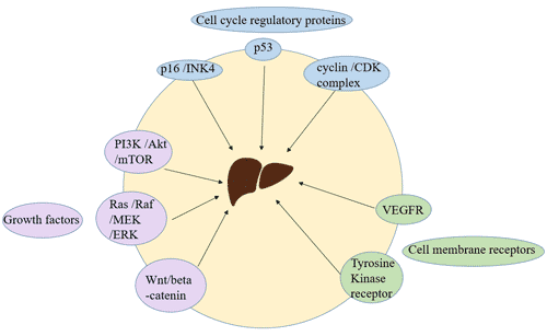
7.2 Antigen Detection

Pathogen detection mainly refers to direct detection of virus or viral genes from host samples by virus isolation and culture, electron microscopic observation, viral antigen detection and gene assay. For clinical diagnosis, antigen detection and RT-PCR (reverse transcription-PCR) are generally used.

7.3 Nucleic Acid Detection

HIV nucleic acid test can be used for the auxiliary diagnosis, course monitoring, treatment program guidance, determination of curative effect and prediction of disease progression, etc.

Commonly used methods for detecting HIV viral load include reverse transcription PCR (RT-PCR), nucleic acid sequence based amplification (NASBA), branched DNA hybridization (bDNA), real-time fluorescence quantitative PCR.

8. Is There A Cure for HIV?

There is currently no effective HIV treatment, but with proper medical care, HIV can be controlled. For example, Magic Johnson was diagnosed with the HIV virus in 1991. After years of adherence to treatment, in March 1997, after examination, the doctor determined that Johnson had overcome the virus and fully recovered.

8.1 AIDS Prevention

If you are HIV-negative, you have several options to protect yourself from HIV infection. The more you take these actions, the safer you will be.

  • Use condoms.
  • Reduce your number of sexual partners.
  • Pre-exposure prophylaxis. It refers to high-risk groups taking HIV drugs daily to reduce the risk of infection. The federal guidelines recommend that consideration should be given to those who are HIV- and have a continuing sexual relationship with HIV positive partners.
  • Post-exposure prophylaxis (PEP) means taking antiretroviral drugs to prevent infection after possible exposure to HIV. PEP should only be used in emergencies and must be used within 72 hours of the most recent possible exposure to HIV.

If you are HIV+, you can take many steps to prevent HIV transmission to HIV-negative partners.

  • Use AIDS drugs to prevent sexual transmission of HIV, this is called treatment as prevention (TasP).This is one of the most effective options for preventing HIV transmission.
  • If HIV carriers can take HIV drugs throughout pregnancy, childbirth and childbirth, the risk of mother-to-child transmission during pregnancy, childbirth and childbirth can be reduced.
  • Choose a less risky sexual behavior
  • Strengthen public HIV education and understanding of government HIV/AIDS policies.

8.2 The Life Cycle of HIV

  • The virions bind to host cell surface receptors and penetrate the cell membrane.
  • The virion remove the protein shell in the cytoplasm of target cells, and release the gene RNA and reverse transcriptase (RT).
  • Viral RNA is reverse transcribed into proviral DNA under the catalysis of reverse transcriptase.
  • The proviral DNA binds to the virally encoded integrase (IN), forms a preinteg ration complex (PIC), and is translocated into the nucleus.
  • Viral DNA is integrated into the chromosome of the host cell by the integrase in the nucleus. Studies on the complete sequencing of the human genome have shown that HIV is preferentially integrated into actively transcribed genes [14], and this feature may further enable them to be copied and transmitted.
  • Replication and translation are conducted by using the existing gene replication and protein expression system of host cells.
  • The protease (PR) of the virus cleaves the polyprotein produced by gene expression into various active structural and functional proteins, and assembles with the replicated genetic material RNA into a mature progeny virus.
  • Once all of the viral components are expressed, new virions assemble at the plasma membrane and bud from the cell surface. To achieve complete budding, the virus hijacks components of the cell’s vesicular transport machinery.

The mechanism of anti-aids drugs is to block one or several key steps in the HIV life cycle

Figure 8 Life cycle of HIV and targets of anti-HIV drug

8.3 HIV Cure

Based on the life cycle of HIV-1 as described above, the anti-aids drugs currently in use or under development mainly include:

  • Entry inhibitor [15]: acts on HIV’s viral attachment, coreceptor interaction, fusion of HIV and cells;
  • Nucleoside reverse transcriptase inhibitors (NRTIs) and non-nucleoside reverse transcriptase inhibitors (NNRTIs): reverse transcription of viral RNA;
  • Integrase inhibitor [16]: integration of proviral DNA;
  • Protease inhibitors (PIs) [17]: transcription of DNA, translation of viral proteins, viral assembly, budding and maturation of HIV Virion [18] [19].

At present, the world’s hottest and most successful new anti-aids drugs with new mechanism of action mainly focus on the two fields of HIV-1 entry inhibitor and HIV-1 integrase inhibitor [20]. This new anti-AIDS drug has brought new therapeutic hopes to more and more drug-resistant patients [21].

In May 2018, a research team used CRISPR/Cas9 editing technology to develop an effective method to destroy the regulatory genes of HIV and successfully inhibit the proliferation of HIV-1 in infected cells [22].

Potential Future Options

There are currently no HIV vaccine to prevent or treat HIV infection. However, scientists are working hard to develop. Currently, a vaccine trial called HVTN702, supported by the National Institutes of Health, is testing whether an experimental vaccine program can safely prevent HIV infection among adults in South Africa.

References

[1] Barrésinoussi F, Chermann J C, Rey F, et al. Isolation of a T-lymphotropic retrovirus from a patient at risk for acquired immune deficiency syndrome (AIDS) [J]. Science, 1983, 220(4599):868-871.

[2] Gallo RC, Sarin PS, Gelmann EP, et al. Isolation of human T-cell leukemia virus in acquired immune deficiency syndrome (AIDS) [J]. Science, 1983, 220(4599):865-867.

[3]Ratner L, Haseltine W, Patarca R, et al. Complete nucleotide sequence of the AIDS virus, HTLV-III [J]. Nature, 1985, 313(6000):277-284.

[4]Sanchez-Pescador R, Power M D, Barr P J, et al. Nucleotide sequence and expression of an AIDS-associated retrovirus (ARV-2) [J]. Science, 1985, 227(4686):484.

[5]Wain-Hobson S, Sonigo P, Danos O, et al. Nucleotide sequence of the AIDS virus, LAV [J]. Cell, 1985, 40(1):9-17.

[6]Sharp P M, Hahn B H. Origins of HIV and the AIDS Pandemic [J]. Cold Spring Harbor Perspectives in Medicine, 2011, 1(1):a006841.

[7]Rhodes T, Singer M, Bourgois P, et al. The social structural production of HIV risk among injecting drug users [J]. Social Science & Medicine, 2005, 61(5):1026-1044.

[8] Smith J H, Whiteside A. The history of AIDS exceptionalism [J]. Journal of the International Aids Society, 2010, 13(1):1-8.

[9] Votteler J, Schubert U. Human Immunodeficiency Viruses: Molecular Biology [J]. Encyclopedia of Virology, 2008, 511:517-525.

[10] Hollingsworth TD, Anderson RM, Fraser C. HIV-1 transmission, by stage of infection [J]. The Journal of Infectious Diseases, 2008, 198(5):687-693.

[11]Moore D J, Masliah E, Rippeth J D, et al. Cortical and subcortical neurodegeneration is associated with HIV neurocognitive impairment [J]. Aids, 2006, 20(6):879-887.

[12]Patel, P, Mackellar, D, Simmons, P, et al. Detecting acute human immunodeficiency virus infection using 3 different screening immunoassays and nucleic acid amplification testing for human immunodeficiency virus RNA, 2006-2008 [J]. Archives of Internal Medicine, 2010, 170(1):66-74.

[13] Petak F, Albu G, Lele E, et al. Cost-effectiveness of a Fourth-Generation Combination Immunoassay for Human Immunodeficiency Virus (HIV) Antibody and p24 Antigen for the Detection of HIV Infections in the United States [J]. HIV Clinical Trials, 2012, 13(1):11-22.
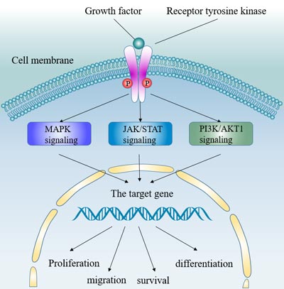
[14] Schröder A R W, Shinn P, Chen H, et al. HIV-1 Integration in the Human Genome Favors Active Genes and Local Hotspots [J]. Cell, 2002, 110(4):521-529.

[15] Tomkowicz B, Collman R G. HIV-1 entry inhibitors: closing the front door [J]. Expert Opin Ther Targets, 2004, 8(2):65-78.

[16] Young S D. Inhibition of HIV-1 integrase by small molecules: the potential for a new class of AIDS chemotherapeutics [J]. Curr Opin Drug Discov Devel, 2001, 4(4):402-410.

[17] Reeves D J D, Piefer A J. Emerging Drug Targets for Antiretroviral Therapy [J]. Drugs, 2005, 65(13):1747-1766.

[18] Temesgen Z, Feinberg J E. Drug evaluation: bevirimat–HIV Gag protein and viral maturation inhibitor [J]. Current Opinion in Investigational Drugs, 2006, 7(8):759.

[19] Li F, Goila-Gaur R, Salzwedel K, et al. PA-457: A Potent HIV Inhibitor That Disrupts Core Condensation by Targeting a Late Step in Gag Processing [J]. Proceedings of the National Academy of Sciences of the United States of America, 2003, 100(23):13555-13560.

[20]Opar A. New HIV drug classes on the horizon [J]. Nature Reviews Drug Discovery, 2007, 6(4):258.

[21] Moore J P, Stevenson M. New targets for inhibitors of HIV-1 replication [J]. Nature Reviews Molecular Cell Biology, 2000, 1(1):40.

[22] Ophinni Y, Inoue M, Kotaki T, et al. CRISPR/Cas9 system targeting regulatory genes of HIV-1 inhibits viral replication in infected T-cell cultures [J]. Scientific Reports, 2018, 8.

Streptococcus pneumoniae, a Primary Cause of Pneumonia

1. What is Streptococcus Pneumoniae?

S. pneumoniae, also known as pneumococcus, are gram-positive, facultative anaerobic member of the genus Streptococcus [1]. In 1881, G. Stermberg in the U.S. and L. Pasteur in France isolated the bacterium, which was originally named Pneumococcus. It was officially named Streptococcus pneumoniaein 1974 because of its close resemblance to Streptococcus. S. pneumoniae is usually shaped like slightly pointed cocci. Individual S. pneumoniae bacterium usually measures between 0.5 and 1.25 micrometers in diameter. Typically, Streptococcus Pneumoniae is completely enclosed by polysaccharide capsules, which makes it such an effective virus. Its cell wall is made up of peptidoglycan, about six layers thick, and lipoteichoic acid which is attached to the membrane by a lipid moiety.

The genome of S. pneumoniae is a closed, circular DNA structure that contains between 2.0 and 2.1 million base pairs depending on the strain. It has a core set of 1553 genes, plus 154 genes in its virulome, which contribute to virulence and 176 genes that maintain a noninvasive phenotype. Among different types of strains, genetic information can vary up to 10% [2].

2. What are The Diseases Caused by Streptococcus Pneumoniae Infection?

S. pneumoniae is extracellular, opportunistic pathogen that colonizes the mucosal surfaces of the human upper respiratory tract (URT), and may be isolated from the nasopharynx of 5–90% of healthy persons, depending on the population and setting. Up to 27–65% of children and <10% of adults are carriers of S. pneumoniae and carriage involves a commensal relationship between the bacterium and the host. Local spread, aspiration or seeding to the bloodstream results in invasive inflammatory diseases [3] [4] [5] (Fig. 1). S. pneumoniae is a leading bacterial cause of a wide range of infections, including otitis media, pneumonia, sepsis and meningitis. Among of them, pneumonia is the most common disease caused by S. pneumoniae infection. Additionally, it can also cause invasive Streptococcus pneumoniae disease in sterile parts of the central nervous system, abdominal cavity, joints, heart valves, and pericardium through blood circulation. As all of these diseases are ‘dead ends’ in the life cycle of the organism, the bacterial factors that cause invasive diseases must also be adaptive for colonization and transmission.

The life cycle of Streptococcus pneumoniae and the pathogenesis of pneumococcal disease

Figure 1. The life cycle of Streptococcus pneumoniae and the pathogenesis of pneumococcal disease
*This figure is derived from the publication on Nat. Rev. Microbiol[6]

Regarding of pneumonia caused by S. pneumoniae infection, it is more common duringa winter and early spring. In tropical climates with dry and rainy seasons, pneumococcal disease tends to occur more in the dry season. Moreover, travelers are more likely to get pneumococcal disease if they spend time in crowded settings or in close contact with children in countries where pneumococcal vaccine is not routinely used.

3. Who is at Risk?

As previously mentioned, The S. pneumoniae is found and survives the mucosal surfaces of the human upper respiratory tract of a person, such as the nose, skin, throat and nasal cavity more precisely the nasopharynx. Although it does not cause any harm to the human body, this bacterium is known to pave the way for various diseases in the people who have weak immune systems. Therefore, certain people are at risk and should watch out for the symptoms of it, including people above 65 years old, infants and toddlers (the infants or children below the age of 2 years), alcoholics, after surgery or serious illness, chain smokers and people with a weak immune system.

4. What are the Symptoms of Streptococcus Pneumoniae Infection?

S. pneumoniae is the primary cause of pneumococcal diseases in people whom have very low immune systems, children and elderly people. These diseases are contagious and spread through contact with people who are ill or who carry the bacteria in their throat. Therefore, it is recommended to watch out for the symptoms of pneumococcal disease. The infections of the pneumococcal disease mostly happen around the sinuses, bloodstream, lungs, middle ear and meninges which is the lining of the spinal cord and brain which ultimately results in meningitis.

Hence, a few of the streptococcus pneumoniae symptoms infection are: cough, chills and fever, difficulty in breathing, rapid breathing, pain in the chest, headache, stiff neck, low alertness, confusion, sensitivity to light, increased heart rate, a sensation of cold and shivering and shaking, discomfort and pain, sweaty skin, short breath, sleepiness, ear pain, the swollen or red eardrum, bloodstained sputum, nausea and vomiting and drowsiness.

5. How to Diagnose Streptococcus Pneumoniae Infections?

Usually, doctors can diagnose pneumonia based on your symptoms, physical exam, lab tests and chest x-ray. If doctors suspect invasive pneumococcal disease, like meningitis or bloodstream infections, they collect samples of cerebrospinal fluid or blood and send them to a laboratory for bacterial cultivation, which is the main technique used to diagnose the causative bacterium in cases of middle ear infection and sinusitis. The bacteria can be identified if neutrophils are present in the sample and if there are more than ten gram-positive diplococci.

If the results of the test are inconclusive, or if further tests are required, the bacteria can be investigated further by being streaked onto blood agar. When this test is performed, the S. pneumoniae should start to exhibit alpha-hemolysis, where the blood agar has areas of green coloring around the colonies of the bacteria that has been streaked onto the agar. However, this test is not always conclusive because other members of the Streptococcus bacteria also exhibit similar reactions and can cause alpha-hemolysis. In order to confirm this test to be conclusive, the streaked organisms should also exhibit sensitivity to optochin or bile, which can more conclusively prove that the bacteria is S. pneumoniae.

6. What are Treatments for Streptococcus Pneumoniae Infection?

Pneumococcus can spread from a person to another. You can get S. pneumoniae from respiratory droplets from the nose or mouth of an infected person. It is common for people, especially children, whom carry the bacteria in their throats without being sick. Moreover, accumulating evidence has suggested that factors such as Ply (CUSABIO provides ply with the species of serotype 4 and serotype 2) that contribute to the disease state by enhancing inflammation also promote the transmission of S. pneumoniae [7]. Antibiotic is the most common treatment for pneumococcal disease. The symptoms of pneumococcal pneumonia usually go away within 12 to 36 hours after you start taking medicine. However, some bacteria such as S. pneumoniae have become resistant to some of the antibiotics used to treat these infections. Such antibiotic resistance is increasing worldwide because these medicines have been overused or misused.

Antibiotic treatment for invasive pneumococcal infections typically includes ‘broad-spectrum’ antibiotics until results of antibiotic sensitivity testing are available. Broad-spectrum antibiotics work against a wide range of bacteria. Once the sensitivity of the bacteria is known, a more targeted antibiotic may be selected.

7. How to Protect Yourself from Streptococcus Pneumoniae Infection?

Prevention of a person from S. pneumoniae is getting them vaccinated with a vaccine which is known as Pneumonia vaccine. This kind of vaccine is recommended to children below 2 years of age, adults above 65 years of age and everyone else who have weak immune systems.

Taking a pneumonia vaccine shot helps to prevent the harmful, life-threatening and contagious pneumococcal disease that this bacteria cause. It thus also helps in preventing a person from all the other diseases that follow pneumococcal diseases.

There are two kinds of pneumonia vaccines available that helps to prevent Streptococcus pneumoniae bacteria, including conjugate pneumococcal vaccine (PCV13) and polysaccharide pneumococcal vaccine (PPV23). So how to choose pneumococcal vaccine, and what are the differences between PCV13 and PPV23? You can find the answers to the above questions on the following table.

PCV13 PPV23
Different ingredients PCV13 is a polysaccharide protein-binding vaccine and effectively stimulate the immune system of infants and young children to produce sufficient protective antibodies, and have immune memory; PPV23 is a polysaccharide vaccine, contains only pneumococcal capsular polysaccharide antigen, without carrier protein and can’t stimulate effective immune response in babies under 2 years old
Other name 13-valent pneumococcal conjugate vaccine 23-valent pneumococcal polysaccharide vaccine
Different serotypes of preventable pneumococcal bacteria PCV13 is a vaccine that protects against 13 strains serotypes of pneumococcal bacteria: 1, 3, 4, 5, 6A, 6B, 7F, 9V, 14, 18C, 19A, 19F and 23F. PPV23 is a vaccine that protects against 23 strains serotypes of pneumococcal bacteria: 1, 2, 3, 4, 5, 6B, 7F, 8, 9N, 9V, 10A, 11A, 12F, 14, 15B, 17F, 18C, 19F, 19A, 20, 22F, 23F, and 33F.
Different vaccination age It is suitable for babies from 6 weeks to 2 years old. It is suitable for high-risk groups over 2 years old and elderly over 50 years old.
Different vaccination procedure The routine vaccination procedure is one dose of each for basic immunization at 2, 4, and 6 months, and one dose for booster immunization at 12-15 months. A total of 4 doses are given. Just one dose are given.
Salt content Into a muscle Into a muscle or under the skin
Produced by different companies It is made by Wyeth Pharmaceuticals and marketed by Pfizer Inc. It is made by Merck & Co., Inc.

In terms of PCV13, you must note that the first dose can be given as early as 6 weeks of age, followed by doses 2 and 3 every 4-8 weeks, and 3 doses of basic immunization can be completed at the latest before 7 months of age. Besides, you may ask whether you still need to be vaccinated if you have already had pneumonia? Why do you still get pneumonia after being vaccinated?

Actually, there are many pathogens that can cause pneumonia, and S. pneumoniae is the most common one. Moreover, S. pneumoniae is a big family, more than 90 kinds have been discovered. Even if you have had pneumonia before, you are only infected with one or more of them at most, which does not mean you are resistant to the remaining pneumococcus. In another words, pneumonia vaccine can only prevent pneumonia caused by S. pneumoniae, but it can’t help other diseases such as viral pneumonia and mycoplasma pneumonia.

References

[1] Ryan KJ, Ray CG, eds. Sherris Medical Microbiology. McGraw Hill. 2004, ISBN 978-0-8385-8529-0.

[2] van der Poll T, Opal SM. Pathogenesis, treatment, and prevention of pneumococcal pneumonia [J]. Lancet. 2009, 374 (9700): 1543–56.

[3] Abdullahi, O. et al. The prevalence and risk factors for pneumococcal colonization of the nasopharynx among children in Kilifi District, Kenya [J]. PLoS ONE. 2012, 7, e30787.

[4] Yahiaoui, R. Y. et al. Prevalence and antibiotic resistance of commensal Streptococcus pneumoniae in nine European countries [J]. Future Microbiol. 2016, 11, 737–744.

[5] Bogaert, D., De Groot, R. & Hermans, P. W. Streptococcus pneumoniae colonisation: the key to pneumococcal disease. Lancet Infect. Dis. 2004, 4, 144–154.

[6] Jeffrey N. Weiser, Daniela M. Ferreira and James C. Paton. Streptococcus pneumoniae: transmission, colonization and invasion [J]. Nat. Rev. Microbiol. 2018.

[7] Lipsitch, M. & Moxon, E. R. Virulence and transmissibility of pathogens: what is the relationship [J]? Trends Microbiol. 1997, 5, 31–37.

What Is Tomato Mosaic Virus

1. What Is Tomato Mosaic Virus?

ToMV is a positive-sense, single-stranded RNA virus belonging to the Virgaviridae family, Tobamovirus genus. It is a kind of plant virus with worldwide distribution and has a wide range of hosts, which can not only infect Solanaceae, Cruciferae, and other vegetable crops, but also infect flowers and seedlings, etc. ToMV causes mosaic disease of many important crop plants, especially threatening tomato production worldwide.

2. Structure of Tomato Mosaic Virus

ToMV has a rod-shaped structure with a 300 nm length and an 18 nm radius. Its genome encodes four different proteins: 180kDa/RNA dependent RNA polymerase, 130kDa/Methyltransferase/Helicase, 30kDa/Movement protein (MP) and 18kDa/Coat protein (CP) [2]. The 130KDa and 180KDa proteins are encoded by the genomic RNA and involved in viral replication. The MP and CP proteins are translated from the respective sub-genomic mRNAs and participate in the viral package and movement. Studies have demonstrated that ToMV particles are very stable and remain infectious for many years after extraction.

Crystal structure of the ToMV-Hel and Tm-1 (431) complex

Figure: Crystal structure of the ToMV-Hel and Tm-1 (431) complex.
*The picture is cited from http://www.naro.affrc.go.jp/archive/nias/eng/research/h26/nias2014e-04.htm


ToMV and tobacco mosaic virus (TMV) are quite closely related in the genus Tobamovirus and are both spread from plant to plant by mechanical means. They are distinguished on the basis of genetic, protein, and host-range differences.

3. The Transmission of Tomato Mosaic Virus

ToMV is a particularly stable virus that can survive for several years in soil or other substrates, especially in the disinfected remains of leaves and roots, which is the main source of transmission and spread of the virus [3]. It is spread mainly by mechanical friction, and seeds, leaf and root debris also carry the virus [3]. ToMV can also be carried on the mouthparts of chewing insects or animals. However, there is no true insect vector for ToMV. Seeds are the main factor in the long-distance transmission of ToMV. Infected seeds can pass the virus to the plant, but the disease is usually introduced and spread primarily through human activity. The virus can easily be transferred from workers’ contaminated hands, tools, and clothing to plants through cultural operations such as plant tying, removing of suckers, and harvest. This leads to infected plants being frequently distributed in a line in the rows worked.

4. Symptoms of Tomato Mosaic Virus Infection

ToMV may attack plants at any growth stage and any part of the plants. Different infectious parts may show distinct symptoms. The intensity of these symptoms are associated with the nature of the strains, cultivar, age of the infected plant, temperature, intensity of light, and nitrogen and boron soil content [4].

ToMV disease mainly damages the leaves, stems, and fruits of plants [4]. The leaves of the infected plants are yellow and green, with a mosaic and mottled appearance. Sometimes, the leaves are stunted. Heavily infected plants may appear like ferns, with leaves in some areas becoming dark green and raised. There are dark brown patches on the stem and tendrils, and the discoloration is only limited to the epidermal tissue, but not deep into the stem. Some plants also infect fruits. The fruits may have yellow or brown strips on the surface and be slightly concave and show internal browning just under the skin, reducing fruit number and size. Plants seedlings infected with ToMV can cause plant growth retardation and dwarf. Symptoms may be inhibited during cool temperatures. Consequently, infected seedlings may not display symptoms until moved to a warm environment.

5. Control of Tomato Mosaic Virus Infection

ToMV is one of the main factors severely impacting tomato production and cultivation. Therefore, several integrated control measurements for ToMV have been proposed [4] [5].

5.1 Plant Antiviral Varieties

There are several tomato varieties resistant to ToMV. Studies have found that tomatoes containing the Tm-22 gene could specifically resist to ToMV strains ToMV-0, ToMV-1 and ToMV-2.

5.2 Use Certified Virus-free Seeds

ToMV is transmitted from plant to plant through vegetative propagation, grafting, and seeds. It is significantly important to ensure that any seeds planted are virus-free. This blocks ToMV from being spread to healthy plants through mechanical activities.

5.3 Take Sound Sanitation Measurements

Sanitation is the most important measurement to practice when controlling ToMV. Tools and infected materials should be sanitized and disinfected carefully. Washing hands with soap and water before and during the handling of plants to reduce potential transmission among plants. Destroy any seedlings that appear stunted or distorted and then decontaminate tools and hands. Keep the area around the tomatoes weeded and free of plant detritus to minimize areas the disease can harbor. Control insects as well to lessen the chances of contamination.

6. ToMV-related Products

The impact of ToMV on crops is quite significant. So scientists have been studying ToMV to find more effective solutions for preventing or controlling ToMV infection. In order to promote the research of ToMV, CUSABIO has developed several related products, including the MP and CP of ToMV.

Products
Recombinant ToMV Movement protein (MP) (TMV strain tomato) Recombinant ToMV Capsid protein (CP) (strain S-1)
Recombinant ToMV Movement protein (MP) (TMV strain K2) Recombinant ToMV Capsid protein (CP) (strain Korean)
Recombinant ToMV Movement protein (MP) (TMV strain K1) Recombinant ToMV Capsid protein (CP) (TMV strain K1)
Recombinant ToMV Movement protein (MP) (strain LII) Recombinant ToMV Replicase large subunit, partial (TMV strain K2)
Recombinant ToMV Movement protein (MP) (strain LIIa) Recombinant ToMV Replicase large subunit, partial (TMV strain tomato)
Recombinant ToMV Capsid protein (CP) (TMV strain K2) Recombinant ToMV Replicase large subunit, partial (TMV strain K1)
Recombinant ToMV Capsid protein (CP) (TMV strain tomato)

References


2019 Novel Coronavirus

2019 Novel Coronavirus

Based on the fact that the number of infected people in China has increased and outbreaks have occurred in many countries. On January 30, 2020, the World Health Organization announced that the 2019 Novel Coronavirus (SARS-CoV-2) was listed as Public Health Emergency of International Concern (PHEIC). At this point, the SARS-CoV-2 disease (COVID-19) disease is not only a battle in China, but also a global battle.

CUSABIO paid close attention to the progression of SARS-CoV-2. Once obtaining the virus gene sequence information, we quickly developed and produced SARS-CoV-2 related products. Targets include the Spike protein (S), N protein (N) and ACE2. Moreover, we also provide several products about coronavirus to meet the needs of scientific research.

The following is a list of reagents and information for SARS-CoV-2 research. Click the links, you’ll reach the corresponding parts.

SARS-CoV-2 Protein Reagents

Target Name Product Name Code Species Source
E & M Recombinant Severe acute respiratory syndrome coronavirus 2 Envelope small membrane protein & Membrane protein(E & M),partial CSB-EP3402GND SARS-CoV-2 E.coli
N Recombinant Human Novel Coronavirus Nucleoprotein(N) CSB-EP3325GMY SARS-CoV-2 E.coli
N Recombinant Human Novel Coronavirus Nucleoprotein(N) CSB-YP3325GMY SARS-CoV-2 Yeast
N Recombinant Human Novel Coronavirus Nucleoprotein(N) CSB-MP3325GMY SARS-CoV-2 Mammalian cell
N Recombinant Human Novel Coronavirus Nucleoprotein(N) CSB-YP3325GMYa4 SARS-CoV-2 Yeast
N Recombinant Human Novel Coronavirus Nucleoprotein(N),partial CSB-EP3325GMY1 SARS-CoV-2 E.coli
N Recombinant Human Novel Coronavirus Nucleoprotein(N),Biotinylated CSB-EP3325GMY-B SARS-CoV-2 E.coli
N Recombinant Human Novel Coronavirus Nucleoprotein(N)(D3L,R203K,G204R,S235F) (Active) CSB-EP3325GMY(M1) SARS-CoV-2 E.coli
N Recombinant Human Novel Coronavirus Nucleoprotein(N)(P80R) (Active) CSB-EP3325GMY(M3) SARS-CoV-2 E.coli
N Recombinant Human Novel Coronavirus Nucleoprotein(N)(P13L) (Active) CSB-EP3325GMY(M4) SARS-CoV-2 E.coli
N Recombinant Human Novel Coronavirus Nucleoprotein(N)(D103Y) (Active) CSB-EP3325GMY(M5) SARS-CoV-2 E.coli
N Recombinant Human Novel Coronavirus Nucleoprotein(N)(S202N) (Active) CSB-EP3325GMY(M8) SARS-CoV-2 E.coli
N Recombinant Human Novel Coronavirus Nucleoprotein(N)(L230F) (Active) CSB-EP3325GMY(M9) SARS-CoV-2 E.coli
N Recombinant Human Novel Coronavirus Nucleoprotein(N)(I292T) (Active) CSB-EP3325GMY(M10) SARS-CoV-2 E.coli
N Recombinant Human Novel Coronavirus Nucleoprotein(N)(Q384H) (Active) CSB-EP3325GMY(M11) SARS-CoV-2 E.coli
N Recombinant Human Novel Coronavirus Nucleoprotein(N)(R203K,G204R) (Active) CSB-EP3325GMY(M12) SARS-CoV-2 E.coli
N Recombinant Human Novel Coronavirus Nucleoprotein(N)(R203M,D377Y) (Active) CSB-EP3325GMY(M13) SARS-CoV-2 E.coli
N Recombinant Human Novel Coronavirus Nucleoprotein(N)(D63G,R203M,D377Y) (Active) CSB-EP3325GMY(M14) SARS-CoV-2 E.coli
NSP3 Recombinant Severe acute respiratory syndrome coronavirus 2 Non-structural protein 3(nsp3),partial CSB-EP3398GND SARS-CoV-2 E.coli
NSP5 Recombinant Severe acute respiratory syndrome coronavirus 2 3C-like proteinase(NSP5) CSB-EP3389GND SARS-CoV-2 E.coli
Nsp9 Recombinant Severe acute respiratory syndrome coronavirus 2 Non-structural protein 9(nsp9) CSB-MP3388GND SARS-CoV-2 Mammalian cell
Nsp9 Recombinant Severe acute respiratory syndrome coronavirus 2 Non-structural protein 9(nsp9) CSB-EP3388GND SARS-CoV-2 E.coli
S (RBD) Recombinant Human Novel Coronavirus Spike glycoprotein(S), partial CSB-YP3324GMY2 SARS-CoV-2 Yeast
S (RBD) Recombinant Human Novel Coronavirus Spike glycoprotein(S), partial CSB-MP3324GMY1 SARS-CoV-2 Mammalian cell
S (RBD) Recombinant Human Novel Coronavirus Spike glycoprotein(S), partial CSB-MP3324GMY1b1 SARS-CoV-2 Mammalian cell
S (RBD) Recombinant Human Novel Coronavirus Spike glycoprotein(S) (K417N),partial CSB-MP3324GMY1(M7) SARS-CoV-2 Mammalian cell
S (RBD) Recombinant Human Novel Coronavirus Spike glycoprotein(S) (K417N),partial CSB-MP3324GMY1(M7)h8 SARS-CoV-2 Mammalian cell
S (RBD) Recombinant Human Novel Coronavirus Spike glycoprotein(S) (E484K),partial CSB-MP3324GMY1(M8) SARS-CoV-2 Mammalian cell
S (RBD) Recombinant Human Novel Coronavirus Spike glycoprotein(S) (E484K),partial CSB-MP3324GMY1(M8)h8 SARS-CoV-2 Mammalian cell
S (RBD) Recombinant Human Novel Coronavirus Spike glycoprotein(S), partial CSB-YP3324GMY1 SARS-CoV-2 Yeast
S (RBD) Recombinant Human Novel Coronavirus Spike glycoprotein(S) (V367F), partial CSB-MP3324GMY1(M1) SARS-CoV-2 Mammalian cell
S (RBD) Recombinant Human Novel Coronavirus Spike glycoprotein(S) (W436R), partial CSB-MP3324GMY1(M2) SARS-CoV-2 Mammalian cell
S (RBD) Recombinant Human Novel Coronavirus Spike glycoprotein(S) (G476S), partial CSB-MP3324GMY1(M4) SARS-CoV-2 Mammalian cell
S (RBD) Recombinant Human Novel Coronavirus Spike glycoprotein(S) (N501Y), partial CSB-MP3324GMY1(M6) SARS-CoV-2 Mammalian cell
S (RBD) Recombinant Human Novel Coronavirus Spike glycoprotein(S) (V483A), partial CSB-MP3324GMY1(M5) SARS-CoV-2 Mammalian cell
S (RBD) Recombinant Human Novel Coronavirus Spike glycoprotein(S), partial,Biotinylated CSB-MP3324GMY1-B SARS-CoV-2 Mammalian cell
S (S1) Recombinant Human Novel Coronavirus Spike glycoprotein(S), partial CSB-YP3324GMYa4 SARS-CoV-2 Yeast
S (S1) Recombinant Human Novel Coronavirus Spike glycoprotein(S), partial CSB-MP3324GMY SARS-CoV-2 Mammalian cell
S (S1) Recombinant Human Novel Coronavirus Spike glycoprotein(S) (D614G), partial CSB-MP3324GMY(M1) SARS-CoV-2 Mammalian cell
S (S1) Recombinant Human Novel Coronavirus Spike glycoprotein(S)(N501Y,P681H), partial CSB-MP3324GMY(M2) SARS-CoV-2 Mammalian cell

SARS-CoV-2 Antibody Reagents

Target Name Product Name Code Species
N Antibody CSB-RA33255A0GMY SARS-CoV-2 ELISA, GICA
N Antibody CSB-RA33255A1GMY SARS-CoV-2 ELISA, WB, GICA
N Antibody CSB-RA33255A2GMY SARS-CoV-2 ELISA
N Antibody, Biotin conjugated CSB-RA33255D1GMY SARS-CoV-2 ELISA
N Antibody, FITC conjugated CSB-RA33255C1GMY SARS-CoV-2 ELISA
N Antibody, HRP conjugated CSB-RA33255B1GMY SARS-CoV-2 ELISA
S Antibody CSB-RA33245A0GMY SARS-CoV-2 ELISA, GICA
S Antibody CSB-RA33245A1GMY SARS-CoV-2 ELISA, GICA, Neutralising
S Antibody, Biotin conjugated CSB-RA33245D1GMY SARS-CoV-2 ELISA
S Antibody, FITC conjugated CSB-RA33245C1GMY SARS-CoV-2 ELISA
S Antibody, HRP conjugated CSB-RA33245B1GMY SARS-CoV-2 ELISA
SARS-CoV-2 RBD Nanobody CSB-RA33245A2GMY SARS-CoV-2 ELISA, GICA, Neutralising
SARS-CoV-2 RBD Nanobody, Biotin conjugated CSB-RA33245D2GMY SARS-CoV-2 ELISA
SARS-CoV-2 RBD Nanobody, FITC conjugated CSB-RA33245C2GMY SARS-CoV-2 ELISA
SARS-CoV-2 RBD Nanobody, HRP conjugated CSB-RA33245B2GMY SARS-CoV-2 ELISA
SARS-CoV-2 Spike RBD Antibody Pair 1 CSB-EAP33245 SARS-CoV-2 S-ELISA
SARS-CoV-2 N Antibody Pair 1 CSB-EAP33255A1 SARS-CoV-2 S-ELISA
SARS-CoV-2 N Antibody Pair 2 CSB-EAP33255A2 SARS-CoV-2 S-ELISA
SARS-CoV-2 N Antibody Pair 3 CSB-EAP33255A3 SARS-CoV-2 S-ELISA
SARS-CoV-2 N Antibody Pair 4 CSB-EAP33255A4 SARS-CoV-2 S-ELISA

SARS-CoV-2 Immune Antibody Reagents

Product Name Code Tested Applications Raised in
S Monoclonal Antibody CSB-MA33245A0m GICA Mouse
S Monoclonal Antibody CSB-MA33245A1m GICA Mouse
S Monoclonal Antibody CSB-MA33245A2m GICA Mouse
S Antibody CSB-PA33245YA01GMY GICA Rabbit
S Antibody CSB-PA33245YA11GMY GICA Rabbit
N Antibody CSB-PA33254A0Rb GICA Rabbit
N Antibody CSB-MA33255A0m GICA Mouse
N Antibody CSB-MA33255A1m GICA Mouse
N Antibody CSB-MA33255A2m GICA Mouse

SARS-CoV-2 ELISA Kit Reagents

Target Code Source Tag Info
SARS-CoV-2 N ELISA Kit CSB-EL33251 serum, plasma, swabs Quantitative
Human SARS-CoV-2 S RBD Ab (IgG) ELISA Kit CSB-EL33241HU serum, plasma Qualitative
Human SARS-CoV-2 S RBD Ab (IgM) ELISA Kit CSB-EL33242HU serum, plasma Qualitative
Human SARS-CoV-2 Neutralizing Antibody ELISA Kit CSB-EL33243HU serum, plasma Qualitative
Human SARS-CoV-2 N Ab (IgG) ELISA Kit CSB-EL3325HU serum, plasma Qualitative
Human SARS-CoV-2 N/S1 Ab (IgG) ELISA Kit CSB-EL3326HU serum, plasma Qualitative
SARS-CoV-2 S1 RBD ELISA Kit CSB-EL33244 serum, plasma, swabs Quantitative

Other Coronavirus Related Reagents

Protein Related Products
Target Name Product Name Code Species Source
HE Recombinant Bovine coronavirus Hemagglutinin-esterase(HE) CSB-YP323648BJK BCoV Yeast
N Recombinant Bovine coronavirus Nucleoprotein(N) CSB-EP320768BJK BCoV E.coli
Orf1ab Recombinant Middle East respiratory syndrome-related coronavirus Orf1ab,partial CSB-EP2883GJE MERS-CoV E.coli
S Recombinant Human coronavirus OC43 Spike glycoprotein(S) ,partial CSB-EP336163HIY HCoV-OC43 E.coli
S Recombinant Human coronavirus OC43 Spike glycoprotein(S) ,partial CSB-YP336163HIY HCoV-OC43 Yeast
S Recombinant Bovine coronavirus Spike glycoprotein(S) ,partial CSB-EP333052BJO BCoV E.coli
S Recombinant Bovine coronavirus Spike glycoprotein(S),partial CSB-EP322803BJK BCoV E.coli
SARS-COV-RBD Recombinant Human SARS coronavirus Spike glycoprotein(S) ,partial CSB-MP348663HQE SARS-CoV Mammalian cell


Antibody Related Products
Product Name Code Species Reactivity Tested Applications
ACE2 Antibody CSB-PA866317LA01HU Human ELISA, IHC, IF
ACE2 Antibody CSB-PA001150GA01HU Human, Mouse, Rat ELISA,WB
APN Antibody CSB-PA001827LA01HU Human, Mouse ELISA, WB, IHC, IF
APN Antibody CSB-PA001827GA01HU Human,Mouse,Rat ELISA,WB
CD147 antibody CSB-PA11759A0Rb Human ELISA, WB, IHC
CTSB Antibody CSB-PA06974A0Rb Human, Mouse ELISA, WB, IHC, IF
DPP4 Antibody CSB-PA06229A0Rb Human, Rat ELISA, WB, IHC, IF
DPP4 Antibody CSB-PA007139GA01HU Human,Mouse,Rat ELISA,WB,IHC
NRP1 Antibody CSB-PA016091ESR1HU Human ELISA, IHC


ELISA Kit Products
Product Name Code Sample Types Detection Range
Human ACE2 ELISA Kit CSB-E04489h serum, plasma, cell culture supernates 0.156 ng/mL-10 ng/mL
Human APN ELISA kit CSB-EL001827HU serum, plasma, tissue homogenates 31.25 pg/mL-2000 pg/mL
Human CD147 ELISA Kit CSB-E12994h serum, plasma 12.5 pg/mL-800 pg/mL
Human CTSB ELISA kit CSB-E13450h serum, plasma, tissue homogenates 0.312 ng/mL-20 ng/mL
Human NRP1 ELISA kit CSB-EL015709HU serum, plasma, cell culture supernates, tissue homogenates 31.25 pg/mL-2000 pg/mL

What is The Coronavirus?

Before introducing the SARS-CoV-2, let us understand what is a coronavirus. Coronaviruses are a large family of viruses that can cause respiratory illnesses such as the common cold. Almost everyone gets infected with coronaviruses at least once in their life, but symptoms are typically mild to moderate. Most coronaviruses are not dangerous, but some are. Those that cause Middle East respiratory syndrome (MERS) or severe acute respiratory syndrome (SARS) can be deadly.

The Typical Structure of Coronavirus

Figure 1. The Typical Structure of Coronavirus

The coronavirus genome encodes a spike protein (S), an envelope protein, a membrane protein, and a nucleoprotein in this order. Among them, spike protein is the most important surface membrane protein of coronavirus.

What is The SARS-CoV-2?

The SARS-CoV-2 was discovered because of Wuhan Viral Pneumonia cases in 2019, and was named by the World Health Organization on January 12, 2020. It belongs to the beta genera of the Coronaviridae family in 2003, together with SARS coronavirus (SARS CoV) in 2003 and MERS coronavirus (MERS CoV) in 2012. The alignment between SARS-CoV-2 and 2002 SARS CoV has about 70% sequence similarity and 40% sequence similarity with MERS CoV. There is currently no specific treatment, but many symptoms can be managed, and need to be treated according to the clinical situation of the patient.

CUSABIO paid close attention to the progression of SARS-CoV-2. Once obtaining the virus gene sequence information, we quickly developed and produced SARS-CoV-2 related products. Targets include the Spike protein (S), N protein (N) and ACE2. Moreover, we also provide several products about coronavirus to meet the needs of scientific research.

How does SARS-CoV-2 Spread?

The virus is reportedly spreading from person-to-person in many parts of China and in some other countries by interacting with ACE2 from mucous membrane of eye, mouth and nose. On Jan. 30, the Centers for Disease Control and Prevention (CDC) identified the first case of person-to-person spread in the United States.

In terms of how one would catch the virus, the CDC says that human coronaviruses are most commonly spread between an infected person and others via:

  • The air (from viral particles from a cough or sneeze);
  • Close personal contact (touching or shaking hands);
  • An object or surface with viral particles on it (then touching your mouth, nose or eyes before washing your hands);
  • And rarely from fecal contamination.

What are The Symptoms of SARS-CoV-2 Infection?

As the Figure 2 shows, the typical symptoms of the SARS-CoV-2 infection include:

  • Main symptoms are fever, fatigue, and dry cough;
  • Nasal congestion, runny nose and other upper respiratory symptoms are rare;
  • Approximately half of the patients experienced dyspnea after one week, and the severe cases progressed rapidly to acute respiratory distress syndrome, septic shock, difficult to correct metabolic acidosis, and coagulopathy.

It is worth noting that in the course of severe and critically ill patients, there can be moderate to low fever, even without obvious fever. Some patients have mild onset symptoms and no fever. They usually recover after 1 week. Most patients have a good prognosis, and a few patients are critically ill and even die.

The Sympotoms of SARS-CoV-2

Figure 2. The Sympotoms of SARS-CoV-2

Where Does SARS-CoV-2 Come From?

Currently, the origin of SARS-CoV-2 also isn’t clear. But experts suppose that the origin still is wildlife like the other coronaviruses. Coronaviruses originate in animals-like camels, civets and bats-and are usually not transmissible to humans. But occasionally a coronavirus mutates and can pass from animals to humans and then from human to human, as such the case with the SARS epidemic in the early 2000s. China’s National Health Commission confirmed that 15 health care workers have become infected, indicating that the virus can spread from human to human.

Writing here, you may confuse that should I be concerned about pets (such as dog and cat) carring SARS-CoV-2? Exactly, there is no evidence suggested coronavirus in dogs and other pets. But, CDC recommends that people traveling to China avoid animals both live and dead.

How to Treat 2019 Novel Coronavirus?

In terms of SARS-CoV-2 treatment, there are no specific treatments for coronavirus infections and most people will recover on their own. So treatment involves rest and medication to relieve symptoms. A humidifier or hot shower can help to relieve a sore throat and cough. If you are mildly sick, you should drink a lot of fluids and rest.

Although there is no vaccine for the new coronavirus, researchers at the U.S. National Institutes of Health confirmed they were in preliminary stages of developing one. Officials plan to launch a phase 1 clinical trial of a potential vaccine within the next three months. Moreover, researchers are also working on gathering samples of the virus to design a therapy that will train patients’ immune cells to detect and destroy the virus.

How to Protect Yourself From Coronavirus?

Although there are no specific treatment for SARS-CoV-2 infection, we can take these measures to prevent us from it:

  • Wash your hands: wet your hands with clean, running water and apply soap.
  • Cover your mouth and nose with a tissue when you cough or sneeze, then throw the tissue and wash your hands. If you do not have a tissue to hand, cough or sneeze into your elbow rather than your hands.
  • Face masks offer some protection as they block liquid droplets.
  • Seek early medical help if you have a fever, cough and difficulty breathing, and share your travel history with healthcare providers.
  • If you have returned from an affected area in the last two weeks, stay indoors and avoid contact with other people for 14 days. This means not going to work, school or public areas.

The Latest Progression of SARS-CoV-2 Research

In this section, we collect several latest progression of SARS-CoV-2 research, including the mechanism of SARS-CoV-2 infection, the detection methods of SARS-CoV-2, the targets of SARS-CoV-2 invasion, etc.

  • As you know, the SARS-CoV-2 uses an RNA-dependent RNA polymerase (RdRp) for the replication of its genome and the transcription of its genes. On April 27, 2020, researchers led by Patrick Cramer of the Max Planck Institute for Biophysical Chemistry in Göttingen, Germany published a study on the bioRxiv entitled “Structure of replicating SARS-CoV-2 polymerase”. They determined the three-dimensional structure of the SARS-CoV-2 polymerase. The results of this study will allow for a detailed analysis of the inhibitory mechanisms used by antivirals such as remdesivir, which is currently in clinical trials for the treatment of coronavirus disease 2019 (COVID-19).
  • For the term of SARS-CoV-2-host interactome targets, except for ACE2 and TMPRSS2, on May 14, 2020, researchers from Central South University and China Agricultural University published a study entitled “The MERS-CoV receptor DPP4 as a candidate binding target of the SARS-CoV-2 spike” online on iScience. They revealed that SARS-CoV-2 spike (hereinafter referred to as SARS-CoV-2-S) receptor-binding domain (RBD) has a potentially high affinity to DPP4 by bioinformatics methods combining crystal structure-based human-viral interaction prediction and protein docking. And SARS-CoV-2-S/DPP4 binding shares key DPP4 residues with that of MERS-CoV-S/DPP4. In addition, compared with SARS-CoV-S, E484 insertion and adjacent substitutions are critical for the DPP4-binding ability of SARS-CoV-2-S. This potential utilization of DPP4 as a binding target for SARS-CoV-2 may provide new insights into the viral pathogenesis and help develop monitoring and treatment strategy for addressing the challenges of COVID-19.
  • On May 26, 2020, two latest studies completed by Chinese scientists were published on Nature. The two studies are about isolated highly active neutralizing antibodies specific for SARS-CoV-2 from patients.One of the studies was completed in cooperation with the Institute of Microbiology, Chinese Academy of Sciences, Wuhan Institute of Virology, Chinese Academy of Sciences and Beijing Ditan Hospital and entitled “A human neutralizing antibody targets the receptor binding site of SARS-CoV-2”, In this study, the researchers reported the isolation of two specific human monoclonal antibodies (MAbs) from a convalescent COVID-19 patient: CA1 and CB6. The researchers found that CA1 and CB6 showed strong SARS-CoV-2 specific neutralizing activity against SARS-CoV-2 in vitro. Moreover, structural studies have shown that CB6 can recognize epitopes that overlap with the ACE2 binding site in the SARS-CoV-2 receptor binding domain (RBD), thereby interfering through steric hindrance and direct interface-residue competition Virus/receptor interaction.Another study was completed in collaboration with multiple research teams at Tsinghua University and entitled “Human neutralizing antibodies elicited by SARS-CoV-2 infection”. In this study, the researchers reported the isolation and identification of 206 RBD-specific monoclonal antibodies from single B cells of 8 SARS-CoV-2 infected individuals. And the researchers identified antibodies with potent anti-SARS-CoV-2 neutralizing activity that are related to ACE2’s ability to compete with RBD.
  • On June 6, 2020, researchers from multiple institutions in China jointly published a study titled “Development of an inactivated vaccine candidate, BBIBP-CorV, with potent protection against SARS-CoV-2” on Cell, including Beijing Biological Products Institute Co., Ltd., China Center for Disease Control and Prevention, Peking Union Medical College, National Food and Drug Administration and Tsinghua University. They reported the pilot-scale production of an inactivated SARS-CoV-2 vaccine candidate (BBIBP-CorV) that induces high levels of neutralizing antibodies titers in mice, rats, guinea pigs, rabbits and nonhuman primates to provide protection against SARS-CoV-2. In addition, BBIBP-CorV showed efficient productivity and good genetic stability in vaccine manufacture. These results support for the further evaluation of BBIBP-CorV to enter a clinical trial.

What You Have to Know about Cytokine Storm and Virus Infection

What You Have to Know about Cytokine Storm and Virus Infection

1. What is Cytokine Storm?

Cytokine storm, also known as high cytokine disease, is the systemic expression of a healthy and vigorous immune system resulting in the release of more than 150 inflammatory mediators such as TNF-α, IL-1, IL-6, IL-12, IFN-α, IFN-β, IFN-γ, MCP-1, and IL-8. Both pro-inflammatory cytokines and anti-inflammatory cytokines are elevated in the serum, and the fierce and often lethal interplay of these cytokines is referred to as a “Cytokine Storm”.

It is a severe excessive immune response caused by a positive feedback cycle between cytokines and immune cells. The symptoms are high fever, redness, swelling, extreme fatigue and nausea, which can be fatal in some cases. Cytokine storm is an important cause of acute respiratory distress syndrome and multiple organ failure (Figure 1).

Cytokine Storm Causes Direct Organ Injury

Figure 1. Cytokine Storm Causes Direct Organ Injury

2. Why Does The Virus Invasion Cause Cytokine Storm?

After the virus enters the host target cells through the receptor-mediated endocytosis to proliferate in large numbers, it uses budding or induces programmed cell death to release more virus. These released virus are recognized by the pattern recognition receptors on the body’s immune cells. Then the immune cells release a large number of cytokines through a series of signal transduction to activate more immune cells, which participate in the elimination of the virus.

However, as you know, immunity is a double-edged sword. When the body’s immune activation attempts to remove exotic pathogenic microorganisms, it also leads to the body’s own damage. If the amount of virus is large and the immunity is strong, a more severe local struggle will occur, causing local inflammation and cell damage, that is, a cytokine storm. At this time, the condition may worsen.

Taking Ebola virus as an example, it escapes the recognition of the immune system by lurking in host immature dendritic cells, macrophages, etc. After a large number of replications and rapid spread, it causes necrosis and decomposition of multiple organ cells. When the Ebola virus is released with the decomposed cells, the immune system will quickly recognize it. Due to the large amount of virus, the immune system is fully fired, releasing a large number of pro-inflammatory cytokines, tissue factors and vasoactive peptides. These released factors lead to penetrate the blood vessel wall and release a large amount of nitric oxide, causing secondary damage to the blood vessel wall and damaging the coagulation system. These reaction will cause the body to fever, internal bleeding, and eventually lead to multiple organ failure (Figure 2).

Abnormal Regulation of Innate Immune Cells in Ebola Virus Infection

Figure 2. Abnormal Regulation of Innate Immune Cells in Ebola Virus Infection

3. Virus and Cytokine Storm

Cytokine storm is common in viral infections, certain medications, and hematopoietic diseases. Cytokine storm is an important cause of acute respiratory distress syndrome (ARDS) and multiple organ failure. It is one of the important causes of death in many diseases, and it recently attractes more and more attention. In this article, we primarily focus on cytokine storm caused by virus infection. In addition to Ebola virus, dengue virus, highly pathogenic avian influenza virus (H5N1, H1H1, etc.), megavirus, smallpox virus, SARS virus, and MERS virus can cause cytokine storms.

3.1 SARS Virus and Cytokine Storm

SARS, also known as severe acute respiratory syndromes, is a major symptom of fever, dry cough, and chest tightness caused by SARS coronavirus infection. It began in December 2002. In terms of SARS-induced cytokine storm, Dr. Peiris of the University of Hong Kong found in autopsy that patients with severe SARS had significant damage to lung tissue, multinucleated giant cells in the alveoli, and alveolar macrophages that increased nuclear amphoteric staining cytoplasm.. These cells can secrete TNF, IL-1, TGF and other cytokines, and then induce fibroblast activation, extracellular matrix deposition and alveolar epithelial damage repair. Phagocytosis of red blood cells in the lung tissue suggests that cytokine regulation is out of control. It is speculated that SARS infection caused the release of many inflammatory mediators.

Zhiying Liu detected the presence of high concentrations of inflammatory and anti-inflammatory factors in cured and dead SARS patients. The high of IL-6, IL-8, and IL-10 suggest that the three cytokines play an important role in the progression of patients’ disease. In the treatment of SARS patients, steroid hormones are commonly used to inhibit the release of cytokines, reduce the response and reduce mortality.

As mentioned on the article entilted 2019 Novel Coronavirus, the alignment between SARS-CoV-2 and 2002 SARS CoV has about 70% sequence similarity and 40% sequence similarity with MERS CoV. SARS-CoV-2 is a new strain of coronavirus that has never been found in humans before. Common signs of a person infected with a coronavirus include respiratory symptoms, fever, cough, shortness of breath, and dyspnea. In more severe cases, infection can cause pneumonia, severe acute respiratory syndrome, kidney failure, and even death. 2019-nCoV infection leads to a severe pneumonia epidemic. Most of the infected people are healthy people and young adults. The symptoms are similar to SARS. Some patients will suddenly change from mild to severe. It cannot be ignored because of the “cytokine storm”.

3.2 Influenza Virus and Cytokine Storm

Influenza virus induce mouse glial cell cytokine storm. Zhou Jianxiang et al. isolate and culture glial cells from the cerebral cortex of newborn mice and infect astrocytes with influenza viruses H1N1 and H3N2 in vitro at 6h and 24h respectively. The supernatant was harvested and the influenza virus were removed by ultrafiltration molecular retention. Conditional supernatants at different cultural time were used to stimulate astrocytes and microglia, respectively. RNA was extracted and reverse transcribed after 24 hours. Real-Time PCR was used to detect the pro-inflammatory factors (TNF-α, IL-1β, and IL- 6) and chemokines (IP-10, MCP-1, and MIP-1) .

This study indicated that the condition supernatant at different cultural time upregulates the proinflammatory factors TNF-α, IL-1β, IL-6 and chemokines IP-10, MCP-1, MIP-1 in normal glial cells. Up-regulation to varying degrees produces a cytokine cascade effect. The cytokine cascade effect caused by influenza viruses H1N1 and H3N2 infected astrocytes may be related to immunopathological damage.

3.3 EBV and Cytokine Storm

EBV-associated lymphocytic proliferative diseases are associated with cytokine storms. CAEBV was nominated as EBV+LPD on a EBV-LPD seminar held at the National Institutes of Health on September 8-9, 2008. EBV+LPD is divided into B cell type (EBV+B-LPD) and T / NK cell type (EBV+T / NK-LPD). It is also called hemophilic syndrome (HPS). Kimura et al. Analyzed the virological differences and cytokine secretion in 39 patients with T-CAEBV and NK-CAEBV. And they found that IL-1β, IL-10 and IFN-γ are highly expressed in both serum, but the levels of IL-13 in patients with NK cell-type infection were higher than that of T-cells, and the serum of some patients who progressed to HPS expressed IL-1β and IFN-γ. In addition, the transcription of cytokines such as IL-1α, IL-1β, IL-10, IL-12p35, IL-13, IL-15, TNF-α, and IFN-γ in peripheral blood mononuclear cells of 19 CAEBV patients were increased.

Moreover, Ishii et al. found that monocytes can promote EBV-related NK / T cell proliferation and secrete LMP1 through IL-15 on the membrane, demonstrating the important role of IL-15 in the progression of EBV-LPD disease. SNK6 cell line highly expresses IFN-γ, IL-9 and IL-10. IL-10 indirectly promotes cell proliferation by promoting the secretion of LMP1 and CD25. Different types of EBV-LPD express different cytokines.

4. Is There Any Treatments of Cytokine Storm?

Currently, the treatments of cytokine storm primarily conclude four measures:

  • Anti-shock therapy (treatment of symptoms, life-saving first): infusion to ensure blood volume, application of vasoactive drugs, mechanical ventilation if necessary, protection of important organ functions;
  • Support and symptomatic treatment (restoration of physical strength): routine infusion, maintaining water, electrolyte and acid-base balance, nutrition support, etc .;
  • Inhibit excessive immune cell activation and cytokine production (curative, seek recovery): clinically, hormone therapy (adrenal corticosteroids, etc.) with appropriate doses and treatments is often used, and nonsteroidal anti-inflammatory drugs and free radical scavengers ( A lot of vitamin C, vitamin E) and so on.
  • Antibody-neutralizing cytokine storm (precise treatment): neutralizing monoclonal antibodies against elevated cytokines to prevent severe disease and death.

5. Is There Any Medicines of Cytokine Storm?

According to the mechanism of the cytokine storm, the thinkings of drug development derive from the following principles:

  • Reduce activating cytokines, such as IL-12, IFN-γ, TNF-α, etc .;
  • Increase inhibitory cytokines, such as IL-10, TGF-β, etc .;
  • Reduce the infiltration of leukocytes into inflammatory tissues, such as 1-phosphate sphingosine (S1P).

Researchers have done a lot of work from the above three ideas, and studied various corticosteroids, PPAR agonists, S1P1 receptor agonists, COX inhibitors, anti-TNF antibodies, ACEI / ARB inhibitors, and OX40 inhibitors against cytokine storms. Of the above therapies, the most effective pneumonia-related cytokine storm therapy in animal experiments is the 1-phosphate sphingosine receptor 1 agonist (S1PR1).

Sphingosine-1-phosphate (S1p), a signaling sphingolipid with five subtypes of its receptors, are mainly expressed in vascular endothelial cells and lymphocytes in lung tissue. S1P1 agonists (CYM-5442 and RP-002) have been reported to protect mice from death caused by severe influenza infection through attenuating cytokines and inhibiting infiltration of innate immune cells.

In a mouse model infected with the 2009 H1N1 pandemic influenza, the S1P1 receptor agonist alone reduced deaths from lethal infections by more than 80%. In addition, the combination of oseltamivir can reduce mouse mortality by 96%. This is by far the best effect of using immune regulation strategies in the treatment of virus-induced cytokine storm.

At present, Novona’s Siponimod, one of Spingosine-1-receptor modulator drugs, has been approved for the treatment of multiple sclerosis. So, can Siponimmod be used in severe pneumonia caused by SARS-CoV-2 virus infection? Compared to remdesivir, the approved cibonimod has better safety, and compared with the use of a large number of hormones in critically ill patients, the targeted cibonimod has smaller side effects, will it work better with antivirals like lopinavir / ritonavir? Let’s wait and see for the next scientific research.

References

[1] Huang C, Wang Y, et al. Clinical features of patients infected with 2019 novel coronavirus in Wuhan, China [J]. Lancet, 2020 Jan 24.
[2] Parsi M, Dargan K. Hemophagocytic Lymphohistiocytosis Induced Cytokine Storm Secondary to Human Immunodeficiency Virus Associated Miliary Tuberculosis[J]. Cureus 2020 Jan 07;12(1).
[3] Benjamin G. Chousterman, et al. Cytokine storm and sepsis disease pathogenesis[J]. Seminars in Immunopathology. July 2017, Volume 39, Issue 5, pp 517–528.
[4] Huang KJ, Su IJ, et al. An interferon-gamma-related cytokine storm in SARS patients [J]. J. Med. Virol. 2005 Feb;75(2).
[5] Sun Y, Jin C, et al. Host cytokine storm is associated with disease severity of severe fever withthrombocytopenia syndrome[J]. INFECT DIS. 2012, 7 (206).
[6] IshiiH, Takahara M, et al. Monocytes enhance cell pro-liferation and LMP1 expression of nasalnatural killer/T-cell lympho-ma cells by cell contact-dependent interactionthrough membrane-bound IL-15 [J]. Int J Cancer. 2011, 130 (1): 48-58.

ACE2, The Hottest Target of SARS-CoV-2 Invasion

ACE2, The Hottest Target of SARS-CoV-2 Invasion

CD147, a New Target of SARS-CoV-2 Invasion

CD147, a New Target of SARS-CoV-2 Invasion

As of March 25, 2020, SARS-CoV-2 (formally named 2019-nCoV) has raged in more than 40 countries and regions, with a cumulative diagnosis of more than 338,924 people worldwide, and nearly 15,568 deaths. Previously, many studies found that SARS-CoV-2 and SARS-CoV can invade the human body through the same receptor-ACE2 protein, and it is the main way for SARS-CoV-2 to invade the human body.

Recently, with the gradual development of the COVID-19 (a pneumonia caused by SARS-CoV-2 infection) research, on March 14, Chen Zhinan et al, from the Fourth Military Medical University reported the second route of SARS-CoV-2 invasion, namely SARS-CoV-2 invasion through the new CD147 route. The research was published on BioRxiv and entitled “SARS-CoV-2 invades host cells via a novel route: CD147-spike protein”. So what exactly is CD147? How did it become a target for SARS-CoV-2 to invade the human body?

1. What is CD147?

The immunoglobulin superfamily (IgSF) consists of proteins with at least one Ig domain and plays an important role in inter-cellular communication. CD147, also known as Basigin or EMMPRIN, is a highly glycosylated single-transmembrane protein with 50kDa-60kDa and belongs to the immunoglobulin superfamily. According to the source of CD147, it is also named differently. In humans, it is called basilgin, leukocyte activation-related M6 antigen, and liver cancer cell antigen Hab18G; in mice, it is called basilin or gp42; in rats, it is called OX47 ; and in chickens, it is called HT7 or neurothelin.

CD147 recognizes molecules in the same cell, especially molecules in the same membrane (Cis recognition) and molecules located outside the cell (Trans recognition). As shown in Figure 1, in Cis recognition, BSG or BSG1 (the two subtypes of CD147) can bind to proteins in the same cell (especially the same membrane), such as GLUT1 and CD44; in Trans recognition, GPV1 And RdCVF can be combined with BSG or BSG1 [1] [2].

The two manners of recognizing various molecules of BSG or BSG1

Figure 1. The two manners of recognizing various molecules of BSG or BSG1

2. What is The Structure of CD147and Subtypes?

The gene of CD147 is located at 19p13.3 and consists of 10 exons. According to the cloned cDNA sequence, the length of the mRNA of CD147 molecule is about 1.7kb, and about 115 nucleotides before the N-terminal start are non-coding regions. The coding region encodes 269 amino acids, 21 amino acids are signal peptides, and 185 are in the middle which constitute the extracellular domain. A total of 24 amino acids from 206-229 are transmembrane regions, which are highly conserved between species and members of the BSG family [3] [4]. The 39 amino acids at the C-terminus are intracellular domains, the transmembrane region includes 3 leucines and a phenylalanine, and appears every 7 amino acids, which is a typical leucine zipper structure.

CD147 has two subtypes in humans (Basigin-1 and Basigin-2), which are caused by different splicing and different transcription start sites. As shown in Figure 2, Basigin-1 (BSG1) has three Ig domains and is a retinal-specific form. Basigin-2 (BSG2) is a common form with two Ig domains. Because of its widespread distribution, we usually also refer to BSG2 as BSG. The Ig domain is divided into V region, C1 and C2 regions, and I region. The I area is usually located between the V and C areas. Related studies have determined the spatial structure of the extracellular part of BSG by X-ray crystallography and NMR spectroscopy [5] [6]. The Ig domain of the BSG is allocated as follows: D0, I area; D1, C2 area; D2, I area.

Schematic presentation of two BSG isoforms

Figure 2. Schematic presentation of two BSG isoforms

3. What is The Function of CD147?

CD147 is widely distributed in the body. Different types of CD147 produced by the same gene are caused by different forms of glycosylation, while CD147 of a heterologous gene is caused by a different N-terminal sequence in the DNA encoding it. CD147 existing in different systems of the body can participate in a variety of different physiological processes and has a variety of different physiological functions.

The study found that CD147 molecule is expressed in many normal cell types, including adult and embryonic tissues, and the expression levels are different. However, EMMPRIN/CD147 is expressed in various tumor tissues, especially malignant tumors, such as breast cancer, lung cancer, kidney cancer, and lymphoma, and the expression is significantly higher than corresponding normal tissues. EMMPRIN can stimulate fibroblasts to produce MMPs in tumor cells, such as MMP-1, MMP-2, MMP-3, and MMP-9. Moreover, these MMPs produced by mesenchymal cells can greatly accelerate tumor progression in vivo [7].

In the central nervous system, CD147 is often called neurothelin, which mainly expresses blood-brain barrier endothelial cells, choroidal epithelial cells and retinal pigment epithelium. It is one of the markers of blood-brain barrier formation. Cell maturation is consistent and related to the function of the normal blood-brain barrier of the human body. Because the blood-brain barrier forms an anatomical barrier between the central nervous system and the body’s immune system, neurothelin may be involved in cell recognition. In addition, CD147 molecule can participate in the interaction between neurons and glial cells [8].

In addition, related research shows that CD147 is also expressed in the basal layer of epidermal cells, outer root sheath cells of hair follicles, sperm head, blood system, digestive system and urinary system. Therefore, CD147 is not only closely related to tumorigenesis and development, but also participates in tissue reconstruction, lymphocyte response, spermatogenesis, viral infection, and neurological function regulation in early development. In Figure 3, we summarize the biological functions of CD147 and the corresponding interacting molecules.

The collection of CD147 biological function

Figure 3. The collection of CD147 biological function

4. How does The Virus Invade The Human through CD147?

CD147 plays an important role in the infection of various viruses on the human body, such as HIV, HBV, HCV, KSHV and so on. Related studies have shown that during HIV-1 infection, cyclophilin A (CyPA) of the host cells incorporates the nascent virus through interaction with HIV-1 Gag protein. As the virus matures to release the Gag protein, CyPA redistributes on the surface of the virus, and mediates HIV-1 adhesion to target cells by interacting with protein receptors expressed by host cells. The combination of CD4 and chemokine receptors on the host cell promotes the fusion of the virus and the cell membrane, and eventually causes the virus to invade the host cell. The CD147 molecule of the host cell can promote the infection of the host cell by the HIV-1 virus through interaction with the virus-associated CyPA.

During the severe acute respiratory syndrome (SARS) coronavirus invasion into host cells, CD147 molecule can mediate its similar mechanism in HIV-1 invasion through interaction with CyPA. CD147-antagonist peptide 9 has a high binding rate to HEK293 cells and has an inhibitory effect on SARS-CoV. Since SARS coronavirus and SARS-CoV-2 have similar characteristics, Chen Zhinan’s research team investigated the possible role of CD147 in the latter’s invasion of host cells. Experiments have shown that blocking CD147 on host cells has an inhibitory effect on SARS-CoV-2, and that CD147 plays an important role in promoting the invasion of host cells by the virus. In addition, surface plasmon resonance analysis confirmed the interaction between CD147 and S (Spike glycoprotein)[9].

5. CD147and Disease

CD147 is a transmembrane glycoprotein widely existing on the surface of cell membranes. It belongs to the immunoglobulin superfamily. It can be widely involved in normal physiological metabolism and pathophysiological processes by combining with various factors. In this section we focus on several studies that are relatively hot for CD147-related diseases.

  • CD147 and Malignant Tumors

CD147 is expressed at high levels in a variety of malignant tumors. In the development of tumors, CD147 promotes tumor growth, invasion and metastasis by inducing the production of matrix metalloproteins (MMPs). The development of tumors is a dynamic process involving the interaction of different cellular and non-cellular components of the tumor microenvironment (TM) [10].

The infiltration and metastasis of malignant tumor cell needs to cross the basement membrane and tissue gap mechanism. The high expression of CD147 can increase the expression and activity of MMPs, thereby degrading the essential components of the basement membrane, destroying the tissue mechanical barrier, and promoting tumor infiltration and metastasis. The effect of CD147 on MMPs can be accomplished by activating MMPs or MMPs activators. CD147 participates in cell-matrix adhesion by forming complexes with α3β1 and α6β1, and promotes the spread and metastasis of malignant tumor cells. In addition, the changes in the C-terminal structure of CD147 cells affect the aggregation and disaggregation of skeletal proteins in the cells, and participate in the movement of cells and the formation of pseudopods [11].

  • CD147 and Acute Kidney Injury

In general, acute kidney injury (AKI) accounts for 1-2% of hospitalized patients, while more than 40% of patients enter the intensive care unit, and the mortality rate of patients with AKI and multiple organ failure in the ICU exceeds 50%. currently, the mechanism of AKI is unclear.

Previous studies have shown that in cecal ligation and puncture (CLP) -induced organ dysfunction, differential gel electrophoresis (DIGE) confirmed that after CLP and sepsis-induced renal insufficiency, CyPA increased, but CyPA decreased after injection of CD147 antibody. The result indicates that CyPA receptors are inhibited by intraperitoneal anti-CD147 antibodies. In addition, The concentrations of TNF-α, IL-6, and IL-10 in serum were significantly reduced after CLP in 24 hours, suggesting that injection of CD147 antibody significantly reduced the production of pro-inflammatory and anti-inflammatory cytokines. This also confirms that anti-CD147 can prevent AKI [12].

Several studies have shown that extracellular CyPA can exert as a pro-inflammatory factor through CD147, while anti-CD147 antibodies have an anti-proinflammatory effect (Figure 4). The pathophysiological relevance of CyPA-CD147 interactions to inflammatory processes has been studied in many animal models. Studies on synovial macrophages in patients with rheumatoid arthritis have found that the expression of CyPA and CD147 can be detected, and the stimulation of CD147 can induce the production of MMP-9 and pro-inflammatory cytokines, and promote macrophage cell migration. Therefore, in a collagen-induced arthritis model, blocking the interaction between CD147 and CyPA by antibodies can significantly reduce the symptoms of arthritis [13].

However, in terms of ischemic kidney injury, CD147 may be a double-edged sword in the disease process, because upregulation of CD147 can increase the production of MMP and induce the adaptation of leukocytes in ischemic tissues, thereby destroying the tissues [14]. Studies have found that CD147 interacts with E-selectin and can promote renal inflammation in renal ischemia/reperfusion injury by increasing the adaptability of neutrophils to the renal tubule interstitium.

A proposed mechanism implicated in CyPA/CD147-mediated cell response in AKI

Figure 4. A proposed mechanism implicated in CyPA/CD147-mediated cell response in AKI
  • CD147 and Other Diseases

As mentioned, CD147 plays an important role in many physiological and pathological processes as a highly glycosylated transmembrane adhesion molecule. Recent studies have shown that oxidized low-density lipoprotein can promote the expression of CD147 in platelets, and CD147 can degrade the extracellular matrix through MMPs, leading to rupture or instability of atherosclerotic cherry plaques. This lays a theoretical foundation for the role of CD147 molecules in cardiovascular disease.

In the researches of human endometrial epithelial cell, CD147 levels in patients with endometriosis were higher than normal levels. WB results showed that in the experimental group containing CD147 antibodies, Bax and Caspase3 involved in apoptosis were significantly up-regulated. Causes increased endometrial epithelial cell apoptosis and decreased cell viability.

In addition, in the rabbit hydraulic injury model, it was found that the content of CD147 in the injured area of brain tissue was significantly increased and the expression of MMP9 was increased, which indicates that CD147 has an important role in the inflammatory response after traumatic brain injury.

6. The Latest Progression of CD147 Research

In this section, we collect several latest progression of CD147 research as follows:

  • On March 24, 2020, Chen Zhinan et al. from the Air Force Military Medical University published a study on the pre-printed platform medRxiv online without peer review entitled “Meplazumab treats COVID-19 pneumonia: an open-labelled, concurrent controlled add-on clinical trial”. Meclizumab can competitively bind to CD147, block the binding of SARS-CoV-2 S (Spike glycoprotein) to CD147, and prevent the virus from continuing to infect new cells.

    The study found that humanized anti-CD147 antibody meplazumab (metapzumab) is safe and effective in the treatment of patients with COVID-19 pneumonia. The results of this study support the large-scale clinical study of meplazumab (meplezumab) as a drug for the treatment of COVID-19 pneumonia [15].

  • On 07 April 2020, Xinling Wang et al. published a study on Cellular & Molecular Immunology entitled “SARS-CoV-2 infects T lymphocytes through its spike protein-mediated membrane fusion”. This study demonstrated that SARS-CoV-2 can infect T cells via its spike protein-mediated membrane fusion, resulting in lymphocytopenia. Moreover, besides ACE2, the results of this study also suggested that other receptors mediate the entry of SARS-CoV-2 into T cells, such as CD147, present on the surface of T lymohocytes [16].
  • On April 21, 2020, Ulrich H and Pillat MM from University of São Paulo published a review on Stem Cell Rev Rep entitled “CD147 as a Target for COVID-19 Treatment: Suggested Effects of Azithromycin and Stem Cell Engagement”. This review have collected the studies of CD147 for COVID-19 treatment.

    As you know, CD147, a receptor on host cells, is a novel route for SARS-CoV-2 invasion. Thus, drugs that interfere in the spike protein/CD147 interaction or CD147 expression may inhibit viral invasion and dissemination among other cells. Studies suggest beneficial effects of azithromycin in reducing viral load of hospitalized patients, possibly interfering with ligand/CD147 receptor interactions; however, its possible effects on SARS-CoV-2 invasion has not yet been evaluated. Moreover, resident lung progenitor/stem are extensively differentiated into myofibroblasts during pulmonary fibrosis, a complication observed in COVID-19 patients. This process, and the possible direct viral invasion of progenitor/stem cells via CD147 or ACE2, could result in the decline of these cellular stocks and failing lung repair [17].

References

[1] Miyauchi, T., Kanekura, T., et al. Basigin, a new, broadly distributed miber of the immunoglobulin superfamily, has strong homology with both the immunoglobulin V domain and the b-chain of major histocompatibility complex class II antigen [J]. J. Biochi. 1990, 107, 316-323.
[2] Takashi Muramatsu. Basigin (CD147), a multifunctional transmibrane glycoprotein with various binding partners [J]. J. Biochi. 2016, 1-10.
[3] Curtin, K.D., Meinertzhagen, I.A., and Wyman, R.J. Basigin (iMPRIN/CD147) interacts with integrin to affect cellular architecture [J]. J. Cell Sci. 2005, 118, 2649-2660.
[4] Miyauchi, T., Masuzawa, Y., and Muramatsu, T. The basigin group of the immunoglobulin superfamily: complete conservation of a segment in and around transmibrane domains of human and mouse basigin and chicken HT7 antigen [J]. J. Biochi. 1991, 110, 770-774.
[5] Schlegel, J., Redzic, J.S., et al. Solution characterization of the extracellular region of CD147 and its interaction with its enzyme ligand cyclophilin A [J]. J. Mol. Biol. 2009, 391, 518-535.
[6] Redzic, J.S., Armstrong, G.S., et al. The retinal specific CD147 Ig0 domain: from molecular structure to biological activity [J]. J. Mol. Biol. 2011, 411, 68-82.
[7] Lijuan Xiong, Carl K. Edwards, et al. The Biological Function and Clinical Utilization of CD147 in Human Diseases: A Review of the Current Scientific Literature [J]. Int. J. Mol. Sci. 2014, 15.
[8] Dana P, Saisomboon S, et al. CD147 augmented monocarboxylate transporter-1/4 expression through modulation of the Akt-FoxO3-NF-κB pathway promotes cholangiocarcinoma migration and invasion [J]. Cell Oncol (Dordr). 2019 Nov 15.[9] Ke Wang, Wei Chen, et al. SARS-CoV-2 invades host cells via a novel route: CD147-spike protein [J]. BioRxiv.[10] Alexandra Landras, Coralie Reger de Moura, et al. CD147 Is a Promising Target of Tumor Progression and a Prognostic Biomarker [J]. Cancers (Basel).2019, 11(11).
[11] Fei F, Li S, et al. The roles of CD147 in the progression of gliomas [J]. Expert Rev Anticancer Ther. 2015, 15(11):1351-9.
[12] X i n Qu, Chunting Wang, et al. The Roles of CD147 and/or Cyclophilin A i n Kidney Diseases [J]. Mediators Inflamm.2014, 2014():728673.
[13] J. Y. Kim, W. J. Kim, et al., The stimulation of CD147 induces MMP-9 expression through ERK and NF-kappaB in macrophages: implication for atherosclerosis [J]. Immune Network. 2009, 9(3):90–97.
[14] X. Zhu, Z. Song, et al. CD147: a novel modulator of inflmmatory and immune disorders [J]. Current Medicinal Chiistry. 2014, 21(19): 2138–2145.
[15] Chen Zhinan, Zhu Ping, et al. Meplazumab treats COVID-19 pneumonia: an open-labelled, concurrent controlled add-on clinical trial [J]. medRxiv, 2020.
[16] Xinling Wang, Wei Xu, et al. SARS-CoV-2 infects T lymphocytes through its spike protein-mediated membrane fusion [J]. Cell Mol Immunol. 2020.
[17] Ulrich H and Pillat MM. CD147 as a Target for COVID-19 Treatment: Suggested Effects of Azithromycin and Stem Cell Engagement [J]. Stem Cell Rev Rep. 2020 Apr 20.

Nanobody, what a Powerful Novel Antibody!

Nanobody, what a Powerful Novel Antibody!

As the COVID-19 outbreaks in the world, we urgently need safe and effective antiviral antibodies to as new drugs and treatment options for combating the new coronavirus (SARS-CoV-2). When it comes to therapeutic antibodies, monoclonal antibodies (mAb) will come to our mind. Indeed, monoclonal antibodies (mAbs) are the largest and fastest growing fields in the pharmaceutical industry. During the SARS and MERS outbreaks, some neutralizing monoclonal antibodies were developed and confirmed their potential in treating coronavirus infections. However, their clinical utilization is still hindered by the time-consuming and expensive antibody production process of eukaryotic systems. Here, we focus on a new type of antibody, which is an antibody developed against the shortcomings of mAb. It is a single-domain antibody made of camel immunoglobulin, also known as VHH or Nanobody. So what is Nanobody? And what are the advantages of Nanobody?

1. How was Nanobody Discovered?

Nanobodies (Nbs) are characteristic by small molecular weight, and their unique molecular structure. These features make them suitable for many fields such as disease diagnosis and treatment.

Nanobodies were discovered in the 1980s. Two college students complained to Hamers (a professor of immunology from the Free University of Brussels in Belgium) that the results of the experimental courses arranged by the school were known and not challenging. So Professor Hamers gave the remaining half a liter in the refrigerator to study the camel blood of sleeping sickness to Muyldermans etc. and told them to try to purify the camel antibody from it. Muyldermans and others were surprised to find that some of the antibodies purified from camel blood did not belong to the standard type of all vertebrates, but were a completely new, simpler variant antibody. This result puzzled everyone. Hamers immediately set up a research team to conduct special research on this antibody. From then on, the camel antibody has rapidly evolved from a student’s experiment to the main project studied by Hamers and colleagues. In 1993, Hamers et al. reported for the first time on Nature that there is a heavy-chain antibody (HcAb) that naturally lacks light chains in camelids and sharks [1]. Compared with conventional monoclonal antibodies, except for the lack of a light chain, there is no CH1 region between the heavy chain variable region and the hinge region, only one heavy chain variable region (VHH) and two conventional CH2 and CH3 regions. The VHH is so-called Nanobody. For the structure of Nanobody, we will introduce in the next section.

2. What is Nanobody?

Nanobodies (Nbs), also known as VHH antibodies, are antibodies with a single variable domain located on a heavy chain, derived from the Alpaca heavy chain IgG antibody (HCAb). Nbs are often seen as an alternative to conventional antibodies, and have significant differences in both production and use that influence their suitability. The main difference between Nbs and conventional antibodies has to do with their structure and their domains. As the figure 1 shows, conventional antibodies have two variable domains, called VH and VL, which offer each other stability and binding specificity. Nbs have VHH domains and lack VL domains, but are still highly stable. Lacking the VL domain also means Nbs have a hydrophilic side.

Structures and schematic representation of antibodies and antibody fragments

Fig 1. Structures and schematic representation of antibodies and antibody fragments
*This diagram is derived from the publication published on Anal Bioanal Chem [2]

The VHH fragment of the heavy chain antibody is different from the VH characteristics of the conventional antibody, but the VHH structure cloned and expressed separately has the structural stability equivalent to the original heavy chain antibody and the binding activity with the antigen, which is currently known to bind the target antigen, the smallest unit [3]. The structural analysis results show that the VHH crystal is 2.5nm wide and 4.8nm long, and its molecular weight is only about 15KDa.

3. What is the Production of Nanobody?

Nanobodies (Nbs) are increasingly becoming popular as reagents for research with a larger number of papers utilizing Nbs appearing in the literature. On September 3, 2018, EMA approved the Sanofi Nanobody drug (Caplacizumab, also called Cablivi) for the treatment of adult acquired thrombotic thrombocytopenic purpura (aTTP). Cablivi became the first specific aTTP therapeutic drug and the first nanobody drug to be marketed.

With the first Nanobody drug approved, how to produce stable Nbs with large scale becomes an urgent problem to be solved. Currently, immune libraries are the most common option for the production of Nbs. As the figure 2 shows, the genetic information can be obtained through active immunization using an immunogen or using non-immunized Camelidae animals (such as camels, dromedaries, llamas, or alpacas) by collection of blood which contains the lymphocytes. Upon the specific sequence is amplified and inserted in a cloning vector, the screening process to isolate the most suitable Nbs is performed by phage display technology, or using other methods like cell surface display, mRNA/cDNA display, or MS spectrometric identification [4]. The most commonly used strategy to carry out this sort of screening is based on phage display selection.

Nanobody production scheme using a phage display library

Fig 2. Nanobody production scheme using a phage display library
*This diagram is derived from the publication published on Anal Bioanal Chem [2]

4. What are the Advantages of Nanobody?

The unique characteristics of Nanobodies (Nbs) have some significant advantages over traditional antibodies. They combine the ideal properties of monoclonal antibodies with some beneficial properties of small molecule drugs.

  • Nbs are very stable in nature and maintain biological activity under high temperature conditions, which are easy to transport and store. Moreover, Nbs are stable under strong acid and alkali conditions, so oral preparations can be prepared, while traditional antibodies are easily decomposed under high temperature, strong acid and alkali conditions, and need to be stored at low temperature [5] [6];
  • Nbs can penetrate deep into the antigen due to its structural characteristics [7], so they can recognize antigens that traditional antibodies cannot access;
  • Nbs are very small, can enter the inside of tumor tissues to completely remove the tumor, and can also enter brain tissues through the blood-brain barrier [8]. The molecular weight of traditional antibodies is ten times that of Nbs, and the tissue permeability is poor;
  • Nbs have a simple structure and can be produced on a large scale in simpler microbial systems such as E. coli and yeast, which significantly reduces R&D and production costs and solves the problem of large-scale preparation of antibody products.

However, Nbs still have some obstacles as in vitro diagnostic reagents. Due to the small molecular weight of Nbs, their structures need to be modified before they can be used in the development of clinical diagnostic kits. However, the commonly used structural modification techniques will negatively affect the biological activity of Nbs, thus seriously restricting the application of Nanobody products.

5. What are the Applications of Nanobody?

Nbs have a number of interesting applications, and many of them are early stages of development. These applications include basic research to clinical therapeutics. These applications are primarily divided into three parts, involving research, diagnosis and therapy.

The Applications of Nanobody in Research

  • Nbs as affinity capture reagents: comparing with larger antibody formats, Nbs have higher capacity binding surfaces and lower nonspecific background binding due to their small size and single domain format [9]. With their monovalent mode of binding, they can be eluted under mild conditions, and their high stability allows repeated use [10].
  • Nbs as crystallization chaperones: Nbs exhibit a good track record as chaperones to crystallize challenging proteins because of their ability to lock proteins in a particular conformation, stabilize flexible domains, and shield aggregating surfaces from solvents [11] [12]. They have been used in a number of protein crystallization studies, and same properties have also been exploited to stabilize amyloid-β protofibrils and prevent formation of mature amyloid fibrils.
  • Intracellular target imaging & immunomodulation: Antibodies acting inside living cells are called “intrabodies”. As Nbs fold surprisingly well into functional entities, even in the reducing intracellular environment, Nbs are expressed inside cells fused with a fluorescent protein which can be used to track the activity of their antigen in living cells [13]. They can also be used to functionally knock out the antigen in the cell, and fused with signal peptides to be targeted to specific subcellular compartments.

The Applications of Nanobody in Diagnosis

  • Nbs as probes in novel biosensors: Nbs can be used in biosensors in the fields of medicine, environment, and food analysis. Their site-specific functional motifs are easy to introduce and their small size allows for a high capacity binding surface, leading to higher sensitivity.
  • Nbs for noninvasive in vivo imaging: Nbs can be used as a tracer for noninvasive molecular imaging to study disease processes due to their small size, which allows for rapid tissue penetration and blood clearance.

The Applications of Nanobody in Therapy

  • Nbs in Antivenom therapy: Polyclonal immunoglobulin fragments are being currently used to produce antivenoms with low potency and unstable effectiveness. Moreover, they also possess severe adverse effects. The small size of Nbs allows them to diffuse through the body with a bio-distribution, which matches that of the small venom toxin, and after the nanobody captures the venom, the complex is still small enough to be rapidly eliminated by the kidneys.
  • Nbs against infections: Nbs can be developed as an agent against bacterial, viral, and parasitic infections. A Phase I trial of Nbs targeting respiratory syncytial virus showed that Nbs could fight infection. Nbs lack the Fc region of a conventional antibody, so they do not neutralize and eliminate the pathogen. However, they have their own inherent neutralizing effect.
  • Nbs in Immune-based therapeutics: Nbs can be used to combat cancer and other diseases by inhibiting ligand-receptor interactions, such as antagonizing anti-von Willebrand factor to block the initiation of thrombosis, or inhibiting anti-TNF-α to treat arthritis.

6. The Latest Progression of Nanobody Research in SARS-CoV-2 Therapy

In this section, we collect several latest progression of Nanobody research in SARS-CoV-2 therapy as follows:

  • On May 5, 2020, Jason S. McLellan, et al. published a study entitled “Structural Basis for Potent Neutralization of Betacoronaviruses by Single-Domain Camelid Antibodies” on the Cell. The study found that he isolation of single-domain antibodies (VHHs) from a llama immunized with prefusion-stabilized coronavirus spikes. These VHHs neutralize MERS-CoV or SARS-CoV-1 S pseudotyped viruses, respectively. The study also showed that cross-reactivity between the SARS-CoV-1 S-directed VHH and SARS-CoV-2 S and demonstrated that this cross-reactive VHH neutralizes SARS-CoV-2 S pseudotyped viruses as a bivalent human IgG Fc-fusion. These results of the study provide a molecular basis for the neutralization of pathogenic betacoronaviruses by VHHs and suggest that these molecules may serve as useful therapeutics during coronavirus outbreaks [14].
  • On May 14, 2020, researchers from Fudan University in China published a paper titled “Identification of Human Single-Domain Antibodies against SARS-CoV-2” online on the Cell Host & Microbe. Researchers have successfully established a phage-displayed human single-domain antibody library. This versatile platform can rapidly isolate human Nanobodies (Nbs) and screen SARS-CoV-2 antibodies. Nbs can not only be used alone but also cooperate with other antibodies; the small size feature has also become an ideal building block for bispecific or multispecific antibodies, effectively preventing the emergence of viral escape mutations and many other advantages. Therefore, these fully human Nanobodies are expected to be developed as effective preventive and therapeutic drugs for clinical treatment of COVID-19 [15].
  • On June 6, 2020, a scientific research team from China published a study entitled “Development of an inactivated vaccine candidate, BBIBP-CorV, with potent protection against SARS-CoV-2” online on the Cell. They reported the pilot-scale production of an inactivated SARS-CoV-2 vaccine candidate (BBIBP-CorV) that induces high levels of neutralizing antibodies titers in mice, rats, guinea pigs, rabbits and nonhuman primates to provide protection against SARS-CoV-2. Moreover, BBIBP-CorV exhibits efficient productivity and good genetic stability for vaccine manufacture. These results support the further evaluation of BBIBP-CorV in a clinical trial [16].

References

[1] Hamers-Casterman, C., T. Atarhouch, et al. Naturally occurring antibodies devoid of light chains [J]. Nature. 1993, 363:446-448.

[2] J.-Pablo Salvador & Lluïsa Vilaplana. Nanobody: outstanding features for diagnostic and therapeutic applications [J]. Bioanal Chem. 2019.

[3] Cui Li, Zhuoran Tang, et al. Natural Single-Domain Antibody-Nanobody: A Novel Concept in the Antibody Field [J]. J Biomed Nanotechnol. 2018. 14:1-19.

[4] Liu W, Song H, et al. Recent advances in the selection and identification of antigen-specific nanobodies [J]. Mol Immunol. 2018, 96:37–47.

[5] Perez JM, Renisio JG, et al. Thermal unfolding of a llama antibody fragment: a two-state reversible process [J]. Biochemistry. 2001, 40(1):74–83.

[6] Dumoulin M, Conrath K, et al. Single-domain antibody fragments with high conformational stability [J]. ProteinSci. 2002, 11(3):500–15.

[7] Conrath KE, Lauwereys M, et al. Beta-lactamase inhibitors derived from single-domain antibody fragments elicited in the Camelidae [J]. Antimicrob Agents Chemother. 2001, 45(10):2807–2812.

[8] McMurphy T, Xiao R, et al. The anti-tumor activity of a neutralizing nanobody targeting leptin receptor in a mouse model of melanoma [j]. PLoS One. 2014, 9(2):e89895.

[9] Verheesen P, Ten Haaft MR, et al. Beneficial properties of single-domain antibody fragments for application in immunoaffinity purification and immuno-perfusion chromatography [J]. Biochim. Biophys. Acta. 2003, 1624(1–3), 21–28.

[10] Gholamreza HassanzadehGhassabeh, Nick Devoogdt, et al. Nanobodies and their potential applications [J]. Nanomedicine. 2013, 8(6): 1013–1026.

[11] Baranova E, Fronzes R, et al. SbsB structure and lattice reconstruction unveil Ca2+ triggered S-layer assembly. Nature. 2012, 487(7405):119–122.

[12] Rasmussen SGF, Choi H-J, et al. Structure of a nanobody-stabilized active state of the beta(2) adrenoceptor [J]. Nature. 2011, 469(7329): 175–180.

[13] Ries J, Kaplan C, et al. A simple, versatile method for GFPbased super-resolution microspcopy via nanobodies [J]. Nat. Methods. 2012, 9(6), 582–584.

[14] Daniel Wrapp, Dorien De Vlieger, et al. Structural Basis for Potent Neutralization of Betacoronaviruses by Single-Domain Camelid Antibodies [J]. Cell. 2020, 181(5): 1004-1015.

[15] Yanling Wu, Cheng Li, et al. Identification of Human Single-Domain Antibodies against SARS-CoV-2 [J]. Cell Host & Microbe. 2020, 27(6):891-898.

[16] Hui Wang, Yuntao Zhang, et al. Development of an inactivated vaccine candidate, BBIBP-CorV, with potent protection against SARS-CoV-2 [J]. Cell. 2020.

TMPRSS2, an Activator for SARS-CoV-2 Invasion

With the COVID-19 spreads around the world, scientists have work hard to explore the mechanism of SARS-CoV-2 invasion. And there are many SARS-CoV-2-Host interactome targets which have been reported, such as ACE2, TMPRSS2 and CD147. The relationship between SARS-CoV-2 and TMPRSS2 was firstly reported in the study entitled “The novel coronavirus 2019 (2019-nCoV) uses the SARS-coronavirus receptor 1 ACE2 and the cellular protease TMPRSS2 for entry into target cells” [1]. Then, more and more studies about TMPRSS2 have been published. So what is the TMPRSS2? And how does SARS-CoV-2 invade human body via TMPRSS2?

1. What is The TMPRSS2?

Transmembrane protease serine 2, also known as TMPRSS2, is a member of transmembrane protease serines (TMPRSSs), which are a family of proteins with conserved serine protease domains located on the cell membrane. The basic structure of TMPRSSs is similar. The C-terminal protease domain is outside the cell, the N-terminal is located inside the cell, and it also has a single transmembrane domain. The difference lies in the backbone region.

As the Figure 1 shows, TMPRSS2 gene is located on human chromosome 21: 41, 464, 551-41, 531, 116. A significant feature of the TMPRSS2 gene is that several androgen receptor elements (AREs) are located upstream of the transcription start site and the first intron [2] [3].

A schematic diagram of TMPRSS2 genomic location

Figure 1. A schematic diagram of TMPRSS2 genomic location
*This diagram is derived from the publication published on Biochimie [2]

The TMPRSS2 protein, encoded by TMPRSS2 gene, consists of 492 amino acids which anchors to the plasma membrane. As the Figure 2 shows, it converts to its form through autocatalytic cleavage between Arg255 and Ile256. After cleavage, the mature proteases are mostly membrane-bound, yet a noticeable portion of them can be liberated into the extracellular milieu. The protease catalytic domain contains a catalytic triad consisting of the amino acid residues His296, Asp345 and Ser441, corresponding to His57, Asp102 and Ser195 of chymotrypsinogen [4].

The location and structure of TMPRSS2 protein

Figure 2. The location and structure of TMPRSS2 protein
*This diagram is derived from the publication published on Biochimie [2]

2. What is The Distribution of TMPRSS2?

The expression of TMPRSS2 has obvious tissue specificity. Human TMPRSS2 is an androgen-regulated, type II transmembrane-bound serine protease that is predominantly expressed in prostate, with relatively lower level of expression in lungs, colon, liver, kidneys and pancreas. According to in situ hybridization analysis of mouse embryos and adult tissues, mouse TMPRSS2 is also expressed in gastrointestinal tract, urogenital tract and respiratory tract epithelial cells, indicating that the expression distribution of TMPRSS2 in mice and humans is very similar. The tissue specific expression also suggests that diseases caused by abnormal TMPRSS2 may prefer to men than women.

3. What is The Function of TMPRSS2?

TMPRSS2 is closely related to prostate cancer. In 2005, most prostate cancers (up to 70%) were found to have fusion of the TMPRSS2 and oestrogen-regulated gene (ERG), both on chromosome 21. Soon thereafter, other members of erythroblast transformation-specific (ETS) variant gene (ETV) family were found to have gene fusions, although at much lower frequencies, including ETV1 (chromosome 7), ETV4 (chromosome 17), ETV5 (chromosome 3), and ETS domain-containing protein gene (ELK4, chromosome 1).

Taking the TMPRSS2 and ERG fusion as an example, gene fusion, also called gene rearrangement, often causes abnormal activation of certain genes. As the Figure 3 shows, the gene rearrangement consists of actual loss of genetic material between two genes on the same chromosome; note the deletion of intervening genes between TMPRSS2 and ERG on chromosome 21. A second form of fusion is due to translocation, when a gene moves to another location on the same chromosome or a different chromosome. Both mechanisms apply for the gene rearrangements in prostate cancer. Given the high prevalence of prostate cancer, this fusion gene is probably the most common fusion gene in human cancer. Further, TMPRSS2-ERG can be used as a diagnostic marker for prostate cancer.

Mechanism of TMPRESS2-ERG fusion (chromosome 21)

Figure 2. Mechanism of TMPRESS2-ERG fusion (chromosome 21)
*This diagram is derived from the publication published on BJU Int. [5]

Additionally, besides its role in prostate cancer, it also related to virus. We will illustrate this function in the next section.

4. How is TMPRSS2 Related to Viruses?

In 2019, the study from Naoko Iwata-Yoshikawa et al. showed that TMPRSS2 activates the spike protein of highly pathogenic human coronavirus, such as severe acute respiratory syndrome-related coronavirus (SARS-CoV) and Middle East respiratory syndrome-related coronavirus (MERS-CoV). In vitro, the activation of TMPRSS2 induces virus-cell membrane fusion at the cell surface. In this study, they examined the role of TMPRSS2 using animal models of SARS-CoV and MERS-CoV infection. The results showed that lack of TMPRSS2 in the airways reduces the severity of lung pathology after infection by SARS-CoV and MERS-CoV [6].

Actually, as early as 2011, Ilona Glowacka and others from the Hannover Medical School in German assessed whether the S (Spike glycoprotein) of SARS-CoV is proteolytically processed by TMPRSS2. In this study, the results of western blot analysis revealed that SARS S was cleaved into several fragments upon coexpression of TMPRSS2 (cis-cleavage) and upon contact between SARS S-expressing cells and TMPRSS2-positive cells (trans-cleavage). cis-cleavage resulted in release of SARS S fragments into the cellular supernatant and in inhibiting antibody-mediated neutralization. trans-cleavage activated SARS S on effector cells to fuse with target cells. It shows that TMPRSS2 may promote viral spread and pathogenesis by reducing viral recognition by neutralizing antibodies and by activating SARS S for cell-cell and virus-cell fusion [7].

5. How does SARS-CoV-2 Invade Human Body via TMPRSS2?

SARS-CoV-2, one type of coronavirus, share the central biological properties and similar structure with SARS-CoV. Regarding the ways of coronavirus to enter cells involving TMPRSS2, two studies reported on two ways back in 2013. One is that the cell surface is mediated by TMPRSS2, and the other is mediated by cathepsin L in the nucleus. Stefanie Gierer et al. found that TMPRSS2 and cathepsins B and L can activate the novel human coronavirus EMC (hCoV-EMC) and fuse with target cells. Therefore, TMPRSS2 and cathepsin have become potential targets for controlling hCoV-EMC [8] [9]. In 2014, Adeline Heurich and others once again proved the role of TMPRSS2 and HAT in coronavirus. They found that these two enzymes can cleave and activate the spike protein of SARS-CoV for membrane fusion. In addition, these enzymes also cleave SARS-CoV receptor ACE2 [10].

In 2020, with the COVID-19 globally broke out, Markus Hoffmann and other further to demonstrate that coronaviruses use their spike proteins to select and enter target cells and insights into SARS-CoV-2 spike (S)-driven entry might facilitate assessment of pandemic potential and reveal therapeutic targets. In their study, they demonstrated that SARS-CoV-2-S uses the SARS-CoV receptor, ACE2, for entry and the cellular protease TMPRSS2 for SARS-CoV-2-S priming. A TMPRSS2 inhibitor blocked entry and might constitute a treatment option. These results revealed important commonalities between SARS-CoV-2 and SARS-CoV infection, which might translate into similar transmissibility and disease pathogenesis [1] [11].

6. The Latest Progression of TMPRSS2 Research

In this section, we list several latest progression of TMPRSS2 and SARS-CoV-2 Research as follows:

  • On April 12, 2021, the team from Stanford University and University of Iowa created a structure-based phylogenetic computational tool named 3DPhyloFold to systematically identify structurally similar serine proteases (TMPRSS2) with known therapeutic inhibitors and demonstrated effective inhibition of SARS-CoV-2 infection in vitro and in vivo [12].
  • On Mar 19, 2021, the team from Ulm University Medical Center reported that the acute phase protein α1AT is an inhibitor of TMPRSS2 and SARS-CoV-2 entry, and may play an important role in the innate immune defense against the novel coronavirus [13].
  • On Mar 11, 2021, the team from Chang Gung University revealed that risk of SARS-CoV-2 infection and COVID-19 disease severity increased by air pollution exposure and underlying IPF. It can be mediated through upregulating ACE2 and TMPRSS2 in pulmonary fibroblasts, and prevented by blocking the IL-8/CXCR1/2 pathway [14].
  • On Jan 26, 2021, the team from Fondazione IRCCS Policlinico San Matteo demonstrated that MSCs derived from different human tissues are not permissive to SARS-CoV-2 infection, support the safety of MSCs as potential therapy for COVID-19 [15].
  • In Jan, 2021, the research data of the team from Hokkaido University were shown to result from their inability to utilize the entry pathway involving direct fusion mediated by the host type II transmembrane serine protease, TMPRSS2. The study demonstrated that the S protein polybasic cleavage motif is a critical factor underlying SARS-CoV-2 entry and cell tropism [16].

7. The drugs Targeting TMPRSS2 of SARS-CoV-2 Treatment

Accumulating evidence has shown that TMPRSS2 is a powerful target in SARS-CoV-2 (COVID 19) treatment. We collect the latest clinic data of drugs targeting TMPRSS2 of SARS-CoV-2 treatment as follows:

Title Types of drugs Indications Phase Sponsors Last update date
Low-dose Hydroxychloroquine and Bromhexine: a Novel Regimen for COVID-19 Prophylaxis in Healthcare Professionals (ELEVATE Trial) Organic heterocyclic drugs Hydroxychloroquine; Antimalarials; Enzyme Inhibitors; Antirheumatic Agents Early Phase 1 Instituto Nacional de Rehabilitacion April 5, 2021
Randomized Trial of Bicalutamide to Block TMPRSS2 in Males With COVID-19 Infection Organic heterocyclic drugs COVID-19 Phase 3 University of Florida January 27, 2021
RECOVER: Phase 2 Randomized, Double-Blind Trial TREating Hospitalized Patients With COVID-19 With Camostat MesilatE, a TMPRSS2 Inhibitor Organic heterocyclic drugs Severe Acute Respiratory Syndrome Phase 2 Alan Bryce January 11, 2021
An Exploratory Analysis of the Expression of Receptors and Activating Proteases Mediating SARS-CoV-2 Entry and the Association Between HSD3B1 Gene Polymorphisms With Outcomes in SARS-CoV-2 Infected Patients  / Covid19 N/A Ricardo Pereira Mestre August 18, 2020
RAndomized Clinical Trial in COvid19 Patients to Assess the Efficacy of the Transmembrane Protease Serine 2 (TMPRSS2) Inhibitor NAfamostat (RACONA Study) Organic heterocyclic drugs COVID19 Phase 2/Phase 3 University Hospital Padova April 20, 2020
Efficacy of Aerosol Combination Therapy of 13 Cis Retinoic Acid and Captopril for Treating Covid-19 Patients Via Indirect Inhibition of Transmembrane Protease, Serine 2 (TMPRSS2) Organic heterocyclic drugs Covid19 Phase 2 Kafrelsheikh University October 26, 2020
 

The Recently Emerged New Swine Influenza Virus -- G4 EA-H1N1

Figure:The schematic of G4 structure

The EA-H1N1 viruses have prevalent in Europe and Asia for decades, and they mainly contain 6 genotypes. 2011-2018 data showed that the EA-H1N1 swine influenza virus continuously appeared in the pig population and that the G1 was initially predominant in the pig population from 2011 to 2013. The G4 virus kept showing up since 2013 and become the dominant virus in the Chinese pig population since 2016. At present, the G4 is the single major genotype circulating in China.

3. The Transmissibility of the New Swine Flu Virus

Laboratory experiments showed that the G4 virus could replicate in the upper respiratory tract of humans through the preferential binding to human SAα2, 6Gal receptors. This is a key prerequisite for infecting human cells. And studies found that the G4 virus is effectively contagious and transmissible in ferrets.

Besides, a total of 13 cases of human infection caused by the EA-H1N1 swine influenza virus have been found in the influenza surveillance network in China Since 2010, including 3 cases of G4 genotype virus infection. It is suggested that EA-H1N1 swine influenza viruses, including G4 genotype viruses, can occasionally infect humans, but they can not effectively propagate between humans.

Furthermore, low antigenic cross-reactivity of human influenza vaccine strains with G4 virus indicate that preexisting population immunity does not defense against G4 viruses. Serological surveillance showed that 35 out of the 338 swine farmers were positive for the G4 virus, accounting for 10.4%. The seropositive rate even reached 20.5% among the 18 to 35-year old participants, indicating that the predominant G4 virus has got increased human infectivity that enhances the opportunity.

4. Why Do Experts Predict the New Swine Influenza Virus A Possible Pandemic?

Results on ferrets uncovered that the G4 virus is highly transmissible between ferrets through direct contact and respiratory droplets, as opposed to the G1 EA HIN1 virus. Ferrets were chosen as experimental animal models because their symptoms were similar to those of humans. And the G4 virus led to more serious clinical symptoms on ferrets, including more serious pathological changes in the lung with obvious multifocal consolidation, hemorrhage & edema, and more severe bronchiolitis & bronchial pneumonia. This suggests that G4 viruses may cause more severe infections in humans. No effective vaccines may also note that the G4 virus is susceptible to human infection.

In addition, the G4 virus has currently confirmed to be widely transmitted in the pig population and bears the possibility of jumping from pigs to humans. Therefore, once the G4 virus can spread from person to person, it could cause a pandemic.

5. Be Alert but Do Not Panic

Related experts agreed that the newly reported G4 swine influenza virus is an H1N1 subtype influenza virus evolved from the 2009 H1N1 swine flu virus and is more common in human seasonal influenza and swine influenza. Influenza viruses are ready to mutate, but most of them are not infectious and virulent to humans and animals. The World Health Organization (WHO) emergency project director Ryan also stressed on July 1 that the G4 virus is not new, and has been continuously monitored by the global influenza monitoring network, WHO cooperation center, and Chinese relevant departments since 2011. The information released recently is only the monitoring results at this stage.

Although the probability of infection among the general public is extremely low, it does not mean there is no possibility of infection. Furthermore, no one is immune to this virus. So we should always be alert but not panic.

In daily life, we should pay attention to maintain good personal and environmental hygiene and minimize contact with livestock, poultry, and wild animals. At the same time, we should actively understand the knowledge of influenza prevention and insist on the annual influenza vaccination. The public should consciously abide by the relevant provisions and not buy or carry pork and its products that have not passed the quarantine inspection. In addition, in the process of raw meat processing and cooking, the basic health habits such as separation of raw meat and cooked meat, thorough cooking, keeping the hand clean and so on, are recommended.

In the next step, the experts will continue to strengthen the monitoring and analysis of the pig population, promptly warning and disposing of situations that may cause major outbreaks in humans and animals. China CDC and WHO have developed a variety of candidate vaccines against closely related strains of various types. If any strain with the possibility of effective transmission appears in the population, relevant vaccines can be rapidly developed.

References

[1] Honglei Sun, Yihong Xiao, et al. Prevalent Eurasian avian-like H1N1 swine influenza virus with 2009 pandemic viral genes facilitating human infection [J]. PNAS. June 29, 2020.
[2] J. Liu et al., Emergence of European avian influenza virus-like H1N1 swine influenza A viruses in China [J]. J. Clin. Microbiol. 2009, 47, 2643–2646.
[3] Castrucci MR, Donatelli I, et al. Genetic reassortment between avian and human influenza A viruses in Italian pigs [J]. Virology. 1993;193:503–6.
[4] Xie Z, Zhang M, et al. Identification of a triple-reassortant H1N1 swine influenza virus in a southern China pig [J]. Genome Announc. 2014;2:e00229-14.
[5] Yang H, Chen Y, et al. Prevalence, genetics, and transmissibility in ferrets of Eurasian avian-like H1N1 swine influenza viruses [J]. Proc Natl Acad Sci U S A. 2016;113:392–7.

New Mechanism, NRP1 Enhances the Ability of SARS-CoV-2 Infection

New Mechanism, NRP1 Enhances the Ability of SARS-CoV-2 Infection

Does Antibody-dependent Enhancement Occur in SARS-CoV-2 Infection?

Does Antibody-dependent Enhancement Occur in SARS-CoV-2 Infection?

Cell Markers

Cell markers, also known as cell surface antigens, serve as monograms to help identify and classify cells. This section includes several types of cell, involving stem cells, macrophages, lymphocytes and so on.pibus leo.

Stem Cell-What a Miraculous Resource in Human Body

1. What is Stem Cell?

Stem cells refer to a type of cells with the potential for proliferation and differentiation, and have the ability to self-renewing and produce highly differentiated functional cells.

In another word, it is a primitive undifferentiated cell with multi-potential differentiation and self-replication ability, and is the original cell that forms the tissues and organs of mammals. Stem cells are morphologically common, usually circular or elliptical, with small cell size, relatively large nucleus, mostly chromosomal nuclei, and high telomerase activity. As the figure 1 shows, the stem cell can differentiate various type of cells, including cardiac cells, enterocytes, fat cells, neuron and chondrocyte, etc.

The diagram of stem cell differentiation

Figure 1. The diagram of stem cell differentiation

Whether stem cells are self-replicating or differentiated functional cells is mainly determined by the state of the cells themselves and micro-environmental factors. These include various cyclic factors (Cyclin) and Cyclin-Dependent Kinase, gene transcription factors, and cytokines that affect asymmetric cell division. Micro-environmental factors, including stem cells and surrounding cells, stem cells and outer matrices, and the interaction of stem cells with various soluble factors.

2. The Main Types of Stem Cell

Currently, there are two classification methods of stem cell. One is based on different developmental phases, it is divided into two groups, embryonic stem cell and adult stem cells. Another is based on differentiation potential, it is divided into three groups, totipotent stem cells, pluripotent stem cells and unipotent stem cells.

2.1 Embryonic Stem Cells

Embryonic stem cells, also known as ESCs, are a type of cells isolated from early embryos or primitive gonads. They have the characteristics of in vitro proliferation, self-renewal and multi-directional differentiation in vitro. ES cells can be induced to differentiate into almost all cell types both in vitro and in vivo.

Actually, for embryonic stem cell research, it always has been a controversial area. Supporters believe that this study can help cure many intractable diseases, because embryonic stem cells can differentiate into multiple functional APSC pluripotent cells, which is a manifestation of scientific progress. However, opponents argue that embryonic stem cell research must destroy the embryo, and the embryo is the life form of the uterus when the person has not yet formed. This has anti-life ethics.

2.2 Adult Stem Cells

Adult stem cells refer to undifferentiated cells that are present in an already differentiated tissue that are self-renewing and capable of specializing in the formation of cells that make up that type of tissue. Adult stem cells are present in various tissues and organs of the body.

Under certain conditions, adult stem cells either produce new stem cells or differentiate according to certain procedures to form new functional APSC pluripotent cells, thereby maintaining the dynamic balance of growth and decline of tissues and organs. Adult stem cells in adult individual tissues are mostly dormant under normal conditions, and can exhibit different degrees of regeneration and regeneration ability under pathological conditions or induced by external factors.

2.3 Totipotent Stem Cells

Totipotent stem cells, also known as TSCs, are cells that can develop into intact individual potentials of various tissues and organs. TSCs refer to all cells before oosperm to 32 cells in the cleavage stage. TSCs are stem cells that have unlimited differentiation potential and can differentiate into all tissues and organs. In other words, it has the potential to form a complete individual differentiation. In further differentiation.

2.4 Pluripotent Stem Cells

TSCs can form various tissue stem cells, also known as pluripotent stem cells. Pluripotent stem cells (Ps) have the potential to differentiate into a variety of cellular tissues, but lose the ability to develop into intact individuals, and their developmental potential is limited. For example, bone marrow pluripotent hematopoietic stem cells can differentiate at least twelve blood cells, but cannot differentiate into cells other than the hematopoietic system.

Recently, one kind of Ps is very popular with researchers named induced pluripotent stem cells, also known as iPS.

iPS are a type of pluripotent stem cell that can be generated directly from adult cells. The iPSC technology was pioneered by Shinya Yamanaka’s lab in Kyoto, Japan, who showed in 2006 that the introduction of four specific genes encoding transcription factors could convert adult cells into pluripotent stem cells, and he was awarded the 2012 Nobel Prize along with Sir John Gurdon “for the discovery that mature cells can be reprogrammed to become pluripotent. The production of iPS follows these steps (as the figure 2 shows):

  • Isolate and culture donor cells.
  • Transduce stem cell-associated genes into the cells by viral vectors. Red cells indicate the cells expressing the exogenous genes.
  • Harvest and culture the cells according to ES cell culture, using mitotically inactivated feeder cells (lightgray).
  • A small subset of the transfected cells become iPS cells and generate ES-like colonies.

A scheme of the generation of IPS cells.

Figure 2. A scheme of the generation of IPS cells.

2.5 Unipotent Stem Cells

As known to all, unipotent stem cells have not received the same attention and research as totipotent and pluripotent stem cells, but they have vast potential to treat serious health conditions. unipotent stem cells refer to a type of cells that can differentiate along only one lineage. A special feature of stem cells is their ability to differentiate and form specialized cells. Another special property is their ability to proliferate, or divide repeatedly.

As mentioned, a unipotent stem cell, in comparison with other types of stem cells such as pluripotent, totipotent or multipotent cells, has the lowest differentiation potential. This means that the cell has the capacity to differentiate into only one type of cell or tissue, which is lower in potential compared to stem cells that give rise to a broad range of cell types. However, their ability to self-renew, does make them a valuable candidate for therapeutic use in treating disease. They are thus able to generate healthy and viable cells for transplant purposes.

Read here, you may be interested in the relationship among the totipotent stem cells, pluripotent stem cells and unipotent stem cells. The relationship among the three groups are illustrated clearly in the figure 3.

Pluripotent stem cells are derived from totipotent stem cells differentiation. Pluripotent stem cells have the potential to differentiate into a variety of cellular tissues, but lose the ability to develop into intact individuals. The third type stem cells, also known as unipotent stem cell, which are further differentiated by pluripotent stem cells. This kind of stem cells can only differentiate into one type or two closely related types of cells.

For example, neural stem cells can be differentiated into various types of nerve cells; hematopoietic stem cells can be differentiated into various blood cells such as red blood cells and white blood cells.

The relationship among three groups of stem cells


Figure 3. The relationship among three groups of stem cells

3. The Markers of Hot Type of Stem Cells

Stem cell markers are genes and their protein products used by scientists to isolate and identify stem cells. Stem cells can also be identified by functional assays. Here, we collect several markers of popular types of stem cells, and hope these information can give an inspiration to your research.

3.1 Cancer Stem Cell Markers

Cancer stem cells (CSCs) are subpopulations of cancer cells (found within tumors or hematological cancers) that can self-renew, generate diverse cells in the tumor mass, and sustain tumorigenesis. For the origin of CSCs, cancer researchers hypothesizes two ways. One is a result of mutational hits on normal stem cells, another theory associates adult stem cells with tumor formation.

Currently, CSCs have been identified in various solid tumors. Markers most frequently used for isolating CSCs from solid and hematological tumors include: CD133 (also known as PROM1), CD90 (THY1), CD44 (PGP1), ALDH1A1, CD34, CD24 (HSA), CD200 (OX-2) and EpCAM.

3.2 Neural Stem Cell Markers

Neural stem cells, also known as NSCs, are stem cells that can self-renew and give rise to differentiated progenitor cells to generate lineages of neurons as well as glia, such as astrocytes and oligodendrocytes in the central nervous system. They belong to pluripotent stem cells.

Currently, the markers used for isolating neural stem cells include ABCG2, BMI1, CDH2, CTNNB1, CXCR4, FABP3, FABP7, FGFR2, etc. the full targets are listed in the table 1 as follows:

Table 1. The Markers of Neural Stem Cells

ABCG2 BMI1 CDH2 CTNNB1 CXCR4 FABP3 FABP7 FGFR2
FGFR4 ID2 METRN NES NFE2L2 NOG NOTCH1 NOTCH2
OTX2 PAX6 PROM1 RUNX1 SFRP2 SLC2A1 SMARCA4 ASCL1
CALCR CDCP1 FOXD3 FZD9 GATA2 GNL3 HOXB1 LRTM1
MSI1 MSI2 MSX1 NEUROD1 NR2F1 NUMB PAX3 PRKCZ
ROR2 SLAIN1 SOX11 NSOX21 SOX9 TRAF4

3.3 Embryonic Stem Cell Markers

Embryonic stem cell (ESC) markers are molecules specifically expressed in ES cells. Understanding of the functions of these markers is critical for characterization and elucidation for the mechanism of ESC pluripotent maintenance and self-renewal. Here, we list the embryonic stem cell markers in the table 2 as follows:

Table 2. The Markers of Embryonic Stem Cell

CD15 CD24 CD29 CD31 CD59 CD9 c-kit c-Myc
Cripto E-Cadherin Frizzled 5 Integrin alpha 6 KIF4A Lin28A LIN28B Nanog
SOX2 SSEA-3 SSEA-4 v-Myc

3.4 Mesenchymal Stem Cell Markers

Mesenchymal stem cells are multipotent stromal cells that can differentiate into a variety of cell types, including osteoblasts (bone cells), chondrocytes (cartilage cells), myocytes (muscle cells) and adipocytes (fat cells which give rise to marrow adipose tissue). The markers used for isolating mesenchymal stem cells are presented on the table 3 as follows:

Table 3. Mesenchymal Stem Cell Markers

CD105 CD106 CD11b CD14 CD19 CD200 CD271 CD29
CD31 CD34 CD44 CD45 CD73 CD79a CD90 HLA-DRB1
Wc4


4. Stem Cell Therapy

As mentioned before, stem cells are primitive cells with self-replication and multi-directional differentiation potential, are the origin cells of the body, and are primitive cells that form various tissues and organs of the human body. Under certain conditions, it can differentiate into a variety of functional cells or tissues and organs.

Stem cell therapy is the transplantation of healthy stem cells into a patient or body to achieve the purpose of repairing diseased cells or rebuilding normal cells and tissues. Stem cell therapy is like injecting new vitality into the body and is an effective way to fundamentally treat many diseases.

For stem cells, as you know, once the body needs them, they can differentiate into mature somatic cells by dividing them according to developmental pathways.

Perhaps one day, people suddenly discovered that human organs can be produced in the laboratory as needed and according to the process. The replacement of blood cells, brain cells, bones, cardiomyocytes, liver, nerves, etc. is not a problem, even if they suffer from leukemia, Parkinson’s disease. These incurable diseases of cancer and cancer can also survive. The use of stem cells for clinical treatment is the dream of all experts in the field. However, it remains a long way to go to explore.

Surface Markers That Help You Identify Lymphocytes

1. Why is it necessary to identify different lymphocyte subsets?

The balance of lymphocyte subsets maintains the normal immune function of the body. Abnormal changes in the number and function of lymphocyte subsets will lead to a series of pathological changes and immune dysfunction in the body, resulting in the occurrence of diseases. Studies have shown that lymphocyte subsets play important roles in neoplasia [1] [2] , infectious diseases [3] [4], organ transplantation [5] [6], autoimmune diseases, diabetes, and other processes. Therefore, the identification and monitoring of lymphocyte subsets are of great importance in the clinical.

2. How Do We identify different lymphocyte subsets?

The three types of lymphocytes are basically similar in morphology and need to distinguish different subpopulations by surface marker detection. Lymphocyte surface markers vary according to lymphocyte type and differentiation stage.

3. What are Cell Surface Markers?

Cell surface markers refer to membrane proteins embedded in the lipid bilayer structure of cell membrane, generally including membrane antigens, membrane receptors, and other molecules. Interactions between lymphocytes and other cells and molecules in the surrounding environment, as well as the biological effects of lymphocytes in antigens, including recognizing, activating, assisting, inhibiting and killing, are all related to their surface markers. The use of cell surface markers to distinguish T lymphocytes from B lymphocytes has greatly promoted the analysis of the cellular basis of immunological phenomena [7].

3.1 Type of Surface Marker

Detection of lymphocyte surface markers has been widely used in basic and clinical immunological studies and in the determination of patients immune function. Lymphocyte surface markers are generally classified into three major categories: surface immunoglobulin (SIg), differentiation antigen (or specific antigen), and membrane receptors. CD (cluster of differentiation) molecule is one of the most commonly used cell surface markers.

4. T Lymphocyte and Its Cell Surface Markers

4.1 T Lymphocyte

T cells are antigen-specific lymphocytes derived from hematopoietic tissue and mature in the thymus. This is where their name comes from – thymus dependent lymphocytes. T cells can be divided into helper T cells (Th), cytotoxic T cells (Tc), and regulatory T cells (Treg). Changes in the Th/Tc ratio can alter the immune function of the body [8]. When stimulated by antigen, T lymphocytes are transformed into lymphoblasts and then differentiated into sensitized T lymphocytes, which are involved in cellular immunity. It mainly defenses against intracellular infection, tumor cells, and allogeneic cells.

NKT lymphocytes are T lymphocyte subsets with NK cell characteristics [9], which have both the anti-tumor immune activity of T lymphocytes and the non-MHC limiting tumor-killing features of NK cells [10]. And they are important cells in the body against infection, tumor resistance, and regulation of autoimmune balance [11].

4.2 T Lymphocyte Cell Surface Markers

In T lymphocytes, helper T cells can be further divided into Th0, Th1, Th2, Th3 (TGF-β), Th17 (IL17), etc.

The distinction between Th1 and Th2 can be made using a relatively specific marker CD antigen:

Th1—CD3 +CD4+CD30-

Th2—CD3+CD4+CD30+

Studies have shown that Th1 has an obvious cytotoxic effect and mainly secretes cytokines such as IL2, IFN-γ, or TNF-β to assist cellular immunity or participate in delayed-type hypersensitivity reaction. Th1 cell surface markers also include IL-12β2R [12] and IL-18R [13]. Th2 does not have cytotoxic effect and primarily secretes cytokines such as IL4, IL5, IL-6 or IL10 to assist humoral immunity and involves in rapid type hypersensitivity.

The study found [14] that Th1/Th2 imbalance disorders are involved in a variety of disease processes, such as tumor immunity, transplant immunity and allergies.

In T lymphocytes, cytotoxic T cells (Tc) can be further divided into:

Tc1 (IFN-γ) and Tc2 (IL-4, IL5, IL-10).

The relatively specific CD antigen markers that distinguish Tc1 from Tc2:

Tc1—CD3+CD8+CD30-

Tc2—CD3+CD8+CD30+

The following table is a surface marker for T lymphocytes.

Table 1 Main CD antigens and their distribution on T cell surface

CD antigen Distribution
CD2(ER) All T cells and all NK cells
CD3 Mature T cell
CD4(HIVR) Th cell
CD8 Tc cells, partial NK cells
CD25(IL-2R) Activated T cell
CD28 T cell
CTLA-4 Activated T cell
CD40L Activated T cell

4.2.1 CD2

CD2, also known as LFA2, is an adhesion molecule that binds to sheep red blood cells (SRBC) and is also known as the sheep red blood cell receptor (E receptor). Its ligand is LAF3. It enhances adhesion between T cells and APCs or target cells, and promotes T cell recognition of antigens. Its cytoplasmic domain can be linked to a variety of tyrosine kinases and mediate signaling.

4.2.2 CD3

CD3 binds to the TCR transmembrane region to form a stable TCR-CD3 complex. In this complex, the TCR is responsible for recognizing the antigenic peptide-MHC molecular complex, and the CD3 molecule is responsible for transmitting the activation signal into the cell.

The cytoplasmic domain of the CD3 molecule contains an immunoreceptor tyrosine activation motif (ITAM) related to immune cell activation signal transduction, indicating that it is involved in immune cell activation.

4.2.3 CD4

CD4 is a T helper cell marker, which is a single chain transmembrane protein. The extracellular structure belongs to IgSF, and there are four IgSF domains. The first and second domains can bind to MHC class II molecules. CD4 acts as a co-receptor for the TCR-CD3 complex recognition antigen and participates in signal transduction by binding to the MHC class II molecule, p56lek kinase.

4.2.4 CD8

CD8 is a cytotoxic T cell marker, a heterodimer formed by the linkage of α and β chains by disulfide bonds, and the extracellular structure is an IgSF member. The cytoplasmic region of CD8 molecule can be combined with p56lek kinase to participate in signal transduction. CD8+T lymphocytes, which can specifically kill target cells, have anti-tumor, antiviral and important immunomodulatory effects, and their main function is to inhibit the immune response [15].

CD4 and CD8 molecules divide T cells into two distinct subpopulations. CD4 and CD8 are receptors of MHC class II or MHC class I molecules, respectively, and the changes in the number and ratio of CD4+ and CD8+ cells reflect the immune function status of the body.

structure of CD4 and CD8 coreptor

Figure 2 Structure of CD4 and CD8 coreptor

4.2.5 CD28 and CTLA-4 (CD152)

They are classic costimulatory molecules. Structurally, both are highly homologous and are homologous dimers formed by the joining of two polypeptide chains.

CD28: It is expressed in almost all CD4+ T cells, 50% CD8+ T cells. Plasma cells and partially activated B cells are also expressed.

CTLA-4: It expressed only on activated T cells.

The natural ligand of CD28 and CTLA-4 is B7 (CD80/CD86).

CD28-B7 (CD80/CD86) delivers a second signal of T cell activation (co-stimulatory signal).

CTLA-4 -B7 (CD80/CD86) gives activated T cell inhibition signal.

4.2.6 CD40L (CD154)

It belongs to the type Ⅱ transmembrane protein, mainly expressed in the activation of CD4 + T cells and CD8 + T cell surface. CD40L interacts with CD40 on the surface of B cells, providing synergistic stimulation signals to promote B cell proliferation, differentiation, antibody synthesis, and thymus-dependent antigen (TD-Ag)-induced immune responses.

4.2.7 Virus Receptors

Through these receptors, the virus can selectively infect a subset of T cells. For example, HIV can cause AIDS by infecting helper T cells (CD4+ cells).

4.2.8 Other Surface Markers

Interleukin receptors, integrin receptors, transferrin receptors, and the like.

5. B Lymphocyte and Its Cell Surface Markers

5.1 B Lymphocyte

B cells are antigen-specific lymphocytes derived from hematopoietic tissue. Unlike T cells, B cells differentiate and mature in the bone marrow. After being stimulated by antigen, B lymphocytes are first transformed into plasmablasts, and then differentiated into plasma cells, produce and secrete immunoglobulins (antibodies), and participate in humoral immunity. Its function is to produce antibodies, present antigens, and secrete intracellular factors involved in immune regulation [16]. The detection of B cells and their subsets is an important indicator in the study of autoimmune diseases and immune regulation disorders in diseases.

5.2 B Cell Surface Markers

5.2.1 B-Cell Antigen Receptor, BCR / Surface Membrane Immunoglobulin, SmIg

It is an immunoglobulin embedded in the lipid molecules of cell membrane, and is expressed in mature B cells and most B cell tumor cells. Mature B cells mainly express mIgM and mIgD, and their contents also reflect the degree of cell maturation: IgM>IgD is more immature, IgD> IgM is more mature.

BCR is the most characteristic surface marker of B cells. It can bind to corresponding antigens specifically, and it can also bind to anti-immunoglobulin antibodies specifically.

5.2.2 Complement Receptor (CR)

The complement receptor is expressed on the surface of mature B cells, mainly the C3 receptor.

Complement receptor 1 (CR1), also known as C3b receptor or C3b/C4b receptor, binds to C3b and C4b and promotes B cell activation.

Complement receptor 2 (CD21), an important marker of B cells, also known as C3d receptor, is also a receptor for Epstein-Barr virus.

5.2.3 Differentiated Antigen

CD19, CD20, CD21, and CD40 are often selected for B-cell function analysis.

In addition, it also includes mitogen receptor, Fc receptor, MHC antigen and other surface markers.

6. NK Cell and Its Surface Markers

6.1 NK Cell

Natural killer cells are cells with large particles in the cytoplasm. The exact source of NK cells is not well understood and is generally thought to be derived directly from the bone marrow. Activated NK cells exert antiviral [17], anti-tumor [18] and immunoregulatory effects by secreting cytokines and cytotoxicity [19].

6.2 NK Cell Surface Markers

At present, CD3-CD16+CD56+ is often used as its typical symbol.

CD16 (FcγRIII), CD56 (NCAM-1), CD94, CD158 (KIR), and CD161 (NKR-P1A) are often selected for NK cell function analysis.

In summary, lymphocyte subsets can be distinguished by cell surface markers, as shown in table 2.

In summary, lymphocyte subsets can be distinguished by cell surface markers, as shown in table 2. Lymphocyte subsets can be distinguished by cell surface markers, as shown in table 2.

Table 2 Comparison of T, B and NK cell surface markers

Surface marker T cells B cells NK cells
Surface membrane immunoglobulin (SmIg)
TCR
CD2
CD3
CD19, CD20
CD16, CD56
Complement receptor (CR) part
Fc receptor

The cellular surface markers of T lymphocytes, B lymphocytes, and natural killer cells

Figure 3 The cellular surface markers of T lymphocytes, B lymphocytes, and natural killer cells

6. NK Cell and Its Surface Markers

6. NK Cell and Its Surface Markers

6.1 NK Cell

Natural killer cells are cells with large particles in the cytoplasm. The exact source of NK cells is not well understood and is generally thought to be derived directly from the bone marrow. Activated NK cells exert antiviral [17], anti-tumor [18] and immunoregulatory effects by secreting cytokines and cytotoxicity [19].

6.2 NK Cell Surface Markers

At present, CD3-CD16+CD56+ is often used as its typical symbol.

CD16 (FcγRIII), CD56 (NCAM-1), CD94, CD158 (KIR), and CD161 (NKR-P1A) are often selected for NK cell function analysis.

In summary, lymphocyte subsets can be distinguished by cell surface markers, as shown in table 2.

In summary, lymphocyte subsets can be distinguished by cell surface markers, as shown in table 2. Lymphocyte subsets can be distinguished by cell surface markers, as shown in table 2.

Table 2 Comparison of T, B and NK cell surface markers

Surface marker T cells B cells NK cells
Surface membrane immunoglobulin (SmIg)
TCR
CD2
CD3
CD19, CD20
CD16, CD56
Complement receptor (CR) part
Fc receptor

The cellular surface markers of T lymphocytes, B lymphocytes, and natural killer cells

Figure 3 The cellular surface markers of T lymphocytes, B lymphocytes, and natural killer cells

7. Detection of Lymphocytes

7.1 Immunofluorescence

It includes indirect immunofluorescence and direct immunofluorescence techniques.

The process of indirect immunofluorescence: First, the cells to be detected are treated with an unlabeled antibody to form a complex with a specific antigen, and then fluorescently labeled antibodies of anti-antibodies were used to detect the presence of specific antigens in the cells, which had fluorescence enhancement effect.

B cell surface markers-SmIg can be detected by indirect immunofluorescence.

This method is characterized by strong specificity and can accurately determine the distribution of fluorescence in tissues or cells by microscopic observation.

Indirect immunofluorescence and direct immunofluorescence techniques

Figure 4 Indirect immunofluorescence and direct immunofluorescence techniques

7.2 Immunocytochemistry

This method typically employs an enzyme-linked immunoassay, such as the biotin-streptomycin system. Using a microscope, cells expressing brownish yellow color were judged to be CD antigen-positive cells.

7.3 Erythrocyte Garland Test

Red blood cells labeled with the corresponding antibodies are used as indicators to mix with the cells to be detected.

Sheep red blood cells (SRBC) form spontaneous rosettes with human lymphocytes (E rosetta) [20] [21]. Therefore, it can be used to detect SRBC receptors on T cells. The principle of this method is to use the SRBC receptor on the surface of T cells to form a rosette with SRBC.

The method is simple and the reagent is relatively cheap. It is a classical method for T lymphocyte detection. But its disadvantage is that the result is not very stable, subject to personal subjective influence.

8. Application of Lymphocyte Surface Markers

The analysis of lymphocyte subsets can reveal the balance of immune function in patients.

8.1 Application in Tumor Diseases

The immune function of the body is closely related to the occurrence and development of malignant tumors.

The T lymphocyte subsets were abnormal in the peripheral blood of patients with tumors. The CD3+ cells and CD4+ cells in the patients were significantly reduced, while the CD8+ cells were significantly increased. This immunosuppressive state reduces the patient’s ability to recognize and kill mutant cells.

8.2 Application in Autoimmune Diseases

Lymphocyte subsets are often disturbed in patients with autoimmune diseases. Changes in the proportion of lymphocytes are closely related to the occurrence and development of autoimmune diseases [22].

An increase in the ratio of CD4+/CD8+ leads to autoimmune disease [23].

Lymphocyte surface markers can also be used to study the process and function of lymphocyte differentiation. Changes in the number and function of different groups of lymphocytes may also occur in the occurrence of various diseases. For example, CD4+T cells are significantly reduced or even deficient in AIDS patients.

Therefore, the detection of lymphocyte subsets is of great significance in controlling the occurrence and development of some diseases, understanding the pathogenesis and guiding clinical treatment.

References

[1] Niu J, Chang Y, Lu X, et al. Effect of dendritic cell vaccine therapy on lymphocyte subpopulation in refractory primary brain tumor [J]. Indian J Cancer, 2016, 52(4): 587 -589.

[2] Hsieh CT, Luo YH, Chien CS, et al. Induced pluripotent stem cellconditioned medium suppressed melanoma tumorigenicity through the enhancement of Natural-Killer cellular immunity [J]. J Immunother, 2016, 39(4): 153-159.

[3] Lisse I M, Qureshi K, Poulsen A, et al. T-lymphocyte subsets and eosinophil counts in acute and convalescence chickenpox infection: a household study in Guinea- Bissau [J]. Journal of Infection, 2005, 50(2): 125-129.

[4] Yushchuk N D, Gadzhikulieva M M, Balmasova I P, et al. The role of immune factors in the progression of chronic kidney diseases in HIV infection [J]. Ter Arkh, 2015, 88(3): 56-61.

[5] Bravo Soto JA, Esteban De La Rosa RJ, Luna Del Castillo JD, et al. Effect of mycophenolate mofetil regimen on peripheral blood lymphocyte subsets in kidney transplant recipients [J]. Transplant Proc, 2003, 35(4): 1355-1359.

[6] Cho JH, Yoon YD, Jang HM, et al. Immunologic monitoring of T-Lymphocyte subsets and Hla-Dr-Positive monocytes in kidney transplant recipients: a prospective, observational cohort study [J]. Medicine (Baltimore), 2015, 94(44): e1902.

[7] Papamichail M. T and B Lymphocytes: Origins, Properties and Roles in Immune Responses [J]. Immunology, 1975, 28(1).

[8] Yasutomo K. The cellular and molecular mechanism of CD4/CD8 lineage commitment [J]. Journal of Medical Investigation Jmi, 2002, 49(1-2): 1.

[9] Godfrey D I, Macdonald H R, Kronenberg M, et al. NKT cells: what’s in a name? [J]. Nature Reviews Immunology, 2004, 4(3): 231-237.

[10] Yamaguchi Y, Ohshita A, Kawabuchi Y, et al. Adoptive immunotherapy of cancer using activated Autologous lymphocytes-current status and new strategies [J]. Human Cell, 2010, 16(4): 183-189.

[11] Brigl M, Brenner M B. CD1: Antigen Presentation and T Cell Function [J]. Annual Review of Immunology, 2004, 22(1): 817-890.

[12] Rogge L, Barberis – Manino L, Biffi M, et al. Selective expression of an IL – 12 receptor component by human T helper 1 cells [J]. J Exp Med, 1997 , 185: 825-831.

[13] Xu D,Chan WL,Leung BP, et al. Selective expression and functions of IL – 18 recptor on T helper (Th) type 1 but not Th2 cells [J ]. J Exp Med,1998 ,188 (8): 1485-1492.

[14] Mosmann TR, Cherwinski H, BondMW, et al. Two types of murine helper T cell clone. I. Defination according to profiles of lymphokine activities and secreated proteins [J]. J Immunol, 1986 , 136: 2348-2357.

[15] Appay V, Lier R A W V, Sallusto F, et al. Phenotype and function of human T lymphocyte subsets: Consensus and issues [J]. Cytometry, 2008, 73A(11): 975-983.

[16] Yu Q M, Yu C D, Ling Z Q. Elevated Circulating CD19(+) Lymphocytes Predict Survival Advantage in Patients with Gastric Cancer [J]. Asian Pacific journal of cancer prevention, 2012, 13(5): 2219-2224.

[17] Aura M, Marcel C G, Andrea V, et al. Priming of NK Cell Anti-Viral Effector Mechanisms by Direct Recognition of Human Cytomegalovirus [J]. Frontiers in Immunology, 2013, 0-4.

[18] Krzywinska E, Allende-Vega N, Cornillon A, et al. Identification of Anti-tumor Cells Carrying Natural Killer (NK) Cell Antigens in Patients With Hematological Cancers [J]. EBioMedicine, 2015, 2(10): 1364-1376.

[19] Galazka G, Jurewicz A, Domowicz M, et al. HINT1 peptide/Hsp70 complex induces NK-cell-dependent immunoregulation in a model of autoimmune demyelination [J]. Eur J Immunol, 2014, 44(10): 3026-3044.

[20] Lay W H, Mendes N F, Bianco C, et al. Binding of sheep red blood cells to a large population of human lymphocytes [J]. Nature, 1971, 230(5295): 531-2.

[21] Brain P, Gordon J. Rosette formation by peripheral lymphocytes. II. Inhibition of the phenomenon [J]. Clinical & Experimental Immunology, 1971, 8(3): 441-9.

[22] Gopalakrishnan S, Sen S, Adhikari J S, et al. The role of T-lymphocyte subsets and interleukin-5 blood levels among Indian subjects with autoimmune thyroid disease [J]. Hormones (Athens, Greece), 2010, 9(1): 76-81.

[23] Harrington L E, Hatton R D, Mangan P R, et al. Interleukin 17–producing CD4+ effector T cells develop via a lineage distinct from the T helper type 1 and 2 lineages [J]. Nature Immunology, 2005, 6(11): 1123-1132.

Differentiation of Th17 Cells

1. Discovery of Th17 Cells

Th17 cells were discovered in 2005 by Harrington et al[2]. Th17 cells are named mainly based on the cytokines they secrete. The cells were confirmed by establishing a mouse model of autoimmune encephalitis and a mouse model of collagen-induced arthritis. Th17 cells have important implications in autoimmune diseases and defense responses [3]. The discovery of Th17 cells presents a new target for the treatment of autoimmune diseases.

2. Cell Markers of Th17 Cells

In humans, Th17 cells can be identified using cell surface markers for CD4, CD161 and CCR6. Cell markers of Th17 cells can be divided into two categories: Intracellular markers and extracellular markers.

Intracellular markers: IL-17A, IL-17F, IL-21, IL-22, RORα, RORγt, Stat-3.

Extracellular Markers: CD3, CD4, CD38, CD161, CD194 (CCR4), CD196 (CCR6), IL-1R, TGF-β.

Cell Markers of Th17 Cells

Figure 2 Cell Markers of Th17 Cells

3. Differentiation Regulation of Th17 Cells

The differentiation of Th17 cells is regulated by many cytokines and signaling molecules. CD4+ T cells differentiate into Th17 under the synergistic action of TGF-β and IL-6.

The first step in CD4+ T cell activation is the involvement of the T cell receptor (TCR). The intensity of the TCR signal determines the direction of Th1/Th2 differentiation. However, the effect of TCR signaling on the differentiation of Th17 cells is not clear.

Different factors have different regulatory effects on the regulation of Th17 cell differentiation.

Transforming growth factor beta (TGF-β), IL-6, IL-23 [4], IL-21 and RORγt play an active role in the differentiation and formation of Th17 cells, while interferon gamma (IFN-γ), IL-4, and cytokine signaling 3 (Socs3), Ets-1 and IL-2 inhibited its differentiation.

3.1 Positive Adjustment

Th17 cell differentiation mainly includes three stages: induction, amplification and stabilization.

In the initial differentiation stage, the initial CD4+ T cells in vivo differentiated into Th17 cells under the synergistic effect of TGF-β and IL-6. IL-6 + TGF-β is sufficient condition for its directional differentiation into Th17 cells [5].

Regulatory factors in this stage mainly include the following:

TGF-β

TGF-β plays an important role in the differentiation of Treg cells and Th17 cells. Activated primary CD4+ T cells differentiated into Foxp3+ Treg cells under the action of TGF-β alone; they differentiated into Th17 cells under the combined induction of TGF-β and IL-6.

TGF-β promotes Th17 cell differentiation by up-regulating the expression level of IL-23 receptor (IL-23R). TGF-β also promotes the expression of forkhead box P3 (Foxp3) and RORγt. Foxp3 inhibits the expression of RORγt. Therefore, when the concentration of TGF-β is too high, high expression of Foxp3 is induced to antagonize the differentiation promoting action of the transcription factor RORγt, thereby inhibiting the differentiation of Th17 cells [6]. The role of Foxp3 is inhibited by IL-6 and IL-21. Cytokines such as TGF-β, IL-6 and IL-21 complete the differentiation of Th17 cells through complex regulatory mechanisms.

IL-6

IL-6 is an important immunoregulatory factor and plays an important role in the early stages of various immune responses. IL-6 can directly act on T cells and induce STAT3 activation by signal transduction of tyrosine residues of gp130. STAT3 can induce the expression of Th17 cell-specific transcription factors RORγt and RORα to promote the differentiation of Th17 cells.

The IL-6-gp130-STAT3 pathway is required for the differentiation of Th17 cells. Blocking IL-6-gp130-STAT3 can be an effective measure to control autoimmune diseases induced by Th17 cells.

Furthermore, IL-6 induces IL-23R expression and differentiation of Th17 cells by endogenous TGF-β. In the absence of IL-6, IL-21 can replace IL-6 and TGF-β to induce differentiation of Th17 cells [7] and release IL-21.

IL-9

IL9 is a cytokine produced by Th2. Thl7 cells also express IL9. IL-9 can induce Thl7 cell differentiation in synergy with TGF-β, and its induction efficiency is similar to that of TGF-β+IL-21-induced Th17 differentiation.

IL-1

IL-1 plays a signal-regulating role in the early differentiation of Th17 cells [8]. In the absence of exogenous TGF-β, IL-1 synergizes with IL-6 and IL-23 to promote the differentiation of Th17 cells. IL-1R1 expression is up-regulated during Th17 cell differentiation. IL-1R1 expression was mainly affected by IL-6, and IL-23 and TGF-β had little effect on IL-1R1 expression. IL-1R1 expression is also dependent on STAT3, RORα and RORγt.

IL-21

Amplification phase is mainly mediated by IL-21. The cytokine IL-21 is secreted by Th17 cells themselves, which may promote or maintain the differentiation of Th17 cells by autocrine. The expression of IL-21 is dependent on STAT3. STAT3 can directly bind to the secretory IL-21 promoter, induce Th17 cells to reproduce IL-21, and form the STAT3-Th17-IL-21 autocrine loop [9]. IL-21 can be induced by IL-6 and up-regulates the expression of IL-23 receptor together with IL-6. IL-21 not only promotes the amplification of Th17 cells, but also maintains its phenotypic stability.

IL-23

Stable phase is mainly maintained by IL-23. Although IL-23 is not involved in the early differentiation of Th17 cells, it is an important cytokine that regulates the immune function of Th17 cells, and has a function of promoting the proliferation of Th17 cells and maintaining the stability of cell subsets.

Among the mechanisms of autoimmune diseases, IL-23 is an important effector factor that promotes immunopathological damage caused by Th17 cells, and plays an important role in the induction of autoimmune diseases such as EAE and collagen arthritis.

IL-23, together with TGF-β, IL-6 and IL-21, can up-regulate the expression of IL-23R on the surface of Th17 cells and promote the production of IL-17A, IL-17F and IL-22. IL-23 binds to its receptor and activates the JAK-STAT signaling pathway, which leads to phosphorylation of Jak2 and Tyk2, which in turn promotes the phosphorylation of signal transduction and activator of transcription1 (STAT1), STAT3, STAT4 and STAT5.

The cytokine IL-23 may also up-regulate IL-17 expression by activating the STAT3 signaling pathway [10].

IL-1 also plays an important role in the expansion and stabilization of Th17 cells.

IRF4

IRF4 (Interferon-regulatory factor 4) factor is also a key component of Th17 cell development. Brustle et al[11] found that IRF4 has a positive effect on the differentiation of Th17 cells.

STAT3

Activation of STAT3 is necessary for IL-6 and IL-2 to regulate Th17 differentiation.

Deletion of STAT3 results in a significant decrease in the expression of Th17 cell-specific transcription factors RORγt and RORα, while Foxp3 expression is increased. Foxp3 inhibits RORγt-mediated IL-17 mRNA transcription by directly binding to RORγt, thereby affecting the function of Th17 cells [12].

When STAT3 is hyperactive, the expression of ROR γt is increased, which inhibits the expression of Foxp3, thereby inhibiting the differentiation of CD4+ T cells into Tregs and promoting the proliferation of Th17 cells.

In addition, STAT3 can also enhance the ability of Th17 cells to respond to IL-23 and up-regulate the expression of IL-17 by the inhibitory factor of SOCS3.

RORγt and RORα

RORγt (orphan nuclear receptor gamma t, RORγt) is a Th17 cell-specific transcription factor. RORγt is continuously expressed during the differentiation of Th17 cells and controls the expression of important cytokines such as IL-17. It induces differentiation of primary CD4+ T cells into Th17 cells, and IL-17 expression is also dependent on its presence. Compared with RORγt, RORα had a weak ability to promote Thl7 cell differentiation, which seemed to play a synergistic role in the expression of Th17 cells.

3.2 Negative Adjustment

IL-27

IL-27 is a cytokine that inhibits the differentiation of Th17 cells, and STAT1 is involved in this inhibitory effect. IL-27 deficiency will lead to hyperfunction of Th17 cells and promote inflammatory responses in the central nervous system [13].

IL-2

Recent studies have found that IL-2 is a suppressor of Th17 cell differentiation. The mechanism by which IL-2 inhibits Th17 differentiation requires the involvement of STAT5 [14]. IL-2 can phosphorylate STAT5 and directly bind to the promoter of IL-17 gene to inhibit the expression of IL-17. In addition, IL-2 significantly reduced the expression of RORγt.

STAT1

STAT1 inhibits the differentiation of Th17 cells. On the one hand, STAT1 attenuates STAT3 activity by up-regulating SOCS3 expression; on the other hand, STAT1 inhibits TGF-β-mediated Smad transcriptional activity, thereby inhibiting Th17 cell differentiation.

STAT5

Current experiments have shown different effects of STAT5 on Th17 cell differentiation. Yang et al[15] found that overexpression of active STAT5 did not affect Th17 cell differentiation, while some studies found that STAT5 was involved in mediating the inhibitory effect of IL-2 on Th17 differentiation.

Socs3

Suppressor of cytokine signaling 3 (SOCS3) is a group of proteins that inhibit the signal transduction of Janus kinase (JAK) and STAT signaling in the differentiation of Th17 cells. It acts as a negative regulator.

Mechanism of action of Socs3: Socs3 restricts the phosphorylation of STAT3 and inhibits the binding of STAT3 to the IL-17A/F promoter, thereby inhibiting the production of Th17 cells.

IL-6 and IL-21 can promote the expression of cytokine signaling inhibitor 3 (SOCS3). In contrast, TGF-β inhibits SOCS3.

Ets-l

Moisan J et al[16] showed that Ets-1 is a negative regulator of Th17 cell differentiation. Ets-l inhibits the differentiation of Th17 cells by regulating the expression of IL-2.

IFN-γ

IFN-γ can block the action of Smad3 on TGF-β receptor by inhibiting the phosphorylation of Smad3, thereby interfering with the process of TGF-β-induced Th17 cell differentiation.

Cytokines and transcription factors that affect Th17 cell differentiation

Figure 3 The regulatory process of Th17 cell differentiation


4. Biological Effects of Th17

Th17 cells mediate inflammatory responses mainly by secreted cytokines such as IL-17, IL-21, IL-22, IL-26, and tumor necrosis factor α (TNF-α), and play an important role in the development of extracellular pathogen infection, tumor, transplantation rejection, and autoimmune tissue injury.

Among the cytokines secreted by Th17 cells, the most important effector molecule is IL-17. The cytokine family includes 6 members of IL-17 (A ~ F) and 5 receptors (IL-17RA ~ IL-17RD and SEF).

The main biological effect of IL-17 is to promote inflammatory responses, which play an important role in host immunity against bacterial infection.

Physiological effects of IL-17A: In the early stages of infection or inflammation, IL-17A participates in the pro-inflammatory response by effectively mediating neutrophils;

IL-17A can induce the expression of IL-6, acute phase protein (APP), granulocyte colony-stimulating factor (G-CSF) and prostaglandin E2 (PGE2), and enhance the proinflammatory effect together with TNF-α; IL-17A also increases the growth of vascular endothelial cells, thereby promoting angiogenesis.

IL-17F and IL-17A have the most homology at the amino acid level, and also have overlapping effects in different autoimmune diseases [17].

Biological Effects of Th17 cells

Figure 4 Biological Effects of Th17 cells

5. Th17 Cells and Disease

Th17 cells play an important regulatory role in autoimmune diseases, infectious diseases, and transplant rejection. Numerous studies have shown that IL-17 is significantly associated with autoimmune diseases such as experimental autoimmune encephalitis (EAE), asthma, and rheumatoid arthritis (RA).

5.1 Th17 Cells and Rheumatoid Arthritis

Rheumatoid arthritis (RA) is a chronic autoimmune disease, its pathogenesis remains unclear. In the occurrence and development of rheumatoid arthritis, the activation of Th17 cells and their cytokines play a crucial role [18].

CD4+ T cells can produce a large amount of IL-17 after stimulation with IL-23. IL-17 can stimulate the expression of various chemokines, further inducing neutrophils, macrophages and lymphocytes to accumulate in the synovial tissue. Then, the differentiation, proliferation and functional stability of pathogenic T cells cause synovial tissue hyperplasia and synovial degrading enzymes, which ultimately leads to synovial inflammation in rheumatoid arthritis patients.

5.2 Th17 and Infectious Diseases

Th17 is an important effector cell and target cell of inflammation, which plays an important role in the process of chronic inflammation. The balance of Th17/Treg in the body is an important regulatory mechanism for the occurrence of infection and the severity of infection. Th17 cells are important lymphocytes involved in infectious diseases.

Cytokine IL-23 can induce the development of Th17 and promote the secretion of other factors (such as IL-17, IL-6, IL-8, etc.).

5.3 Th17 and Tumor

Most current studies suggest that Th17 cells can promote tumor development [19]. The study found that [20] IL-17 can promote tumor growth in mice. However, some studies have suggested that Th17 cells can inhibit the development of tumors [21].

References

[1] Park H, Li Z, Yang X O, et al. A distinct lineage of CD4 T cells regulates tissue inflammation by producing interleukin 17 [J]. Nature Immunology, 2005, 6(11): 1133-1141.

[2] Harrington L E, Hatton R D, Mangan P R, et al. Interleukin 17-producing CD4+ effector T cells develop via a lineage distinct from the T helper type 1 and 2 lineages [J]. Nature Immunology, 2005, 6(11): 1123-1132.

[3] Cua D J, Sherlock J, Chen Y, et al. Interleukin-23 rather than interleukin-12 is the critical cytokine for autoimmune inflammation of the brain [J]. Nature, 2003, 421(6924): 744-748.

[4] Croxford A L, Mair F, Becher B. IL-23: One cytokine in control of autoimmunity [J]. European Journal of Immunology, 2012, 42(9): 2263-2273.

[5] Carrier Y, Gao W, Korn T, et al. Reciprocal developmental pathways for the generation of pathogenic effector TH17 and regulatory T cells [J]. Nature (London), 2006, 441(7090): 235-238.

[6] Zhou L, Lopes J E, Chong M M, et al. TGF-beta-induced Foxp3 inhibits T(H)17 cell differentiation by antagonizing ROR gamma t function [J]. Nature, 2008, 453(7192): 236-40.

[7] Monteleone G, Pallone F, Macdonald T T. Interleukin-21: a critical regulator of the balance between effector and regulatory T-cell responses [J]. Trends in Immunology, 2008, 29(6): 0-294.

[8] Chung Y, Chang S H, Martinez G J, et al. Critical Regulation of Early Th17 Cell Differentiation by Interleukin-1 Signaling [J]. Immunity, 2009, 30(4): 576-587.

[9] Wei L, Laurence A, Elias K M, et al. IL-21 Is Produced by Th17 Cells and Drives IL-17 Production in a STAT3-dependent Manner [J]. Journal of Biological Chemistry, 2007, 282(48): 34605-34610.

[10] Mathur A N, Chang H C, Zisoulis D G, et al. Stat3 and Stat4 Direct Development of IL-17-Secreting Th Cells [J]. The Journal of Immunology, 2007, 178(8): 4901-4907.

[11] Floess S, Freyer J, Siewert C, et al. Epigenetic Control of the foxp3 Locus in Regulatory T Cells [J]. PLoS Biology, 2007, 5(2): e38.

[12] Ichiyama K, Yoshida H, Wakabayashi Y, et al. Foxp3 inhibits ROR gamma t-mediated IL-17A mRNA transcription through direct interaction with RORgammat [J]. Journal of Biological Chemistry, 2008, 283(25): 17003-17008.

[13] Tone Y, Furuuchi K, Kojima Y, et al. Smad3 and NFAT cooperate to induce Foxp3 expression through its enhancer [J]. Nature Immunology, 2008, 9(2): 194-202.

[14] Cobb B S, Hertweck A, Smith J, et al. A role for Dicer in immune regulation [J]. Journal of Experimental Medicine, 2006, 203(11): 2519-2527.

[15] Li B, Carey M, Workman J L. The Role of Chromatin during Transcription [J]. Cell, 2007, 128(4): 0-719.

[16] Moisan J, Grenningloh R, Bettelli E, et al. Ets-1 is a negative regulator of Th17 differentiation [J]. Journal of Experimental Medicine, 2007, 204(12): 2825-2835.

[17] Chaudhari S S, Moussian B, Specht C A, et al. Functional Specialization Among Members Of Knickkopf Family Of Proteins In Insect Cuticle Organization [J]. Plos Genetics, 2014, 10(8): e1004537.

[18] Leipe J, Grunke M, Dechant C, et al. Role of Th17 cells in human autoimmune arthritis [J]. Arthritis & Rheumatism, 2014, 62(10): 2876-2885.

[19] Iwahashi. Tumor-infiltrating CD4+ Th17 cells produce IL-17 in tumor microenvironment and promote tumor progression in human gastric cancer [J]. Oncology Reports, 2011, 25(5).

[20] Numasaki M, Fukushi J I, Ono M, et al. Interleukin-17 promotes angiogenesis and tumor growth [J]. Blood, 2003, 101(7): 2620-2627.

[21] Yang L J, Qi Y X, Hu J, et al. Expression of Th17 Cells in Breast Cancer Tissue and Its Association with Clinical Parameters [J]. Cell Biochemistry & Biophysics, 2012, 62(1): 153-159.

How do Th1 and Th2 Cells Differentiate?

1. Th1 and Th2 Cells

Although Th1 and Th2 are differentiated from common precursor cells, there are certain differences between them:

Here, we list part of CUSABIO methylated histone antibodies on the table 1.

Table 1 Differences in Th1 and Th2 cells

Differences Th1 Th2
Secreted cytokines IFN-γIL-2IL-3TNF-αTNF-β IL-4, IL-10IL-5, IL-9, IL-13
Characteristic cytokines IFN-γ IL-4
Adjustment factor IFN-γIL-12 IL-4, IL-13, IL-5

Th1 and Th2 commonly secreted cytokines: GM-CSF, IL-3, TNF

2. The Role of Th1/Th2 Cells

Th1 and Th2 cells play an important role in immunity. Th1 cells stimulate cellular immune response, participate in the inhibition of macrophage activation and stimulate B cells to produce IgM, IgG1.

Th2 stimulates humoral immune response, promotes B cell proliferation and induces antibody production (IL-4). It can also induce the differentiation and proliferation of mast cells (IL-3, IL-4), and the differentiation and proliferation of eosinophilic leukocytes (IL-5).

Under normal circumstances, the differentiation of Thl/Th2 cells is in a balanced state, and once the balance of Thl/Th2 cells is shifted, it will lead to the occurrence of diseases [2]. Overexpression of Th2 can lead to inappropriate immune responses, leading to diseases such as allergies and asthma. Overexpression of Th1 or Th17 can lead to autoimmune diseases such as rheumatoid arthritis and multiple sclerosis [3] [4].

3. Cytokines and Transcription Factors of Th1/Th2 Cell Differentiation

Cytokines and transcription factors involved in the differentiation of Th1 and Th2

Figure 1 Cytokines and transcription factors involved in the differentiation of Th1 and Th2

The differentiation of Th1 and Th2 cells is regulated by many factors, and cytokines play the most important role. Cytokines regulating Th2 cell differentiation were mainly IL-4 and IL-13, while IFN-α, IL-12 and IFN-γ regulated Th1 cell differentiation. In addition, it has recently been found that IL-18 and T1/ST2 products are also involved in Th1/Th2 cell differentiation regulation [5].

3.1 Cytokines in Th1 Cell Differentiation

  • IFN-γ

    IFN-γ promotes Th1 cell differentiation and inhibits Th2 cell differentiation. Studies have found that IFN-γ signaling inhibits STAT6 signaling by preventing STAT6 binding to the IL-4 receptor, suggesting that this is one of the possible mechanisms by which IFN-γ mediates Thl cell typing [6].

    Endogenous IFN-γ production is regulated by the following transcription factors: Nuclear factor of activated T cells (NFAT), NF-κB, IRF-1 family, ERM, YY1 and Hlx.

    The MAPK pathway is also involved in the production of IFN-γ.

  • IL-12

    In general, IL-12 is the most important cytokine that initiates Thl cell differentiation. IL-12 promotes Th1 differentiation by activating STAT4 and subsequently upregulating the expression of IFN-γ [7]. IL-12Rβ2 is only expressed in Th1 cells, but IL-4 can inhibit IL-12Rβ2 expression, resulting in T cells not responding to IL-12, prompting T cells to differentiate into Th2 [8]. Therefore, IFN-γ producing cells that fail to silence IL-4 production potential will destroy Th1 immunity [9].

  • IL-18

    IL-18 is a newly discovered cytokine. IL-18 and IL-12 have strong synergistic effects in IFN-γ production, while IL-18 does not induce Th cell differentiation, but it has an effect on IL-12-induced Th1 cell differentiation [10].

  • STAT4

    STAT4 is involved in Th1 cell differentiation by regulating IL-12 signaling. It is an important regulator of the Jak/STAT signaling pathway. Its activity is affected by the IL-12 receptor signal.

3.2 Cytokines in Th2 Cell Differentiation

  • IL-4

    IL-4 can induce Th2 differentiation through STAT6. The regulation of its activity is affected by the following factors: NFAT1, C-maf, Bcl-6, GATA-3 (NF-κB inhibits the expression of GATA-3 in developing Th2 cells [11]), IL-13, IL-6. MHC class II transform activators on Th1 cells inhibit IL-4.

  • IL-13

    IL-13 and IL-4 share the IL-4R subunit, inducing the same gene expression and inflammatory response.

  • T1 / ST2

    T1 was initially selected and expressed on Th2 cells as a serum and tumor protein-inducible gene, belonging to the IL-1R family. T1/ST2 gene was only selected to be expressed on Th2 cells and was closely related to the production of IL-4.

  • SOCS

    SOCS proteins inhibit signaling in certain signaling pathways. SOCS-1 and SOCS-3 inhibit IFN-γ-induced anti-proliferative and antiviral responses, and SOCS-1 also inhibits IL-4 signaling in B cells and fibroblasts. At the same time, SOCS-1 potentially inhibits the activation of JAK1 and STAT6 by IL-4, but its role in different signals between Th1 cells and Th2 cells has not been confirmed [12].

4. Transcription Factor

In addition to cytokines, many transcription factors are involved in the differentiation of Th1 cells and Th2 cells.

4.1 NFAT

Members of the NFAT family (NFAT1, NFAT2, NFAT3, NFAT4, NFAT5) have highly conserved DNA and calcineurin binding sites. NFAT is phosphorylated by calcineurin and transported into the nucleus. Then it binds to AP-1 under the action of protein kinases such as Ras/Raf-MEK-ERK, and the formed NFAT-AP1 complex binds to DNA and mediates gene transcription.

4.2 C-maf

C-maf is a transcription factor that is specifically expressed in Th2 cells. Activation of C-maf induces IL-4 expression and promotes Th2 differentiation. The researchers found that C-maf also promotes Th2 differentiation via an IL-4-independent pathway. In addition, C-maf is significantly associated with the expression of IL-4R and IL-5R, which may also be an important factor in C-maf regulation of IL-4 gene expression.

4.3 T-bet

T-bet (also known as T-box 21) is a newly discovered Th1-specific transcription factor [13]. In naive CD4+ T cells, T-bet can promote Thl cell differentiation mainly through the following ways:

T-bet stimulates IFN-γ production and promotes Thl cell differentiation [14];

T-bet also upregulates IL-12β2 chain to promote IL-12 response [15].

T-bet can be activated by the IFN-γ-STAT1 pathway, which plays a key role in stabilizing Thl cell phenotype.

In Th2 cells, T-bet can synergize with Runx3 to activate IFN-γ. T-bet also binds to an IL-4 silencer region and inhibits IL-4 expression [16].

4.4 GATA-3

GATA-3 is a GATA family transcription factor that is selectively expressed in Th2 and is up-regulated during Th2 differentiation [17]. It regulates the expression of IL-4, IL-13 and IL-5.

Regulation of GATA-3 expression: In addition to IL-4R/STAT6 signaling, GATA-3 expression can also be expressed by other GATA family members (eg, GATA-1, GATA-3, GATA-4). The TCR signaling system is also important for the expression of GATA-3. Other factors that regulate GATA-3 function include: NF-κB, FOG-1.

5. Signaling Pathway in Th Cell Differentiation

Many signaling pathways are involved in T cell differentiation, and the main ones are as follows:

5.1 T Cell Receptor Signaling Pathway

T cell receptor signaling pathway

Figure 2 T cell receptor signaling pathway

T cell receptor (TCR) signaling and cytokine signaling are both essential for helper T cell differentiation. Specific TCR transcription factors such as activated T cell nuclear factor (NFATs), nuclear transcription factor-κB (NF-κB) and play a key role in T cell differentiation.

Activation of TCR can activate multiple intracellular signal transduction pathways such as mitogen activated protein kinase (MAPK), nuclear factor κB (nuclear kappa B, NF-κB), AP-1 (Fos-Jun) and CaN-NFAT.

The binding of T cell receptor (TCR) to the antigen/histocompatibility complex (MHCⅡ) on the surface of antigen presenting cells (APC) is the first signal required for T cell activation. At this stage, the factors affecting T cell differentiation are mainly the intensity of T cell acquisition signal: weak TCR activation signal can activate Ca2+ flow signal to induce IL-4 synthesis and promote T cell differentiation to Th2; Strong TCR activation signal can activate MAPK pathway to induce IFN-γ synthesis and promote T cells to differentiate to Th1.

In addition, the length of TCR triggering time also affects the differentiation of Th cells: in the presence of IL-12, a transient TCR triggers the initiation of Th1 differentiation, and a long-term TCR trigger initiates Th2 differentiation.

5.2 Notch Signaling Pathway

Notch Signaling Pathway

Figure 3 Notch Signaling Pathway

Currently, four Notch receptors have been found in mammals, namely Notch-1, Notch-2, Notch-3 and Notch-4. Notch ligands include the Jagged family and the Delta-like family. The Jagged family includes Jagged-l and Jagged-2, and the Delta-like family includes DLL-1, DLL-3, and DLL-4 [18] [19].

Delta-like and Jagged ligands induce polarization of T cells to Thl and Th2, respectively, which is independent of IL-4 / STAT6.

5.3 JAK/STAT Signaling Pathway

JAK/STAT signaling pathway in T cell differentiation

Figure 4 JAK/STAT Signaling Pathway

The JAK family protein kinase belongs to the tyrosine protein kinase and contains four members of JAK1, JAK2, JAK3 and TyK2. The STAT family contains seven members of STAT1, 2, 3, 4, 5A, 5B and 6.

JAK-STAT is a major signal transduction pathway mediated by cytokine receptors. IFN-γ activates JAK1, JAK2 and STAT1, IL-12 activates JAK2, TYK2 and STAT4, and IL-4 activates JAK1, JAK3 and STAT6. Blocking of any of the above steps can lead to inhibition of the corresponding Th cell differentiation.

Signal Transduction of Th1 Differentiation

IFN-γR and IL-12R-mediated signal transduction play an important role in the differentiation of Th1.

  • IFN-γR/STAT1 pathway

IFN-γR1 and IFN-γR2 were respectively bound to and tetramerized with IFN-γ. Then IFN-γR1 and IFN-γR2 are phosphorylated by JAK1 and JAK2, respectively, and STAT1 is recruited. The recruited STAT1 is phosphorylated and activated, detached from IFN-γR, dimerized and transferred into the nucleus, and binds to the GAS cis-acting element of the IFN-γ gene, thereby promoting transcription and expression of the IFN-γ gene.

  • IL-12R/STAT4 pathway

IL-12R is a heterodimer composed of β1 and β2 subunits. IL-12Rβ2 is specifically expressed in Th1 cells. Upon binding of IL-12R to IL-12, JAK2 and TyK2 are activated, which in turn phosphorylates tyrosine residues in the cytoplasm of the β1 and β2 subunits. Phosphorylated β1 and β2 subunits bind to and phosphorylate STAT4. STAT4 then forms a homodimer or heterodimer with STAT3 and enters the nucleus, mediating the function of its downstream genes.

Signal Transduction of Th2 Differentiation

During the differentiation of Th2, the JAK1/3/STAT6 signal transduction pathway is mainly mediated by IL-4R and IL-13R.

JAK1/3/STAT6 pathway: IL-4 is a key cytokine that promotes Th2 cell development. The binding of IL-4 activates IL-4R, and JAK1/3 bound thereto is also phosphorylated and activated. The binding of IL-4 activates IL-4R, and JAK1/3 bound to IL-4R is also phosphorylated and activated. Phosphorylated JAK1/3 in turn phosphorylates tyrosine residues on IL-4R, phosphorylated IL-4R recruits and binds to STAT6 monomer. STAT6 monomer undergoes tyrosine phosphorylation under the action of JAK1/3 and detaches from IL-4R to form an active STAT6 dimer. STAT6 dimer nuclear translocation and further initiation of transcription and expression of IL-4 and other genes [20]. Similarly, IL-13 binds to type II IL-4R and promotes transcription and expression of many inflammatory genes via the JAK1/3/STAT6 pathway.

5.4 MAPK Signaling Pathway

The MAPK signaling pathway includes four pathways: extracellular signal-regulated kinase (ERK), c-Jun N-terminal kinase (JNK)/stress-activated protein kinase (SAPK), P38MAPK, and ERK5/BMK1.

The roles of JNK, p38 and ERK in Th1/Th2 differentiation

Figure 5 The roles of JNK, p38 and ERK in Th1/Th2 differentiation

The role of JNK in inducing Thl/Th2 differentiation

JNK is a stress-activated protein kinase (SAPK), including JNK1/2/3. JNK/SAPK can phosphorylate c-Jun, ATF-2 and increase its transcriptional activity, and promote the expression of c-Fos, c-Jun and ATF-2 regulatory genes.

JNK1 can inhibit the differentiation of primary CD4+ T cells into Th2, but does not affect its differentiation into Th1; JNK2 induces differentiation of CD4+ T cells into Th1 and promotes the secretion of IFN-γ in Th1 effector cells, but does not affect Th2 differentiation.

P38

The role of p38 in inducing Th1/Th2 differentiation is bidirectional. The p38MAPK pathway may induce Th1 cell differentiation by activating IFN-γ transcription. P38 can also alter the immunomodulatory function of DC by promoting the secretion of IL-12 in DC, which induces differentiation of CD4+ T cells into Th1 and mediates Th1 type immune response.

ERK

The TCR-induced Ras-ERK/MAPK pathway is capable of inducing Th2 differentiation and mediating the Th2-type immune response.

5.5 Calcium Signaling Pathway

Calcium signaling pathway

Figure 6 Calcium Signaling Pathway

Calcineurin (CaN) is a Ca2+/Calmodulin (CaM)-dependent serine/threonine phosphoprotein phosphatase that catalyzes the dephosphorylation of a variety of already phosphorylated proteins. CaN plays a role in T cell activation, differentiation and proliferation by activating the nuclear factor of activated T cell (NFAT) [21].

In T cells, Ca2+ levels regulate CaN activity, which in turn plays an important role in the activation of NFAT.

The regulation process of Ca2+-CaN-NFAT mainly has two signaling pathways: IP3-Ca2+ and DAC-PKC.

IP3 Ca2+ Signal

IP3 binds to the IP3 calcium channel in the endoplasmic reticulum, promotes the increase of cytoplasmic Ca2+ concentration, and then activates cytoplasmic CaN, which binds to NFAT and leads to its dephosphorylation and activation. After the translocation of the activated NFAT nucleus, it combines with transcription factors such as AP-1 family proteins and other activation factors to form a complex to jointly regulate the expression of cytokines.

DAC-PKC Signal

DAG activates PKC, inhibits PIP2 hydrolysis, and activates IP3 hydrolysis, so that intracellular free Ca2+ does not increase. At the same time, PKC also activates the Ca pump to reduce intracellular free Ca2+. PKC can also activate NFAT, NF-κB, AP-1 and other nuclear factors, and play a synergistic role in the activation and proliferation of lymphocytes and the production of cytokines.

5.6 NF-κB Signaling Pathway

NF-κB Signaling Pathway

Normally, NF-κB and its inhibitor I-κB are present in the cytosol. Activation of upstream kinases (such as NF-κB-inducible kinase or MEKK1) leads to activation of I-κB kinase and phosphorylation of I-κB protein, which separates and degrades phosphorylated I-κB protein from NF-κB. Free NF-κB transfected into the nucleus to activate the target gene. The role of NF-κB in the differentiation of Th cells is just beginning. It has been found that inhibition of NF-κB activity blocks GATA-3.

The Hedgehog signaling pathway is also somewhat related to T cell differentiation. We found that the key Th2 cytokine IL-4 is a novel transcriptional target of Hh signaling in T cells, providing a mechanism for the role of Hh in Th differentiation [22]

References

[1] Seder R A, Paul W E. Acquisition of lymphokine-producing phenotype by CD4+ T cells [J]. Journal of Allergy & Clinical Immunology, 1994, 94: 1195.

[2] Romagnani S. Lymphokine Production by Human T Cells in Disease States [J]. Annual Review of Immunology, 2003, 12(12): 227-257.

[3] Valérie Dardalhon, Korn T, Kuchroo V K, et al. Role of Th1 and Th17 cells in organ-specific autoimmunity [J]. Journal of Autoimmunity, 2008, 31(3): 0-256.

[4] Wan Y Y, Flavell R A. How Diverse-CD4 Effector T Cells and their Functions [J]. Journal of Molecular Cell Biology, 2009, 1(1): 20-36.

[5] Murphy K M, Ouyang W, Farrar J D, et al. Signaling and Transcription in T Helper Development [J]. Annual Review of Immunology, 2000, 18(1): 451-494.

[6] Huang Z, Xin J, Coleman J, et al. IFN-γ Suppresses STAT6 Phosphorylation by Inhibiting Its Recruitment to the IL-4 Receptor [J]. The Journal of Immunology, 2005, 174(3): 1332-1337.

[7] Gately M K, Renzetti L M, Magram J, et al. THE INTERLEUKIN-12/INTERLEUKIN-12-RECEPTOR SYSTEM: Role in Normal and Pathologic Immune Responses [J]. Annual Review of Immunology, 1998, 16(1): 495-521.

[8] Glimcher L H, Murphy K M D A J. Lineage commitment in the immune system: the T helper lymphocyte grows up [J]. Genes Dev, 2000, 14(14): 1693-1711.

[9] Ansel K M, Greenwald R J, Agarwal S, et al. Deletion of a conserved IL-4 silencer impairs T helper type 1-mediated immunity [J]. Nature Immunology, 2004, 5(12): 1251-1259.

[10] O’Garra A. Cytokines induce the development of functionally heterogeneous T helper cell subsets [J]. Immunity, 1998, 8(3): 275.

[11] Das J, Chen C H, Yang L, et al. A critical role for NF-kappa B in GATA3 expression and TH2 differentiation in allergic airway inflammation [J]. Nature Immunology, 2001, 2(1): 45-50.

[12] Losman J A. SOCS-1 is a potent inhibitor of IL-4 signal transduction [J]. J. Immunol. 1999, 162(7): 3770-3774.

[13] Farrar J D, Asnagli H, Murphy K M. T helper subset development: roles of instruction, selection, and transcription [J]. Journal of Clinical Investigation, 2002, 109(4): 31-5.

[14] Szabo S J, Kim S T, Costa G L, et al. A Novel Transcription Factor, T-bet, Directs Th1 Lineage Commitment [J]. CELL, 2000, 100(6): 0-669.

[15] Mullen A C, High F A, Hutchins A S, et al. Role of T-bet in commitment of TH1 cells before IL-12-dependent selection [J]. Science, 2001, 292(5523): 1907-1910.

[16] Djuretic I M, Levanon D, Negreanu V, et al. Erratum: Transcription factors T-bet and Runx3 cooperate to activate IFN-γ and silence IL-4 in T helper type 1 cells (Nature Immunology) [J]. Nature Immunology, 2007, 8(2): 145-153.

[17] Zhang D H, Cohn L, Ray P, et al. Transcription Factor GATA-3 Is Differentially Expressed in Murine Th1 and Th2 Cells and Controls Th2-specific Expression of the Interleukin-5 Gene [J]. Journal of Biological Chemistry, 1997, 272(34): 21597-21603.

[18] Sara González-García, Marina García-Peydró, Alcain J, et al. Notch1 and IL-7 Receptor Signalling in Early T-cell Development and Leukaemia [J]. Current Topics in Microbiology & Immunology, 2012, 360: 47.

[19] Greenwald I, Kovall R. Notch signaling: genetics and structure [J]. Wormbook the Online Review of C Elegans Biology, 2013: 1.

[20] Rosenwasser L J, Zimmermann N, Hershey G K, et al. Chemokines in asthma: Cooperative interaction between chemokines and IL-13 [J]. Journal of Allergy & Clinical Immunology, 2003, 111(2): 227-242.

[21] Gwack Y, Feske S, Srikanth S, et al. Signalling to transcription: Store-operated Ca2+ entry and NFAT activation in lymphocytes [J]. Cell Calcium, 2007, 42(2): 145-156.

[22] Furmanski A L, Saldana J I, Ono M, et al. Tissue-Derived Hedgehog Proteins Modulate Th Differentiation and Disease [J]. The Journal of Immunology, 2013, 190(6): 2641-2649.

Surface Markers of Natural Killer Cells

1. In Vitro Culture of Natural Killer Cells

There are two main methods for in vitro culture of NK cells: trophoblast cell culture and pure factor cell culture.

  • Trophoblast cell culture: trophoblast cells (human tumor cells, i.e., human leukemia K562 cells) are cells that do not divide or proliferate but remain metabolically active. A variety of cytokines stably expressed on the cell membrane surface of the cell promote the targeted activation and expansion of natural killer cells in peripheral blood mononuclear cells.
  • Pure factor cell culture method: The cytokine is used to stimulate the mononuclear cells to develop in the direction of NK cells, and the corresponding cell culture medium is used to make the NK cells proliferate in a large amount. Currently added cytokines are mainly IL-2, IL-7, IL-15, IL-18, of which IL-2 and IL-15 are particularly effective. This method has higher safety than the trophoblast cell culture method.

In vitro culture of natural killer cells

Figure 1 In vitro culture of natural killer cells

2. NK Cell Surface Marker

All cells have their own unique markers, which can be proteins, lipids, glycosylation, etc., which can be used to distinguish between different cell types.

Cellular markers can be expressed either extracellularly or as intracellular molecules. For a long time, NK cells were thought to lack surface antigens and were called “naked lymphocytes.” It is now known that NK cells express a large number of receptors.

There are some differences in surface markers of NK cells in different species. The differences in NK cell expression surface markers in humans and mice are as follows:

Human: CD16(FcγRIII), CD56

Mouse: CD49b (clone DX5)

In addition, up to 80% of NK cells in humans also express CD8. Here are some of the markers for some NK cells.

NK cell markers can be divided into two types: intracellular markers and extracellular markers.

Surface markers of natural killer cells: activating receptors and inhibitory receptors

Figure 2 Surface markers of natural killer cells: activating receptors and inhibitory receptors

2.1 Intracellular Marker

Granulysin

Granzyme B

Perforin

2.2 Extracellular Marker

2.2.1 CD16

CD16 is a surface receptor expressed by natural killer (NK) cells, and CD16 on NK cells is completely transmembrane, rather than linked by GPI anchors, with the cytoplasmic tail of ITAM. It is an immunoglobulin Fc receptor that can trigger cell killing effects, allowing NK cells to mediate antibody-dependent killing (ADCC) [1]. CD16 expression in NK cell is related to signal transduction subunit zeta, and CD16 activation of NK cells leads to zeta tyrosine phosphorylation.

2.2.2 CD56

CD56, also known as a neuronal cell adhesion molecule, is a 200-220 kDa glycoprotein expressed primarily on human NK cells and a small number of MHC-mediated T lymphocytes.

CD56 has five extracellular distal IGSF domains and two proximal fibronectin-like III domains.

CD56 provides an important signal for synaptic growth and neuromuscular interactions. The effect of CD56 on NK cells may be adhesion.

According to the different CD56 density on the surface of NK cells, human NK cells can be divided into two subsets, CD56 dim and CD56 bright.

CD 56 dim has cytotoxic activity to kill target cells, and is the main component of NK cells, accounting for about 90 %-95 % of NK cells. CD56 bright cells mainly secrete immunoregulatory factors, accounting for about 5 %-10 % of NK cells.

2.2.3 CD94

CD94 is displayed on the surface of NK cells as homotypic or heteromorphic to NKG2A. Once the CD94-NKG2A complex binds to the ligand, it has a strong inhibitory effect on NK cells. Although CD94 and dimers have different mechanisms of action, they also have inhibitory capabilities. It may be related to viruses that evade NK cells.

2.2.4 CD160

CD160 exists in the form of GPI or transmembrane, which is mainly expressed in the cytotoxic subsets of NK cells (CD56 dim / CD16 +).

In addition, it is also expressed on γ/δ T cells and a small fraction of CD8 Light αβT containing granzymes and perforin. It can also be expressed in CD8 + T in the intestinal epithelium.

In NK cells, CD160 enhances cell activation and cytotoxicity and activates the secretion of the cytokines IFN-γ, IL-6, IL-8 and TNF-α.

2.2.5 Other Surface Markers of NK Cells

CD2: CD2 is the surface protein of natural killer cells [2]. It is a NK cell trigger receptor with a size of 50-55 kDa and is also expressed in T cells. When bound to ligands on tumor cells, CD2 family members regulate the cleavage activity of natural killer (NK) cells and the production of inflammatory cytokines.

2B4 (CD244): a member of CD2 subset of the immunoglobulin superfamily molecule, expressed in natural killer (NK) cells and other white blood cells [3].

BAT: It distinguishes NK cells from other known subsets of T cells and B cells [4].

CD45: The role of cell surface phosphatase CD45 in NK cell development and activation of receptors in intracellular signaling is a key enzyme in inducing NK cell responses [5].

CD57/HNK1: CD57 is a natural killer (NK) cell marker that reacts with human leukocytes including all natural killer (NK) cells in peripheral blood.

CD69: CD69 is an NK cell activation marker [6]. It forms dimers on the surface of activated T cells, B cells, natural killer cells, neutrophils and platelets, and is a type II membrane glycoprotein associated with the natural killer cell activation antigen family. It plays a key role in NK cell function and helps maintain NK cell activation.

CD107a: A functional marker for identification of natural killer cell activity [7].

CD161: NK cell activation marker [6].

CD335/NKp46: a cell surface receptor activated by NK cells.

CD314 (KLRK1): It expressed in natural killer (NK) cells, activating the innate immune response of NK, leading to cytotoxic activity. It participation in NK cell-mediated bone marrow transplant rejection and may regulate the differentiation and survival of NK cells.
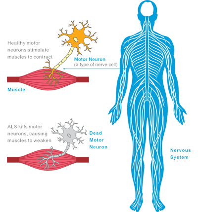
Dipeptidase IV: surface marker of human natural killer cells.

Helix Pomatia Receptors: the first simple and reliable marker detected on NK cells may be useful for purifying this cell type.

LAT (linker for activation of T cells): LAT is a 36-38 kDa complete membrane protein that plays an important role in T cell activation and ACTS as a novel immunohistochemical marker for T cells, NK cells, mast cells, and megakaryotes.

Ly24 (pgp-1): marker expressed on the surface of mouse natural killer cells.
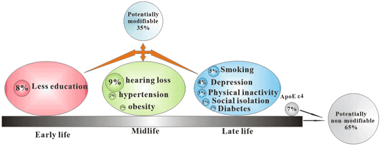
Ly49H: is a NK cell activating receptor unique to C57BL/6 (B6) mice.

NKG2A and NKp80: specific natural killer cell markers in rhesus and pigtail monkeys.

3. The Function of NK Cell Surface Receptor

NK cells are mainly derived from bone marrow lymphoid stem cells, accounting for about 10% of lymphocytes in the body, and mainly distributed in peripheral blood and peripheral lymphoid tissues [8].

Depending on the structure, NK cell surface receptors can be divided into immunoglobulin superfamily and C-type lectin superfamily. According to different functions, NK cell surface receptors can be divided into killer cell activation receptors and killer cell inhibition receptors. The inhibitory receptor expressed on the surface of NK cells maintains the tolerance of NK cells to the host’s own normal tissue cells. The activating receptors expressed on the surface of NK cells can bind to the corresponding ligands on the surface of the target cells, and stimulate the killing effect of NK cells.

The balance of signals mediated by activated receptors and inhibitory receptors affects the killing activity of NK cells on tumors [9].

The different functions of activated and inhibitory receptors

Figure 3 The different functions of activated and inhibitory receptors

4. Anti-Tumor Immune Mechanism

NK cells exert their anti-tumor functions mainly in two ways: immune clearance and immune surveillance.

Immune clearance mainly includes the following three mechanisms:

  • It kills tumor cells by releasing cytotoxic particles. NK cells can release perforin and granzyme, which can induce tumor cell lysis and apoptosis by changing the osmotic pressure of target cells and activating the apoptosis-related enzyme system.
  • The target cell apoptosis system is activated by cell surface synthesized proteins to kill tumor cells. Activated NK cells express FasL and TNF-α, which bind to the corresponding receptor Fas (CD95) and TNFR-1 on the surface of tumor cells to form Fas trimer and TNF-R trimer, and initiate apoptosis system of target cells and kill target cells.
  • Target cells are killed by antibody-dependent cell-mediated cytotoxicity (ADCC). This is the process of targeted NK cell killing target cells with IgG as the intermediate bridge. NK cells can recognize tumor cells that specifically bind to IgG antibodies through surface CD16 molecules, and CD16 interacts with corresponding adaptor proteins containing “immunoreceptor tyrosine activation motifs” in NK cells to phosphorylate adaptor proteins. Thereby initiating intracellular signaling, activating NK cells to release perforin and granzymes to kill target cells.

In addition to the direct killing effect of NK cells themselves on tumor cells, activated NK cells can secrete a variety of cytokines, such as TNF, IFN, etc., which synergistically inhibit or kill tumor cells.

Anti-tumor immune mechanism of NK cells

Figure 4 Anti-tumor immune mechanism of NK cells

5. NK Cells and Tumors

As an important part of the innate immune system, NK cells play an important role in tumor immunity. The expression of activated and inhibited receptors on the surface of NK cells determines their anti-tumor ability. NK cells have certain significance in inhibiting the growth and metastasis of lung cancer, breast cancer, colorectal cancer and other malignant tumors.

5.1 Lung Cancer

NK cells and perforin mediated cytotoxicity play a role in the prevention of lung cancer [10]. Decreased expression of granzyme and perforin is associated with lung cancer [11].

Immunotherapy of NK cells has found that activated NK cells can inhibit the growth of lung cancer cells. The occurrence of lung cancer is closely related to the decrease of NK cell number and activity. Therefore, improving NK cell activity and increasing the number of NK cells are important immunotherapeutic methods to prevent the occurrence and development of lung cancer.

5.2 Breast Tumor

The growth and metastasis of breast tumors are also closely related to the activity of NK cells. NK cell activity was significantly decreased in breast cancer patients, and the expression of surface activated receptors was restricted, while the level of inhibitory receptors was significantly increased [12].

5.3 Leukemia

The expression of inhibitory receptor NKG2A in NK cells of leukemia patients was significantly increased, while the activated receptor NKP46 was significantly decreased, which reduced the NK cell killing activity [13].

By regulating the expression of NK cell surface receptors, its activity can be altered and its immune function enhanced. Therefore, enhancing the immune activity of NK cells by regulating the expression of NK cell receptors is the main research direction of leukemia immunotherapy.

6. Application of NK Cells in Tumor Immunotherapy

According to the anti-tumor immune mechanism of NK cells, tumors can be treated by corresponding methods.

6.1 Antibody-Mediated Cytotoxicity of NK Cells

It is based on antibody-dependent cell-mediated cytotoxicity (ADCC). It can target killing tumor cells or enhance the ADCC effect of NK cells.

For example, CD137 monoclonal antibody can increase the expression of CD137 in NK cells, and as an activation signaling molecule, it significantly enhances the ability of NK cells to degranulate, secrete IFN-γ and anti-tumor activity [14].

CCR4 monoclonal antibody increases the ADCC effect of NK cells [15].

These results indicate that antibodies based on ADCC have a broad prospect of development and application.

6.2 KIR-HLA Mismatch between Donor and Recipient and Anti-Tumor Effect of NK Cells

NK cells can not only produce immune tolerance to normal cells in the body, but also kill tumor cells.

For normal cells, NK cells recognize HLA class I molecules through surface-inhibiting KIR receptors, transduce killing inhibitory signals, and prevent NK cells from killing normal cells.

For tumor cells, the surface lacks HLA-I signaling molecules and is easily recognized and attacked by NK cells. A KIR-HLA mismatch between donors and recipients can cause heterologous reactivity of NK cells, activating donor NK cells to kill recipient cells.

When applied in the treatment of myeloid leukemia and lymphocytic leukemia, this method can effectively enhance the graft anti-leukemia effect [16] and improve the survival rate of patients. In the treatment of kidney cancer, gastric cancer, intestinal cancer, ovarian cancer and other solid tumors, good therapeutic effect has also been achieved [17].

References

[1] Vivier E, Morin P M, O’Brienm C, et al. CD2 is functionally linked to the ζ‐natural killer receptor complex [J]. European journal of immunology, 1991, 21(4): 1077-1080.

[2] Lynn D J, Freeman A R, Murray C, et al. A Genomics Approach to the Detection of Positive Selection in Cattle: Adaptive Evolution of the T-Cell and Natural Killer Cell-Surface Protein CD2 [J]. Genetics, 2005, 170(3): 1189-1196.

[3] Boles K S, Stepp S E, Bennett M, et al. 2B4 (CD244) and CS1: novel members of the CD2 subset of the immunoglobulin superfamily molecules expressed on natural killer cells and other leukocytes [J]. Immunological reviews, 2001, 181(1): 234-249.

[4] Habu S, Hayakawa K, Okumura K, et al. Surface markers on natural killer cells of the mouse [J]. European journal of immunology, 1979, 9(12): 938-942.

[5] Huntington N D, Xu Y, Nutt S L, et al. A requirement for CD45 distinguishes Ly49D-mediated cytokine and chemokine production from killing in primary natural killer cells [J]. Journal of Experimental Medicine, 2005, 201(9): 1421-1433.

[6] Coulam C B, Roussev R G. Correlation of NK cell activation and inhibition markers with NK cytoxicity among women experiencing immunologic implantation failure after in vitro fertilization and embryo transfer [J]. Journal of assisted reproduction and genetics, 2003, 20(2): 58-62.

[7] Alter G, Malenfant J M, Altfeld M. CD107a as a functional marker for the identification of natural killer cell activity [J]. Journal of immunological methods, 2004, 294(1-2): 15-22.

[8] Cheng M, Chen Y, Xiao W, et al. NK cell-based immunotherapy for malignant diseases [J]. Cellular & molecular immunology, 2013, 10(3): 230.

[9] Peruzzi G, Masilamani M, Borrego F, et al. Endocytosis as a mechanism of regulating natural killer cell function: unique endocytic and trafficking pathway for CD94/NKG2A [J]. Immunologic research, 2009, 43(1-3): 210-222.

[10] Frese-Schaper M, Keil A, Yagita H, et al. Influence of natural killer cells and perforin-mediated cytolysis on the development of chemically induced lung cancer in A/J mice [J]. Cancer immunology, immunotherapy, 2014, 63(6): 571-580.

[11] Hodge G, Barnawi J, Jurisevic C, et al. Lung cancer is associated with decreased expression of perforin, granzyme B and interferon (IFN)‐γ by infiltrating lung tissue T cells, natural killer (NK) T‐like and NK cells [J]. Clinical & Experimental Immunology, 2014, 178(1): 79-85.

[12] Mamessier E, Sylvain A, Thibult M L, et al. Human breast cancer cells enhance self tolerance by promoting evasion from NK cell antitumor immunity [J]. The Journal of clinical investigation, 2011, 121(9): 3609-3622.

[13] Stringaris K, Sekine T, Khoder A, et al. Leukemia-induced phenotypic and functional defects in natural killer cells predict failure to achieve remission in acute myeloid leukemia [J]. Haematologica, 2014, 99(5): 836-847.

[14] Lin W, Voskens C J, Zhang X, et al. Fc-dependent expression of CD137 on human NK cells: insights into “agonistic” effects of anti-CD137 monoclonal antibodies [J]. Blood, 2008, 112(3): 699-707.

[15] Kanazawa T, Hiramatsu Y, Iwata S, et al. Anti-CCR4 monoclonal antibody mogamulizumab for the treatment of EBV-associated T-and NK-cell lymphoproliferative diseases [J]. Clinical Cancer Research, 2014, 20(19): 5075-5084.

[16] Cooley S, Weisdorf D J, Guethlein L A, et al. Donor selection for natural killer cell receptor genes leads to superior survival after unrelated transplantation for acute myelogenous leukemia [J]. Blood, 2010, 116(14): 2411-2419.

[17] Re F, Staudacher C, Zamai L, et al. Killer cell Ig‐like receptors ligand‐mismatched, alloreactive natural killer cells lyse primary solid tumors [J]. Cancer, 2006, 107(3): 640-648.

Hematopoietic Stem Cell Surface Marker

1. Type of Hematopoietic Stem Cell

Two types of hematopoietic stem cells have been defined:

Long-term regenerative cells (LTRC) capable of maintaining self-renewal and multilineage differentiation potential throughout life.

Short-term regenerative cells (STRC). They remain pluripotent, but they show more limited potential for self-renewal.

The interval between these two types of cells to reconstruct the myeloid and / or lymphatic system is very short, about 6 weeks.

2. Sources of Hematopoietic Stem Cells

Hematopoietic stem cells are present in adult bone marrow, especially in the pelvis, femur and sternum. They are also found in umbilical cord blood and a small amount of peripheral blood.

2.1 Bone Marrow

Hematopoietic stem cell (HSCs) is a kind of pluripotent stem cell derived from bone marrow.

Hematopoietic stem cells obtained from bone marrow were collected surgically from two posterior iliac crest. About one out of every 100000 cells in bone marrow is a long-term hematopoietic stem cell (LT-HSC).

2.2 Peripheral Blood

Most hematopoietic stem cells come from the bone marrow, and a small number of stem cells and progenitor cells circulate in the blood. In clinical transplantation of human hematopoietic stem cells, donor cells can be collected from peripheral blood. Under the action of hematopoietic growth factors such as granulocyte colony stimulating factor (G-CSF), HSC in bone marrow is mobilized to peripheral blood, and then hematopoietic stem cells are collected by isolation.

2.3 Umbilical Cord Blood (UCB)

Umbilical cord blood is a rich source of hematopoietic stem cells and hematopoietic progenitor cells, and the number of different types of hematopoietic progenitor cells is about 10 times that observed in adult blood. As a special cell source of regenerative medicine, umbilical cord blood also contains many types of stem cells.

2.4 Fetal Hematopoietic System and Embryonic Hematopoietic Stem cells

Fetal hematopoietic system is an important source of hematopoietic stem cells, but it has not been applied in clinic.

Sources of hematopoietic stem cells

Figure 1 Sources of hematopoietic stem cells

3. Biological Characteristics of Hematopoietic Stem Cells

Hematopoietic stem cells are real stem cells because they are pluripotent and self-renewing. Other features include its heterogeneity.

Self-renewal: self-renewal of hematopoietic stem cells means that hematopoietic stem cells produce identical progeny cells. This ability enables hematopoietic stem cells to produce a complete hematopoietic system from a single cell and maintain hematopoiesis throughout an individual’s life.

Pluripotency: It refers to the ability of hematopoietic stem cells to produce major hematopoietic cell types when needed. HSCs can differentiate into various blood cells from myeloid lineages (monocytes and macrophages, neutrophils, basophils, eosinophils, erythrocytes, megakaryocytes / platelets, dendritic cells) and lymphatic lineages (T, B and NK cells).

Hematopoietic stem cells have a very high proliferation potential, so that they can meet the high demand for hematopoiesis in the lifetime of normal adults.

The pluripotency of hematopoietic stem cells

Figure 2 The pluripotency of hematopoietic stem cells

Plasticity: It refers to the ability of hematopoietic stem cells to differentiate into multiple non-hematopoietic tissues such as cardiomyocytes [2].

Hematopoietic stem cell transport: From embryonic origin, HSPC moves from one niche to another. Hematopoietic stem cell niche is the spatial location and physiological microenvironment on which hematopoietic stem cells rely for survival and self-renewal. The transport of hematopoietic stem cells can be divided into homing (describing the trend of cells arriving in a particular environment), retention (the ability of cells to remain in that environment after arrival) and implantation (the ability of hematopoietic stem cells to divide in the environment and form functional offspring).

Heterogeneity: Heterogeneity within hematopoietic stem cells means that hematopoietic stem cells have different physiological characteristics, such as cell cycle state [3] and self-renewal ability [4], have different responses to different external signals, and can form different lineages [5] [6] [7] [8] after transplantation.

The Muller-Sieberg group was one of the first groups to determine HSC heterogeneity. The heterogeneity of HSC can spread stably. How is the heterogeneity within hematopoietic stem cells produced?

The following are possible reasons for HSC heterogeneity:

  • Different sources of hematopoietic stem cells: HSC may come from mesoderm cells, endothelial cells or from anterior hematopoietic stem cells of dorsal aorta.
  • Differences in regulatory factors: the type of HSC may be regulated by different induced developmental tissues (such as AGM, placenta, yolk sac, head) and may change with their migration in circulation, or by developmental niche (possibly blood vessels, liver, nerves or bones), or by different developmental mechanisms (such as EHT).
  • In addition, epigenetic modifications may also explain the existence of different HSC types.

4. HSC and Immune System

HSC niche is the spatial location and physiological microenvironment on which hematopoietic stem cells rely for survival and self-renewal. It maintains the dynamic balance between self-renewal and differentiation of stem cells.

Studies have shown that in vivo, the niche of hematopoietic stem cells provides immune privileged sites for hematopoietic stem cells. Regulatory T cells (tregs) and hematopoietic stem cells are located in the endosteal region of the bone marrow to protect the hematopoietic stem cells from immune attack.

In addition, hematopoietic stem cells themselves have a certain immune immunity potential by regulating the expression of surface immune molecules. When HSCs was activated by strong inflammatory signals and mobilized into circulation, the level of CD47 on the surface of HSCs was significantly up-regulated. CD47 binds to signal regulatory proteins on macrophages to inhibit phagocytosis. The increased expression of CD47 on the surface of mobilized hematopoietic stem cells can protect these cells from phagocytosis.

5. Hierarchy of Human Hematopoietic System

The origin of all blood cells in the hematopoietic system is thought to come from self-renewing hematopoietic stem cells.

Hematopoietic stem cell and progenitor cell (HSPCs) bank can be divided into three types of cells: long-term hematopoietic stem cell (LT-HSC), short-term hematopoietic stem cell (ST-HSPC) and multipotent progenitor cells (MPP).

In the hematopoietic system, long-term hematopoietic stem cells (LT-HSC) are located at the top of all mature blood cells, maintaining a primitive multipotential pool throughout life through their self-renewal and asymmetric cell division potential. In a stable state, progeny derived from hematopoietic stem cells have the ability to rapidly proliferate in peripheral stimulation, replacing the loss of activated, consumed or aged blood cells [9] [10].

St-HSPC and its progenitor cells have the ability to maintain normal hematopoiesis for 6-8 weeks. Loss of normal LT-HSC and ST-HSPC functions is a marker of natural stem cell aging and several hematopoietic diseases, especially related to the development and progression of hematological malignancies [11].

Pluripotent progenitor cell (MPPs) is a pluripotent progenitor cell that has lost its self-renewal potential but can still differentiate into all multiple lines produced by hematopoietic stem cells. MPPs further produce oligopotent progenitors cells, the common lymphoid progenitor (CLPs) and common myeloid progenitor (CMPs).

All these low-potential progenitor cells can differentiate into committed lineages:

CMPs can differentiate into megakaryocyte / erythrocyte progenitor cell (MEPs), granulocyte / macrophage progenitor cell (GMPs) and dendritic cell (DC) progenitor cell.

CLPs can differentiate into T cell progenitor cells, B cell progenitor cells, NK progenitor cells and DC progenitor cells [12] [13].

DC progenitor cells (CD8+DC, CD8-DC and plasma cell-like DC) can be derived from CMPs and CLPs.

Hierarchy of human hematopoietic system

Figure 3 Hierarchy of human hematopoietic system

6. Cell surface Markers of Hematopoietic Stem Cells

Although there are many studies on hematopoietic stem cells, no single molecular marker has been found to be expressed only by hematopoietic stem cells.

Hematopoietic stem cells are called Lin- because of the lack of expression of mature blood cell markers. The identification or isolation of hematopoietic stem cells is generally based on the combination of several different cell surface markers to separate rare hematopoietic stem cells from the surrounding blood cells.

There are many differences between human hematopoietic stem cell and mouse hematopoietic stem cell markers.

Mouse HSC: EMCN+, CD34lo/-, SCA-1+, Thy1.1+/lo, CD38+, C-kit+, lin

Human HSC: EMCN+, CD34+, CD59+, Thy1/CD90+, CD38lo/-, C-kit/CD117+, lin

The markers that distinguish mouse hematopoietic stem cells (LT-HSC) from short-term (ST-HSC) and multipotent progenitor cells (early MPP and late MPP) are as follows.

LT-HSC: CD34-, CD38-, SCA-1+, Thy1.1+/lo, C-kit+, lin-, CD135-, Slamf1/CD150+

ST-HSC: CD34+, CD38+, SCA-1+, Thy1.1+/lo, C-kit+, lin-, CD135-, Slamf1/CD150+, Mac-1 (CD11b)lo

Early MPP: CD34+, SCA-1+, Thy1.1-, C-kit+, lin-, CD135+, Slamf1/CD150-, Mac-1 (CD11b)lo, CD4lo

Late MPP: CD34+, SCA-1+, Thy1.1-, C-kit+, lin-, CD135high, Slamf1/CD150-, Mac-1 (CD11b)lo, CD4lo

6.1 Introduction of Major Markers

6.1.1 CD34

CD34 is one of the most important markers, and is the first widely studied molecule in the isolation and identification of hematopoietic stem cells and their progenitor cells. The expression of CD34 in human umbilical cord blood, bone marrow and peripheral blood was about 0.1-4.9% [14]. CD34 is expressed on 0.55% of human bone marrow cells, and it is also expressed on early progenitor cells, but not on mature bone marrow cells. Other surface markers have been used in conjunction with CD34 to distinguish primordial cell populations.

6.1.2 CD38

CD38, also known as cyclic ADP ribosomal hydrolase, is a glycoprotein present on the surface of many immune cells (leukocytes), including CD4+, CD8+, B lymphocytes and natural killer cells. CD38 markers are used to distinguish hematopoietic stem cell pluripotent progenitor cells (CD38 -) from committed progenitor cells (CD38+).

6.1.3 CD90 (Thy1)

CD90, also known as thy-1, is a 28-30 kDa GPI-linked membrane glycoprotein. CD90 is expressed in hematopoietic stem cells, neurons, thymus cells, peripheral T cells, fibroblasts and stromal cells.

The co-expression of CD90 on CD34 + CD38- cells defines hematopoietic stem cells, while CD34 + CD38-CD90 – defines multipotent progenitor cells.

6.1.4 CD117 (C-Kit)

CD117 is a 145kDa protein tyrosine kinase, also known as c-Kit. CD117 is expressed on pluripotent hematopoietic progenitor cells (about 1.4% of bone marrow cells), mast cells and acute myeloid leukemia cell (AML).

By binding to its ligands, it can induce the phosphorylation of CD117 and stimulate the proliferation and survival of primitive hematopoietic stem cells, red blood cells and monocytes. It plays an important role in gamete formation, melanin formation and hematopoiesis.

6.1.5 CD135 (Flk-2)

CD135, also known as FLK-2, FLT3 and Ly-72, is a type III tyrosine kinase receptor. CD135 is expressed not only in normal CD34+ hematopoietic stem cells, but also in malignant hematopoietic cells, including AML, ALL and CML BC.

The combination of CD135 and FLT3 ligand can regulate the growth of hematopoietic stem cells and promote the survival of primitive hematopoietic progenitor cells with myeloid and B lymphatic potential.

6.1.6 CD150 (SLAM)

CD150, also known as SLAM, type I transmembrane glycoprotein signal transduction lymphocyte activation molecule, is a typical member of SLAM subgroup of CD2 protein family.

SLAM is expressed in thymocytes, T cell subsets, B cells, dendritic cells, macrophages and hematopoietic stem cells.

6.1.7 CD184 (CXCR4)

CD184, is also called Fusin or CXCR4. It is widely expressed in blood and tissue cells, including B cells and T cells, monocytes, macrophages, dendritic cells, granulocytes, megakaryocytes / platelets, lymphoid cells, myeloid precursor cells, endothelial cells, epithelial cells, astrocytes and neurons.

In the bone marrow niche, hematopoietic stem cells express CXCR4. CXCR4 is the receptor of chemokine CXCL12 (SDF-1), which mediates blood cell migration and participates in B lymphocyte and bone marrow production, cardiogenesis, angiogenesis and cerebellar development.

The interaction between CXCR4 and SDF-1 is responsible for the homing and retention of HSC in the niche. The interaction of CXCR4/CXCL12 (SDF-1) in HSC niche is considered to be the mechanism of HSC mobilization [15].

6.1.8 Ly-6A/E (Sca-1)

Ly-6a /E, also known as sca-1, is a member of the ly-6 polygene family, a protein linked to hematopoietic stem cells by glycosylphosphatidylinositol (GPI).

Its expression on pluripotent hematopoietic stem cell (HSC) has been used as a marker of HSC in two kinds of Ly 6 haploid mice. Sca-1 positive hematopoietic stem cells were found in bone marrow, fetal liver, peripheral blood and spleen of adult animals. Ly-6A/E is thought to be involved in the regulation of T and B cell responses.

7. Clinical Application of HSC

7.1 Hematopoietic Cell Transplantation (HCT)

According to the source of stem cells, hematopoietic cell transplantation can be divided into bone marrow transplantation, peripheral blood stem cell transplantation and umbilical cord blood transplantation.

Hematopoietic cell transplantation (HCT)

Figure 4 Hematopoietic cell transplantation (HCT)

7.1.1 Bone Marrow Transplantation

Bone marrow transplantation is a common type of hematopoietic stem cell transplantation. Hematopoietic stem cells play an important role in the re-proliferation of patients’ blood cells in bone marrow transplantation.

According to the relationship between donors and recipients, it can be divided into autologous hematopoietic stem cell transplantation and allogeneic hematopoietic stem cell transplantation.

In addition to solid tumors, bone marrow transplantation is also used to treat hematological diseases such as leukemia and immune system disorders [16].

7.1.2 Peripheral Hematopoietic Stem Cell Transplantation

The concentration of circulating HSC in peripheral blood increased by 100 times after administration of hematopoietic cytokines such as GM-CSF, IL-3 or SCF. Using a cell separator, you can usually obtain the required number of HSCs from peripheral blood.

7.1.3 Umbilical Cord Blood Transplantation

Umbilical cord blood is easily available and does not pose a risk to donors, so it is an attractive source of transplantable hematopoietic stem cells.

Due to the immature immune system, lymphocytes in umbilical cord blood of newborns have been shown to be more tolerant to HLA, so there are fewer acute and chronic graft-versus-host disease (GVHD) in allogeneic transplantation environment.

7.2 Other

Hematopoietic stem cells are also widely used in graft anti-tumor therapy, tolerance induction, gene therapy [17], regenerative medicine and so on.

References

[1] Chotinantakul K, Leeanansaksiri W. Hematopoietic stem cell development, niches, and signaling pathways [J]. Bone marrow research, 2012, 2012.

[2] Chatterjee T, Sarkar R S, Dhot P S, et al. Adult stem cell plasticity: Dream or reality? [J]. Medical journal, Armed Forces India, 2010, 66(1): 56.

[3] Wilson A, Laurenti E, Oser G, et al. Hematopoietic stem cells reversibly switch from dormancy to self-renewal during homeostasis and repair [J]. Cell, 2008, 135(6): 1118-1129.

[4] Ema H, Morita Y, Suda T. Heterogeneity and hierarchy of hematopoietic stem cells [J]. Experimental hematology, 2014, 42(2): 74-82. e2.

[5] Benz C, Copley M R, Kent D G, et al. Hematopoietic stem cell subtypes expand differentially during development and display distinct lymphopoietic programs [J]. Cell stem cell, 2012, 10(3): 273-283.

[6] Dykstra B, Kent D, Bowie M, et al. Long-term propagation of distinct hematopoietic differentiation programs in vivo [J]. Cell stem cell, 2007, 1(2): 218-229.

[7] Sieburg H B, Cho R H, Dykstra B, et al. The hematopoietic stem compartment consists of a limited number of discrete stem cell subsets [J]. Blood, 2006, 107(6): 2311-2316.

[8] Verovskaya E, Broekhuis M J C, Zwart E, et al. Heterogeneity of young and aged murine hematopoietic stem cells revealed by quantitative clonal analysis using cellular barcoding [J]. Blood, 2013, 122(4): 523-532.

[9] Busch K, Klapproth K, Barile M, et al. Fundamental properties of unperturbed haematopoiesis from stem cells in vivo [J]. Nature, 2015, 518(7540): 542.

[10] Sawai C M, Babovic S, Upadhaya S, et al. Hematopoietic stem cells are the major source of multilineage hematopoiesis in adult animals [J]. Immunity, 2016, 45(3): 597-609.

[1] Sun J, Ramos A, Chapman B, et al. Clonal dynamics of native haematopoiesis [J]. Nature, 2014, 514(7522): 322.

[11] Yao J C, Link D C. Concise review: the malignant hematopoietic stem cell niche [J]. Stem Cells, 2017, 35(1): 3-8.

[12] Adams G B, Scadden D T. The hematopoietic stem cell in its place [J]. Nature immunology, 2006, 7(4): 333.

[13] Mosaad Youssef Mohamed,Hematopoietic stem cells: an overview. [J] .Transfus. Apher. Sci., 2014, 51: 68-82.

[14] Pranke P, Hendrikx J, Debnath G, et al. Immunophenotype of hematopoietic stem cells from placental/umbilical cord blood after culture [J]. Brazilian journal of medical and biological research, 2005, 38(12): 1775-1789.

[15] Herbert K E, Levesque J P, Mills A K, et al. How we mobilize haemopoietic stem cells [J]. Internal medicine journal, 2011, 41(8): 588-594.

[16] Rogers I, Casper R F. Umbilical cord blood stem cells [J]. Best Practice & Research Clinical Obstetrics & Gynaecology, 2004, 18(6): 893-908.

[17] Hirschi K K. Hemogenic endothelium during development and beyond [J]. Blood, 2012, 119(21): 4823-4827.

Identify Treg Cells. You Can Do This.

1. Classification of Treg Cells

According to its origin, regulatory T cells can be further divided into natural regulatory T cells (nTreg) and adaptive or inducible regulatory T cells (aTreg or iTreg).

nTreg is mainly CD4+Treg cells, which are differentiated in the thymus by progenitor cells from bone marrow, accounting for about 1% – 3% of the total number of CD4+T lymphocytes, accounting for 5% of the total number of CD4+T lymphocytes in peripheral blood [2]. It plays an immunomodulatory role in peripheral blood, lymphatic organs, inflammatory sites and tumor tissues [3].

aTreg or iTreg include: Th3 (phenotypic characteristic is CD4+CD25low), Tr1 (phenotypic characteristic is CD4+CD25lowCD45RBlow), CD8+ regulatory T cell, natural killer T cell (NKT) and other subtypes. They are closely related to the occurrence of autoimmune diseases and tumors.

iTreg also known as sTreg, is a class of regulatory T cells derived from peripheral mature T cells stimulated by specific antigens and induced by immunosuppressive cytokines (mainly including TGF-β, IL-2, IL-10, IFN-γ, IFN-α, indoleamine 2-3 dioxygenase, and retinoic acid), which accounts for about 4% – 7% of the total number of CD4+T cells.

The development of Treg cells

Figure 1 The development of Treg cells

2. Treg Cell Differentiation

The differentiation, development and function of Treg cells are regulated by a variety of cytokines. Transcription factor forkbox P3 (Foxp3) is involved in the differentiation. Transcriptional activator STAT5 is another important factor involved in the differentiation and survival of Treg cells.

The development of nTreg in the thymus is dependent on the synergistic stimulation of TCR and CD28, which is essential for the steady proliferation and survival of peripheral nTregs.

The development of iTreg requires IL-2 and transforming growth factor (TGF- β), rather than co-stimulation with CD28 [4].

Studies have shown that [5] IL-2 promotes the production of inducible regulatory T cells through STAT5, and IL-2 together with TGF-β induces the naive CD4+ CD25-T cells to transform into CD4+ CD25+ T cells and express Foxp3.

3. Treg Cell Marker

At present, it is believed that CD4+CD25+ Foxp3+ is the main phenotype of Treg cells.

Treg also lowly expressed another specific marker, CD127.

Some receptors are also expressed on the surface of Treg, such as CD5, CD38, CD45, CD62L, CD103, CTLA-4 and inhibitory immune receptor GITR.

The markers expressed by Treg cells can be divided into two categories according to their location:

Cell markers of Treg

Figure 2 Cell markers of Treg

3.1 Intracellular Markers

FOXP3: Fontenot JD et al[6] found that Treg cells highly express forkhead box P3 (Foxp3), which can promote the transformation of immature CD4+T cells into Treg [7].

FOXP3 is a member of the forkhead-like transcription factor family, which is related to the regulation of cell growth and development. FOXP3 is closely related to Treg cells. If FOXP3 gene mutation occurs, it will affect the development and maturation of Treg cells and cause some diseases.

FOXP3 is mainly expressed in lymphoid organs and tissues such as thymus, spleen and lymph nodes. At present, Foxp3 is recognized as the most sensitive marker of Treg cells.

Helios: The gene encodes a member of the Ikaros family of zinc finger proteins and is a hematopoietic specific transcription factor involved in the regulation of lymphocyte development. This protein forms homotype or heteromeric dimers with other members of the Ikaros family and is thought to play a major role in early hematopoietic development.

3.2 Treg Cell Surface Markers

CD4: CD4, also known as T4/leu-3, is a member of the immunoglobulin superfamily. It is a single chain I transmembrane glycoprotein with a molecular weight of 55 kDa.

CD4 β is a part of TCR/CD3 complex and participates in TCR signal transduction.

It is expressed in most thymocytes, helper T cells, type II NKT cells and monocytes / macrophages.

CD25: CD25 is also called IL-2R α. Ly-43, P55 or Tac, is a kind of glycoprotein with molecular weight of 55kDa. It is expressed on activated T and B cells, thymocyte subsets, pre-B cells and Treg cells.

CD39: CD39 (nucleoside triphosphate diphosphohydrolase-1, NTPDase 1) is an extracellular enzyme that can degrade ATP to AMP. It is expressed in B cells, dendritic cells and T cell subsets including regulatory T cells and memory T cells. CD39 is a major member of the immune system, involved in the inhibition of inflammation and the control of platelet activation.

CD62L: CD62L, called L-selectin or LECAM-1, is a single chain type I glycoprotein with a molecular weight of 74 – 95 kDa. It is expressed on most peripheral blood B cells, T cells, NK cell subsets, monocytes, granulocytes and some malignant cells of hematopoietic system. CD62L is very important for immature lymphocytes to homing to high endothelial venules in peripheral lymph nodes and Peyer’s plaques.

CD73: CD73 is a cell surface protein anchored to cells by GPI, with a molecular weight of 69 kDa. In mice, the expression of CD73 in the bone marrow was limited to CD11b+ myeloid cells. In the spleen, it is expressed primarily on T cells.

CD103: CD103, also known as α E integrin or integrin α IEL chain, belongs to the integrin family and is a type I transmembrane glycoprotein. Treg cells highly express CD103. CD103 binds to E-cadherin and mediates lymphocyte homing to intestinal epithelial cells.

CD134: CD134 is a member of the TNF receptor family, also known as OX40 and TNFRSF4, is a 50 kDa type I transmembrane glycoprotein. OX40 was expressed on activated T lymphocytes. The interaction between OX40 and OX40L leads to B cell proliferation and antibody secretion, and regulates primary T cell proliferation and T cell survival. OX40 affects the regulation of tolerance of CD4+T cells.

CD152 (CTLA-4): CD152 is a member of the immunoglobulin superfamily, also known as CTLA-4 or Ly-56, with a molecular weight of 33 kDa. It is expressed on activated T and B lymphocytes.

CTLA-4 negatively regulates cell-mediated immune response, which plays a role in inducing and maintaining immune tolerance, developing protective immunity and regulating thymocytes.

CD194 (CCR4): CCR4 ligands include: CCL17 (TARG) and CCL22 (MDC). CCR4 is expressed in memory T cells, macrophages, platelets, basophils, Th2 cells and Treg cells.

CCR4 and its ligands (CCL17 and CCL22) play an important role in the recruitment of memory T cells in various skin immune diseases.

FR4: Folate receptor 4 (FR4) is the surface receptor of folic acid (vitamin B9). It has high constitutive expression on mouse CD4+ CD25+ natural regulatory T cell (Treg). It binds to CD4 and CD25 and distinguishes Treg from other types of T cells.

GARP: GARP, also known as leucine-rich repeat sequence 32 (LRC32), is a type I membrane glycoprotein with molecular weight of 80kDa. GARP exists on the surface of megakaryocytes, platelets and activated Treg (CD4+, CD25+, FoxP3+ cells), and is a receptor for transforming growth factor-β (TGF-β). GARP may play a role in controlling the inhibitory function of Tregs.

GITR: GITR (glucocorticoid-induced TNFR-related genes), also known as TNFRSF18 and AITR, is a members of the TNF receptor superfamily. It is highly expressed on CD25+CD4+Tregs. The interaction between GITR and its ligands can enhance T cell activation, proliferation and cytokine production, and eliminate the inhibitory function of CD25+CD4+Tregs. The activation of GITR in vivo leads to the development of autoimmune diseases and the restoration of suppressed immune responses.

TGF-β: TGF-β is a potent stimulator of osteoblast formation and plays an important role in bone remodeling.

It can regulate the lineage differentiation of Th17 cells or Treg cells. High concentration is beneficial to the development of Treg cells. The synergistic action of low concentration of TGF- β with IL-6 and IL-21 is beneficial to the differentiation of Th17 cells. It also controls cell proliferation, differentiation and other functions in many cell types.

CD127: CD127, also known as IL-7Rα, is a type I transmembrane glycoprotein with a molecular weight of 60 kDa. The expression of CD127 is down-regulated in Treg cells, and the lack of CD127 is one of the characteristics of Tregs cells. It can be used as a marker of Treg and routine T cell differentiation.

4. What do Treg Cells Do?

The general physiological functions of Treg mainly include the following aspects:

Regulatory T Cells and Immune Tolerance: By inhibiting self-reactive T cells, Treg enables the body to develop active tolerance to its own antigens, preventing the occurrence of autoimmune diseases. In tumors, Treg makes the body produce antigenic tolerance to tumors through immunosuppression, which makes tumor cells escape the immune killing of the body.

Promote Chronic Inflammatory Response: When pathogens invade, effector T cells clear pathogens through a series of immune responses, while Treg plays an opposite role with other immune cells in the body [8]. It exerts inhibitory functions by the secretion of cytokines such as IL-4, IL-10 and TGF-β. It can prevent the occurrence of pathological immune response that causes tissue destruction, but at the same time, it also makes it difficult to remove pathogens and prolong the course of chronic infection.

Immunosuppression: the main function of Treg is to negatively regulate the immune response of the body, so Treg plays a vital role in regulating immune homeostasis and preventing the occurrence of autoimmune diseases. Through immunosuppression, Treg promotes tumor immune escape [9], so it is also regarded as a kind of immune cell that helps tumor survive and promote its growth.

Treg cells regulate immune function and participate in the aging process of the human body. The immune function of mice decreased during aging.

Immunosuppression of Treg cells

Figure 3 Immunosuppression of Treg cells

5. How do Treg Cells Work?

Treg exerts its immunomodulatory function in two ways:

5.1 Direct Cell-to-Cell Contact

Some chemokines cause Treg to gather around immune cells and play a role through a cell-to-cell contact-dependent mechanism. Treg can directly bind to the corresponding receptors on the target cells through CTLA-4, TGF-β and GITR, and inhibit the proliferation of immune cells such as CD4+T, CD8+T, dendritic cells and antigen presenting cells [10].

Treg inhibits the immune response by regulating the number and activity of dendritic cells to invalidate their antigen presentation.

5.2 Secretion of Inhibitory Cytokines

Treg negatively regulates immunity by secreting inhibitory cytokines, such as IL-4, IL-10, IL-35 and TGF- β [11] [12].

Regulatory mechanism of Treg cellsRegulatory mechanism of Treg cells

Figure 4 Regulatory mechanism of Treg cells

6. Regulatory T Cells in Cancer

Tumor microenvironment plays a very important role in the occurrence and development of tumor. Immune cells can affect tumor progression by affecting tumor microenvironment.

Generally speaking, most immune cells can play the role of anti-tumor immunity. Such as helper T cells and cytotoxic T cells. However, Treg plays an opposite role in tumor microenvironment. The immunosuppressive effect of Treg can not only prevent the occurrence of autoimmune diseases, but also promote the immune escape of tumor cells, indirectly accelerate the proliferation of tumor cells and enhance the infiltration ability of tumor cells.

Treg cells can inhibit the development and activation of effector cells and play an important role in mediating tumor immune tolerance. Studies have shown that the number of Treg cells is negatively correlated with the prognosis of tumors [13].

Tumor immune tolerance induced by Treg cells is realized by controlling primary T cells and memory T cells.

Treg cells can also influence CD4+ (Th1, Th2, Th17, NK) and other immune cells through TGF-β induce host immune tolerance.

The immune tolerance induced by Treg cells is also related to the role of dendritic cells (DC) [14].

7. Treg Cells and Immunotherapy

Treg cells play an immunosuppressive role in the immunity of the body, which is favored in the treatment of bone marrow transplantation.

In patients with multiple myeloma, low lymphatic status can improve the success rate of bone marrow transplantation.

Treg cells mediate the tolerance of antigens in the body. The decrease of its number will decrease the tolerance of the body to some of its own antigens and increase the rejection of grafts. Therefore, stimulating the recovery and regeneration of Treg cells in the human body can significantly reduce the rejection reaction of patients to the graft and greatly improve the success of bone marrow transplantation.

The immunosuppressive effect of Treg cells is related to immune escape and tolerance of tumor antigens. Therefore, in tumors, how to reduce the function of Treg is the key.

IL-2 is essential for the development of Treg cells [15]. IL-21 is a kind of cytokine which is similar to IL-2, but has no immunomodulatory function. Replacing IL-2 with IL-21 can prevent Treg cells from developing into sTreg cells. In addition, the combination of anti-CD25+ monoclonal antibody and anti-CD4+ monoclonal antibody can block Treg cells in vivo and eliminate the effect of Treg cells to the maximum extent. Experiments in mice have shown that the more thoroughly the Treg cells are removed, the better the effect of tumor immunotherapy is, and the longer the survival time of mice is [16].

Another idea of tumor immunotherapy is to reverse the immune tolerance induced by Treg cells to tumor cells.

Kiniwa et al[17] found that TLR8 ligand (human Toll-like receptor 8 ligand) can reverse the immune anergy of Treg cells to tumor, eliminate the immune escape of tumor, and improve the efficiency of effector cells.

Other costimulatory factors B7.1 and B7.2 can also reverse the immune tolerance of Treg cells to tumor, eliminate the inhibition of effector cells, and improve the killing effect. Studies have found that when there is a lack of costimulatory factors B7.1 and B7.2, tumor cells will escape the immune surveillance of the body, making T cells in a state of incompetence or inducing their apoptosis, resulting in unlimited tumor growth.

References

[1] Sakaguchi S, Sakaguchi N, Asano M, et al. Immunologic self-tolerance maintained by activated T cells expressing IL-2 receptor alpha-chains (CD25). Breakdown of a single mechanism of self-tolerance causes various autoimmune diseases [J]. The Journal of Immunology, 1995, 155(3): 1151-1164.

[2] Gavin M A, Rasmussen J P, Fontenot J D, et al. Foxp3-dependent programme of regulatory T-cell differentiation [J]. Nature, 2007, 445(7129): 771.

[3] Feuerer M, Hill J A, Mathis D, et al. Foxp3+ regulatory T cells: differentiation, specification, subphenotypes [J]. Nature immunology, 2009, 10(7): 689.

[4] Cassis L, Aiello S, Noris M. Natural versus adaptive regulatory T cells [M]. Kidney Transplantation: Strategies to Prevent Organ Rejection. Karger Publishers, 2005, 146: 121-131.

[5] Zheng S G, Wang J, Wang P, et al. IL-2 is essential for TGF-β to convert naive CD4+ CD25− cells to CD25+ Foxp3+ regulatory T cells and for expansion of these cells [J]. The Journal of Immunology, 2007, 178(4): 2018-2027.

[6] Trzonkowski P, Szmit E, Myśliwska J, et al. CD4+ CD25+ T regulatory cells inhibit cytotoxic activity of T CD8+ and NK lymphocytes in the direct cell-to-cell interaction [J]. Clinical immunology, 2004, 112(3): 258-267.

[7] Li Z, Li D, Tsun A, et al. FOXP3+ regulatory T cells and their functional regulation [J]. Cellular & molecular immunology, 2015, 12(5): 558.

[8] Klabusay M. The role of regulatory T-cells in antitumor immune response [J]. Klinicka onkologie: casopis Ceske a Slovenske onkologicke spolecnosti, 2015, 28: 4S23-7.

[9] Halvorsen E C, Mahmoud S M, Bennewith K L. Emerging roles of regulatory T cells in tumour progression and metastasis [J]. Cancer and Metastasis Reviews, 2014, 33(4): 1025-1041.

[10] Schlößer H A, Theurich S, Shimabukuro-Vornhagen A, et al. Overcoming tumor-mediated immunosuppression [J]. Immunotherapy, 2014, 6(9): 973-988.

[11] Collison L W, Workman C J, Kuo T T, et al. The inhibitory cytokine IL-35 contributes to regulatory T-cell function [J]. Nature, 2007, 450(7169): 566.

[12] Sakaguchi S, Wing K, Onishi Y, et al. Regulatory T cells: how do they suppress immune responses? [J]. International immunology, 2009, 21(10): 1105-1111.

[13] Beyer M, Schultze J L. Regulatory T cells in cancer [J]. Blood, 2006, 108(3): 804-811.

[14] Banerjee D K, Dhodapkar M V, Matayeva E, et al. Expansion of FOXP3high regulatory T cells by human dendritic cells (DCs) in vitro and after injection of cytokine-matured DCs in myeloma patients [J]. Blood, 2006, 108(8): 2655-2661.

[15] Frumento G, Piazza T, Di Carlo E, et al. Targeting tumor-related immunosuppression for cancer immunotherapy [J]. Endocrine, Metabolic & Immune Disorders-Drug Targets (Formerly Current Drug Targets-Immune, Endocrine & Metabolic Disorders), 2006, 6(3): 223-237.

[16] El Andaloussi A, Han Y U, Lesniak M S. Prolongation of survival following depletion of CD4+ CD25+ regulatory T cells in mice with experimental brain tumors [J]. Journal of neurosurgery, 2006, 105(3): 430-437.

[17] Kiniwa Y, Miyahara Y, Wang H Y, et al. CD8+ Foxp3+ regulatory T cells mediate immunosuppression in prostate cancer [J]. Clinical Cancer Research, 2007, 13(23): 6947-6958.

Surface Markers of Mesenchymal Stem Cells

1. Origin and Distribution of Mesenchymal Stem Cells

MSCs were discovered by Friedenstein et al[1] in 1987 from bone marrow stromal cells obtained by natural adhesion method. The concept of MSCs was first proposed by Caplan in 1988.

The heterogeneity of this group of MSCs cells is relatively large, so the developmental origin of MSCs cannot be determined. For example, dental pulp and skin belong to ectoderm origin, while bone marrow, adipose tissue and perinatal tissue belong to mesoderm origin.

MSCs exist widely in human tissues and can be derived from bone marrow, pancreas, skin, lung and other organs and tissues. MSCs are also found in many other tissues.

The origin of mesenchymal stem cells

Figure 1 The origin of mesenchymal stem cells

2. Definition of Mesenchymal Stem Cells

After the concept of MSCs was proposed in 1991, a surge in the number of organizational types of MSCs has been reported over the next decade.

This requires a clear standard for the definition of MSCs.

In 2006, the International Society for Cellular Therapy (ISCT) developed the basic standard for the definition of MSCs, which is also the lowest identification standard for MSCs:

  • Under standard in vitro culture, it keeps attached state.
  • More than 95% of the cells expressing CD105, CD73 and CD90, and cells expressing CD45, CD34, CD14, CD11b, CD79a, CD19 or HLA- class II molecules should not exceed 2 per cent of the total [2].
  • It has the ability to differentiate into osteoblasts, chondrocytes and adipocytes under in vitro induction conditions.

The flaw in the standard is that there is no requirement on whether it can really differentiate in vivo, nor does it test whether its stemness can be maintained.

The molecular characteristics of MSCs from different sources vary greatly [3]. Caplan suggests to change it to medicinal signaling cells (MSCs) [4].

3. Biological Characteristics of Mesenchymal Stem Cells

MSCs are a typical group of cells capable of self-renewal and multidirectional differentiation.

In vitro and in vivo, it can be induced to differentiate into adipose tissue cells, cartilage tissue cells, connective tissue cells, bone tissue cells and neural stem cells. In addition, studies have shown that MSCs may also be induced to differentiate into endodermal cells (lung cells, muscle cells and intestinal epithelial cells) and ectodermal cells (epithelial cells and neurons) [5] [6].

MSCs have unique cytokine secretion function, such as IL-6, IL-7, IL-8, IL-11, stem cell growth factor, granulocyte-macrophage colony stimulating factor, TGF-β and so on.

In addition to the differentiation potential, MSCs have low immunogenicity and immunoregulatory effects.

The immunomodulatory mechanism of mesenchymal stem cells can be mediated by the secretion of soluble cytokines such as IL-10, TGF-β1, prostaglandin E2, hepatocyte growth factor, IL-2 and so on. CD8+ regulatory T cells were induced to play an immunosuppressive role.

The self-renewal and multidirectional differentiation potential of MSCs

Figure 2 The self-renewal and multidirectional differentiation potential of MSCs

4. Immunomodulatory Effect of Mesenchymal Stem Cells

The effect of MSCs on immune system is mainly negative regulation.

  • Monocyte: Monocytes can differentiate into M1 Mφ for pro-inflammatory effect or M2 Mφ for anti-inflammatory effect. The secretion of IL-1RA, IL-10 and CCL-18 by MSCs promotes the transformation of M1 to M2, which plays an anti-inflammatory and negative role in regulating the function of T cells by affecting the formation of Treg.
  • Neutrophils: In vivo, MSCs can help clear bacteria by strengthening the antibacterial ability of neutrophils. In addition, it can also inhibit neutrophil apoptosis and prolong the life span of neutrophils.
  • Dendritic cells (DC): As the main professional antigen presenting cells in the body, dendritic cells can effectively stimulate the activation of T cells and B cells and stimulate the immune response of the body. It has confirmed in vitro experiments that MSCs can significantly reduce the differentiation of monocytes into dendritic cells and maintain the immature state of dendritic cells, so that DC cannot effectively activate the initial T cells and stimulate T cell proliferation.
  • T cells: MSCs inhibited T cell function mainly by releasing soluble factors, direct contact between cells, and inducing Treg generation, which showed a dose-dependent effect.
  • B cells: MSCs secretes a cytokine that inhibits B cell proliferation. MSCs achieves its goal by stagnating the cell cycle of B cells in G0 / G1 phase. MSCs can produce chemokine receptor CXCR4/CXCR5/CXCR7 to change the chemotactic ability of B cells.

Immunomodulatory effect of mesenchymal stem cells

Figure 3 Immunomodulatory effect of mesenchymal stem cells

5. Cell Surface Markers of Mesenchymal Stem Cells

Specific functions of cells are related to their surface markers, which can reflect some basic characteristics of cells [7]. MSCs belong to a hybrid cell group, and their surface antigens are also nonspecific, expressing surface markers of mesenchymal cells, endothelial cells and epidermal cells. At present, the positive markers on the surface of human MSCs are CD10, CD13, CD29, CD90 and CD1, and the negative markers are CD14, CD34 and CD45 [8] [9].

Positive and negative markers of mesenchymal stem cells (MSCs)

Figure 4 Positive and negative markers of mesenchymal stem cells (MSCs)

CD29: CD29, also known as integrin β1, VLA-β chain or gpIIa, is the receptor of a variety of extracellular matrix proteins. It is a single-stranded type I glycoprotein with a molecular weight of 130 kDa and belongs to the integrin family. It is widely expressed in most hematopoietic and non-hematopoietic cells, including leukocytes, platelets, fibroblasts, endothelial cells, epithelial cells and mast cells. As a fibronectin receptor, CD29 participates in a variety of cell-cell and cell-matrix interactions and regulates a variety of important biological functions, including embryonic development, wound repair, hemostasis and prevention of programmed cell death. Its expression is related to the migration of MSCs.

CD44: CD44, also known as Hermes, Pgp1, H-CAM or Hutch, is an 80-95 kDa glycoprotein. It is expressed in leukocytes, endothelial cells, hepatocytes and mesenchymal cells. CD44 is highly expressed in the memory stage of B and T cells and is considered to be a valuable marker of memory cell subsets.

CD44 is an adhesion molecule. It is involved in a variety of cellular functions, including lymphocyte activation, recycling and homing, hematopoiesis and tumor metastasis.

CD54: CD54, also known as ICAM-1, is a type I transmembrane protein with molecular weight 85-110 kDa, which is a member of the immunoglobulin superfamily. It is expressed in activated endothelial cells, high endothelial venules, T, B cells, monocytes / macrophages, granulocytes and dendritic cells. CD54 can be induced by IL-1 and TNF-α and expressed by vascular endothelial cells, macrophages and lymphocytes. CD54 plays a role in cell adhesion and participates in inflammation and leukocyte extravasation. CD54 has also been shown to be the main cellular receptor of rhinovirus.

CD73: CD73 is a 5-nucleotide exonuclease, also known as NT5E. It is a 69 kDa GPI anchored surface protein. CD73 is widely expressed in a variety of cells, including lymphocytes, endothelial cells, smooth muscle cells, epithelial cells and fibroblasts. CD73 can not only participate in the remedial synthesis of purine nucleotides, but also participate in transmembrane signal transduction and cell adhesion as an important immune signal molecule. The stable expression of CD73 on the surface of MSCs is one of the important surface markers to identify MSCs.

CD90 (Thy1): CD90, also known as Thy-1, is a GPI-anchored protein of 25-35 kDa. It belongs to the immunoglobulin superfamily. Human CD90 is expressed in nerve cells, CD34+ cell subsets, fetal hepatocyte subsets, fetal thymocyte subsets, fibroblasts, activated endothelial cells and some leukemic cell lines. Thy-1 is related to cell adhesion, differentiation and cell-cell interaction. It is a marker for the activation of human microvascular endothelial cells and is related to the formation of neovascularization [10]. It is also one of the important markers to identify human MSC [11].

CD105 (Endoglin)CD105, also known as endoglin, is a 90 kDa type I transmembrane glycoprotein from the zona pellucidin (ZP) family.

Endoglin is highly expressed in vascular endothelial cells, chondrocytes and syncytiotrophoblast cells of term placenta, but less expressed in hematopoietic stem cells, mesenchymal stem cells and neural crest stem cells, activated monocytes and lymphoid and myeloid leukemia cells. In angiogenic tissues, such as tumors, wound healing or dermal inflammation, the expression of CD105 on activated endothelial cells is increased. Endoglin is a type III receptor of TGF β superfamily ligands. CD105 plays an important role in the genesis and development of blood vessels. It can maintain the integrity of blood vessels [12].

CD106 (VCAM-1): CD106, also known as vascular cell adhesion protein 1 (VCAM1), INCAM-100 and L1CAM. The proteins encoded by CD106 are sialic glycoproteins with a molecular weight of 110 kDa, and are activated by cytokines such as IL-1 and TNF.

It is a member of the immunoglobulin superfamily and is expressed in inflammatory vascular endothelial cells, macrophage-like cells and dendritic cells, as well as normal and inflammatory tissues. Its expression is related to the stemness maintenance of MSCs [13].

CD166 (ALCAM): CD166, also known as CD6 ligand or activated leukocyte adhesion molecule (ALCAM), belongs to the immunoglobulin superfamily and is a transmembrane glycoprotein with molecular weight of 100-105 kDa.

It is expressed in activated T cells, activated monocytes, epithelial cells, fibroblasts and neurons. ALCAM/CD6 interaction may be involved in the development and regulation of T cells.

In addition, the interaction between ALCAM/CD6 and ALCAM/NgCAM may play a role in the nervous system. The expression of ALCAM is up-regulated in highly metastatic melanoma cell lines and may play a role in tumor migration. It can also participate in the development of embryonic hematopoietic system and the formation of capillaries, and plays an important role in maintaining the multi-directional differentiation potential of MSCs [14].

CD349 (Frizzled-9): CD349, also known as FZD9, is a member of the “Frizzled” gene family, and these proteins are receptors for Wnt signaling proteins. The loss of heterozygosity of FZD9 gene may be related to the phenotype of Williams syndrome. FZD9 is mainly expressed in brain, testis, eyes, skeletal muscle and kidney.

STRO-1: Stro-1 is also a common marker in studies, but has not been mentioned in many studies [15].

As part of the experimental study on the search for reliable markers of mesenchymal stromal / MSCs, Stro-1 antibodies were produced by one of several hybridoma strains produced by intrasplenic immunization of human CD34+ bone marrow cells. Stro-1 is considered to be the most famous MSCs marker, and its antibodies are mainly used in flow cytometry and possible MSCs staining.

TNAP: TNAP (tissue nonspecific alkaline phosphatase) antigen can react with W8B2 antibody. TNAP was selectively expressed on bone marrow MSCs.

CD34: CD34 is a transmembrane salivary mucin, which may be related to adhesion and anti-adhesion. It is one of the negative markers of mesenchymal stem cells, which has always been controversial [16].

The controversial point is that the isolated fresh cells (such as endothelial cells) express CD34, but CD34 gradually disappears with the increase of passage times, and a similar situation exists in hematopoietic stem cells [17]. In other words, the fact that MSCs does not express CD34 is caused by cell culture, which does not accord with its real state in vivo, so whether CD34 should be used as a negative marker of MSCs should be reconsidered.

5.1 Comparison of MSC Markers from Different Sources

At present, bone marrow-derived MSC (BM-MSC), umbilical cord-derived MSC (UC-MSC) and umbilical cord blood-derived MSC (UCB-MSC) are widely used in clinic. Different sources of MSCs not only have some commonalities, but also have some different characteristics.

The expression of most immune markers of UC-MSC was similar to that of BM-MSC, except that the expression of HLA-ABC and CD106 in UC-MSC was lower than that in BM-MSC.

This suggests that UC-MSC has lower immunogenicity than BM-MSC. The low expression of CD106 may be one of the distinguishing points between UC-MSC and BM-MSC.

The cell surface markers of UCB-MSC and BM-MSC were consistent. They all expressed cell adhesion molecules such as CD29, CD44 and CD105, but did not express CD13, CD14, CD34 and CD45.

6. Application of Mesenchymal Stem Cells

The characteristics of MSCs: Self-renewal, multi-directional differentiation potential. In addition, it has low immunogenicity.

Therefore, MSCs can be used as carrier cells for gene therapy to repair various tissues and organs, such as bone, cartilage, tendons, skin, nerve tissue and myocardium, as well as organ or tissue transplantation.

At present, it is widely used in the repair of bone, cartilage and joint injuries, hematopoietic stem cell transplantation in the treatment of graft-versus-host disease, autoimmune diseases, spinal cord injury and nervous system diseases.

References

[1] Friedenstein A J, Chailakhyan R K, Gerasimov U V. Bone marrow osteogenic stem cells: in vitro cultivation and transplantation in diffusion chambers [J]. Cell proliferation, 1987, 20(3): 263-272.

[2] Dominici M, Le Blanc K, Mueller I, et al. Minimal criteria for defining multipotent mesenchymal stromal cells. The International Society for Cellular Therapy position statement [J]. Cytotherapy, 2006, 8(4): 315-317.

[3] Mendicino M, Bailey A M, Wonnacott K, et al. MSC-based product characterization for clinical trials: an FDA perspective [J]. Cell stem cell, 2014, 14(2): 141-145.

[4] Caplan A I. Mesenchymal stem cells: time to change the name! [J]. Stem cells translational medicine, 2017, 6(6): 1445-1451.

[5] Kopen G C, Prockop D J, Phinney D G. Marrow stromal cells migrate throughout forebrain and cerebellum, and they differentiate into astrocytes after injection into neonatal mouse brains [J]. Proceedings of the National Academy of Sciences, 1999, 96(19): 10711-10716.

[6] Pittenger M F, Mackay A M, Beck S C, et al. Multilineage potential of adult human mesenchymal stem cells [J]. science, 1999, 284(5411): 143-147.

[7] Lu L L, Liu Y, Yang S G, et al. Isolation and characterization of human umbilical cord mesenchymal stem cells with hematopoiesis-supportive function and other potentials [J]. haematologica, 2006, 91(8): 1017-1026.

[8] Fakhry M, Hamade E, Badran B, et al. Molecular mechanisms of mesenchymal stem cell differentiation towards osteoblasts [J]. World journal of stem cells, 2013, 5(4): 136.

[9] Mohr S, Portmann-Lanz C B, Schoeberlein A, et al. Generation of an osteogenic graft from human placenta and placenta-derived mesenchymal stem cells [J]. Reproductive sciences, 2010, 17(11): 1006-1015.

[10] Saalbach A, Hildebrandt G, Haustein U F, et al. The Thy-1/Thy-1 ligand interaction is involved in binding of melanoma cells to activated Thy-1-positive microvascular endothelial cells [J]. Microvascular research, 2002, 64(1): 86-93.

[11] He J, Liu Y, Zhu T, et al. CD90 is identified as a candidate marker for cancer stem cells in primary high-grade gliomas using tissue microarrays [J]. Molecular & Cellular Proteomics, 2012, 11(6): M111. 010744.

[12] Gerlach J C, Johnen C, Ottoman C, et al. Method for autologous single skin cell isolation for regenerative cell spray transplantation with non-cultured cells [J]. The International journal of artificial organs, 2011, 34(3): 271-279.

[13] Phelps E A, García A J. Engineering more than a cell: vascularization strategies in tissue engineering [J]. Current opinion in biotechnology, 2010, 21(5): 704-709.

[14] Laschke M W, Harder Y, Amon M, et al. Angiogenesis in tissue engineering: breathing life into constructed tissue substitutes [J]. Tissue engineering, 2006, 12(8): 2093-2104.

[15] Kolf C M, Cho E, Tuan R S. Mesenchymal stromal cells: biology of adult mesenchymal stem cells: regulation of niche, self-renewal and differentiation [J]. Arthritis research & therapy, 2007, 9(1): 204.

[16] Lin C S, Ning H, Lin G, et al. Is CD34 truly a negative marker for mesenchymal stromal cells? [J]. Cytotherapy, 2012, 14(10): 1159-1163.

[17] Gangenahalli G U, Singh V K, Verma Y K, et al. Hematopoietic stem cell antigen CD34: role in adhesion or homing [J]. Stem cells and development, 2006, 15(3): 305-313.

SUPPORT

outstanding technical support

PRODUCT

we offer a full product guarantee

DELIVERY

we offer free delivery to UK universities and non profit organisations