Cell Biology

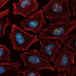
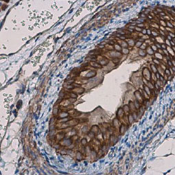
Cell biology is the study of cell structure and function. This includes the most general properties shared by all cells to the specific, highly complex functions unique to specialized cells.

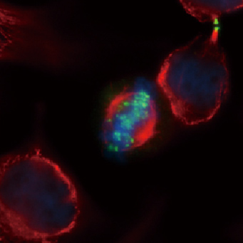
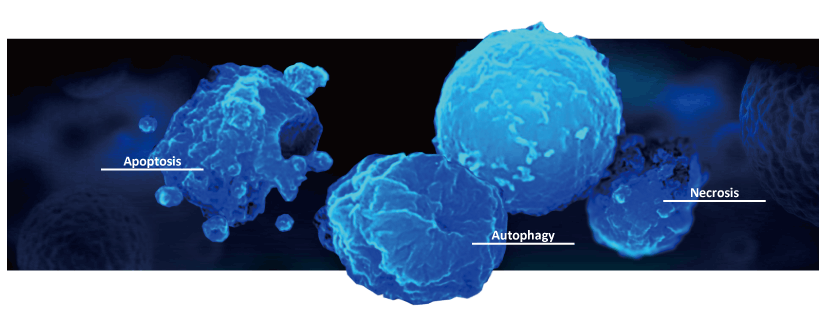
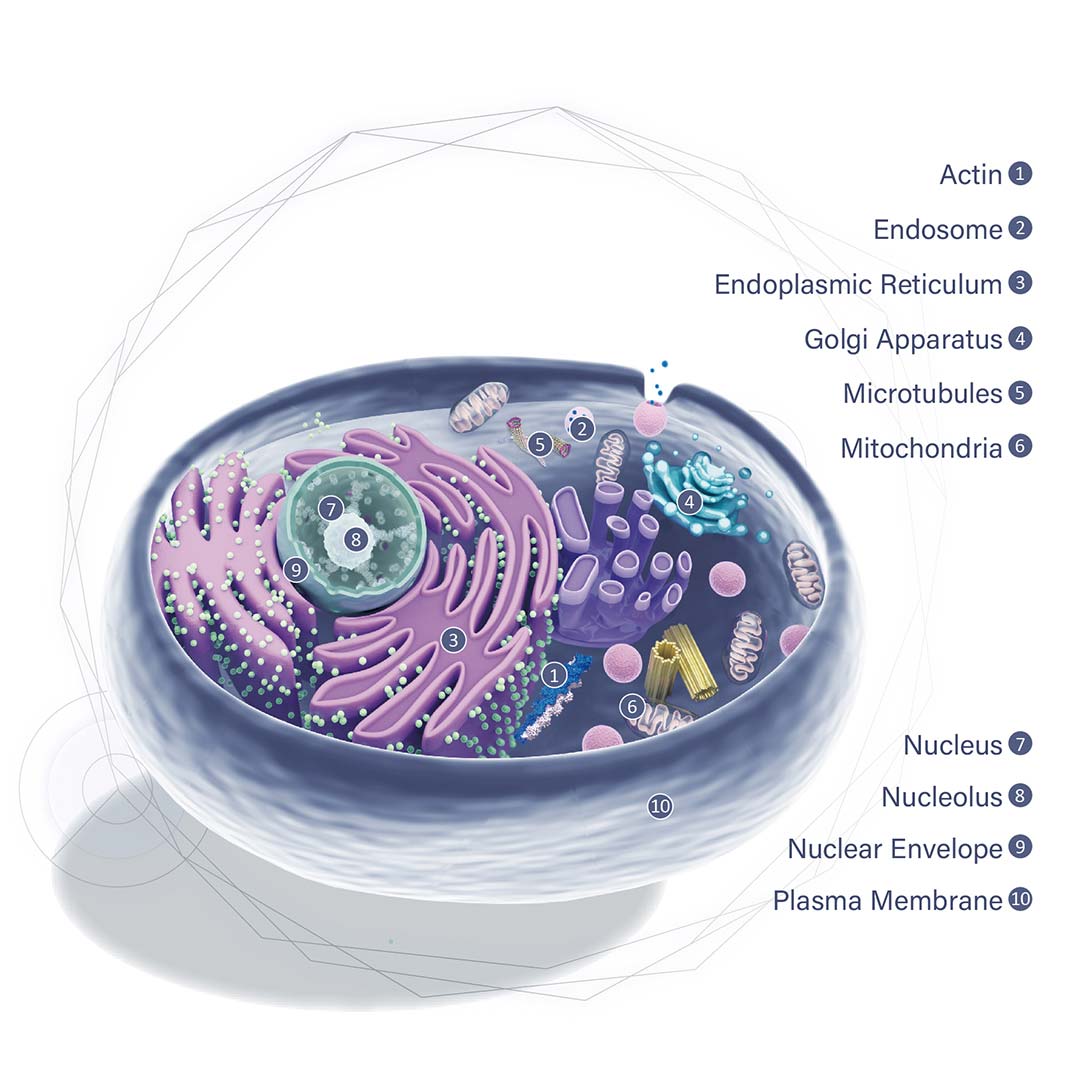
There are an array of research subspecialties within the cell biology field. One is the study of cell energy and the biochemical mechanisms that support cell metabolism. Another focuses on the structure of cell components, known as subcellular compartments or organelles. A third looks at the cell cycle, the progression of cell phases beginning and ending with cell division separated by periods of growth and DNA replication. Finally, there is great interest in the mechanisms associated with cell death, including apoptosis, necrosis, and autophagy. Defining how the molecular mechanisms characteristic of one of these fields influences the others allows scientists to analyze cellular behaviors in more complex ways.

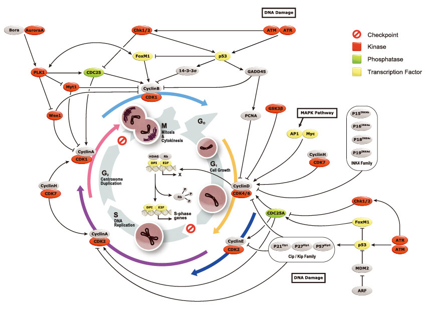
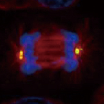
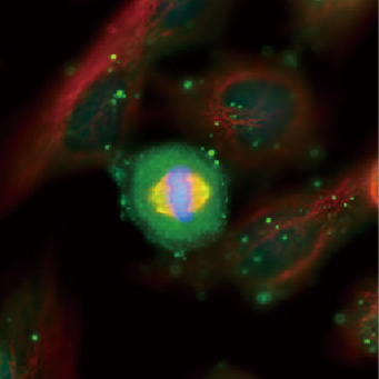
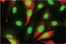
The cell cycle is an ordered set of events by which cells undertake growth and division into two daughter cells. In eukaryotes, the cell cycle is described by five phases: G0, G1, S, G2, and M. In G0 phase, cells have entered a quiescent state or are not dividing. During G1 period, the cells grow and accumulate nutrients for the subsequent S phase, where DNA replication occurs. The gap between S and M phases is termed G2 period, during which the cells continue to grow and prepare for mitosis that occurs in M phase. All these processes are elaborately controlled to ensure proper cell development and renewal. The key molecules that program cell cycle are cyclins and cyclin-dependent kinases (CDKs), whereas inhibitors that prevent improper cell division are two families of genes, the Cip / Kip family and the INK4 family. Notably, these inhibitor genes are also termed tumor suppressor genes as they are instrumental in the prevention of tumor formation.

Highlighted Products


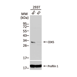
CDK5 antibody [C2C3],
C-term
(GTX108328)

p21 Cip1 antibody [GT1032]
(GTX629543)

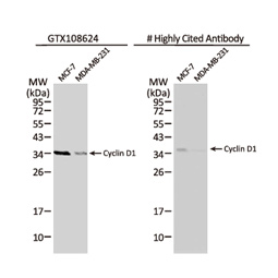
Cyclin D1 antibody
(GTX108624)

gamma Tubulin antibody
(GTX113286)

Aurora A antibody [C3], C-term
(GTX104620)

Aurora B antibody
(GTX132702)

Cell death occurs when a cell is no longer able to sustain essential life functions. Cells can die through one of several biochemically distinct pathways, with apoptosis and necrosis being the two most commonly studied mechanisms. While the term “autophagic cell death” would suggest death by autophagy, this form of cell killing occurs only in specific cases, as autophagy is generally considered a pro-survival process. Aside from these three modalities, other forms of cell death described by terms such as “mitotic catastrophe” or “excitotoxicity” are present in the literature. However, more research will have to be done to determine whether these and other named processes truly represent distinct death pathways.

Apoptosis
    

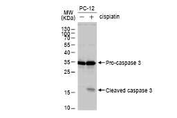
Caspase 3 antibody (GTX110543)


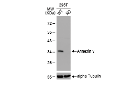
Annexin V antibody (GTX103250)


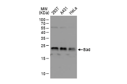
Bad antibody (GTX130108)

  
Necrosis
  

C Reactive Protein antibody (GTX101262)

LDHA antibody (GTX101416)

HMGB1 antibody (GTX101277)

  
Autophagy
     

LC3B antibody
(GTX127375)


SQSTM1 antibody (GTX629890)


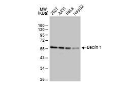
Beclin 1 antibody (GTX133555)


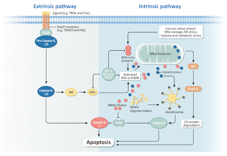
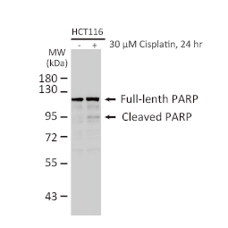
Apoptosis is programmed cell death and is crucial for development and tissue homeostasis. It can be initiated by various stimuli and can progress through a receptor-mediated extrinsic pathway or an intrinsic pathway that starts with mitochondrial events. Both processes involve the activation of caspases. Apoptotic cells undergo distinct changes in cell morphology such as cell rounding, plasma membrane blebbing, and nuclear fragmentation. A spectrum of key players is involved, including transcription factors such as the tumor suppressor p53 and FOXO3a that drive expression of pro-apoptotic genes, effectors such as Caspase 3 and ROCK1, and factors like PARP and AIF that function in a caspase-independent pathway.


Highlighted Products
    

p53 antibody
(GTX112864)

Caspase 3 antibody
(GTX110543)

Caspase 8 antibody
(GTX101070)

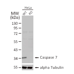
Caspase 7 antibody
(GTX123679)

CDKN2A / p14ARF antibody
(GTX129903)

TNF alpha antibody
(GTX110520)

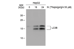
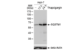
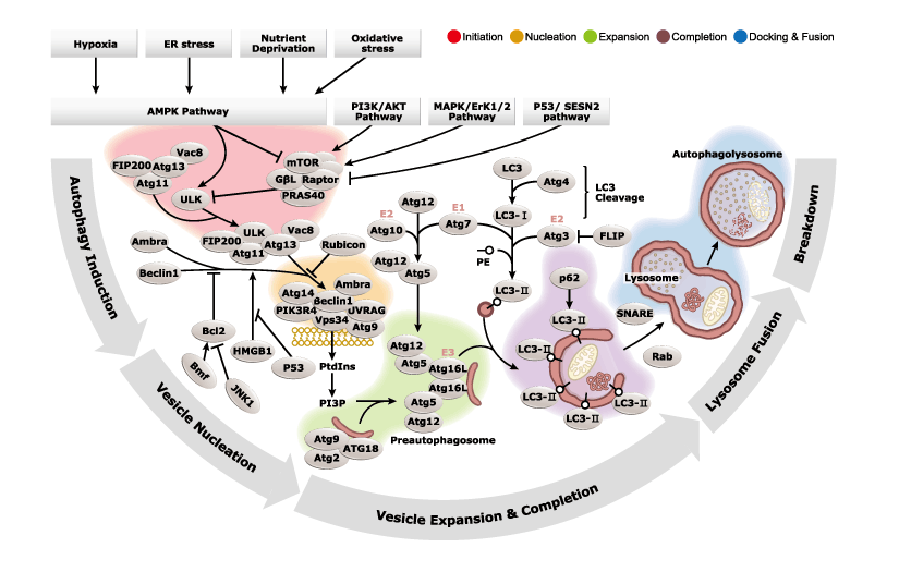
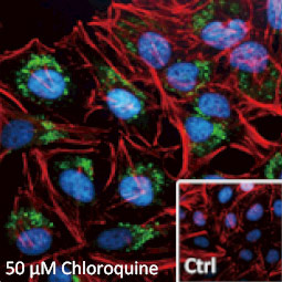
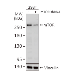
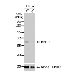
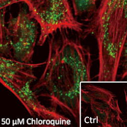
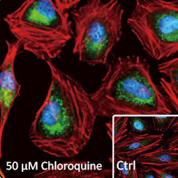
Autophagy has been implicated in many different diseases, including neurodegeneration, cardiac myopathy, autoimmune disease and cancer. The role of autophagy in cancer is complex and paradoxical. While autophagic deficiency has been shown to promote tumorigenesis in animal models, autophagy may actually support tumor growth by enhancing cancer cell survival in the face of nutrient depletion or accumulation of toxic molecules. GeneTex is proud to introduce our antibodies for autophagy research.


Highlighted Products
    

LC3B antibody (GTX127375)

mTOR antibody [C3], C-term (GTX101557)

Beclin 1 antibody (GTX133555)

SQSTM1 / P62 antibody [N3C1], Internal
(GTX100685)

NDP52 antibody  (GTX115378)

HMGB1 antibody (GTX101277)

DFNA5 antibody

Cat No. GTX64590

DFNA5 antibody, N-term

Cat No. GTX81693

DFNA5 antibody, C-term

Cat No. GTX46489

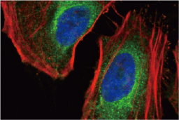
The subcellular location of a protein may suggest potential roles for that factor in one or more cellular processes. Organelle protein-specific antibodies are essential for establishing colocalization of a particular protein of interest with an organelle, thereby contributing crucial insight into its possible function(s). In addition, these organelle marker antibodies can often be used in cell fractionation studies analyzed by western blot alone or after immunoprecipitation.


Cytosol

Endoplasmic Reticulum (ER)
  

GRP94 antibody
(GTX103232)

Calnexin antibody
(GTX109669)

Grp78 antibody
(GTX113340)

Golgi Apparatus
  

GOLPH2 antibody
(GTX116154)

GOLGA5 antibody
(GTX104255)

GM130 antibody
(GTX130351)

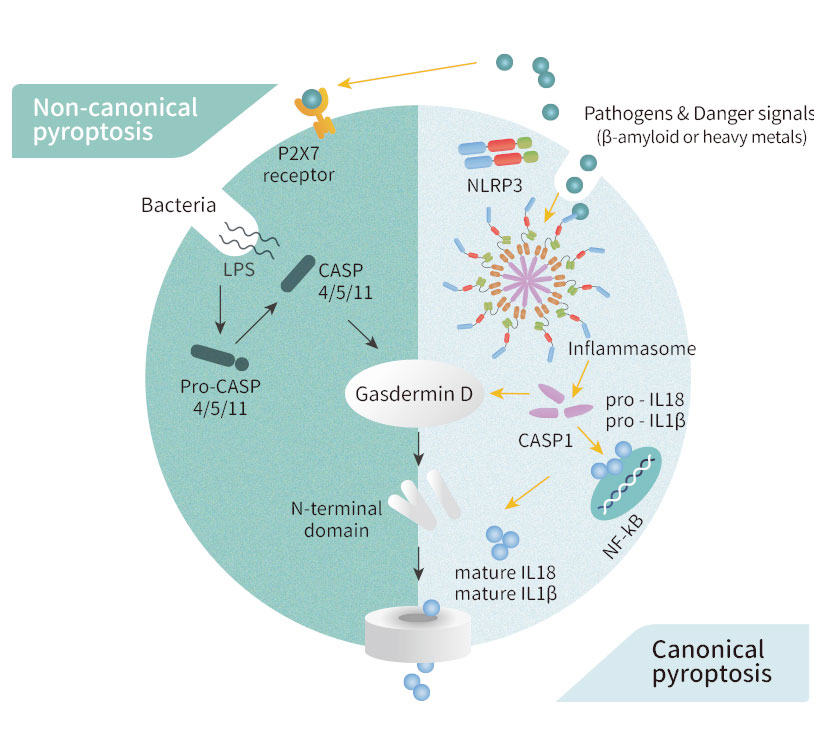
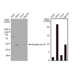
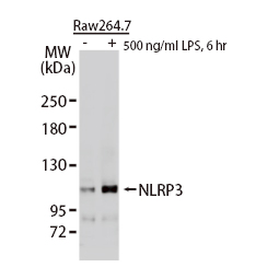
Pyroptosis is a programmed cell death process executed by inflammatory caspases upon initiation of canonical or non-canonical mechanisms. It is triggered by specific inflammatory caspases (caspase-1, -4, -5, -11) that are distinct from those responsible for apoptosis. Both the canonical and non-canonical pathways lead to the activation of gasdermin D (GSDMD), which forms pores that cause cellular leakage and lysis. The canonical sequence involves pathogen-associated molecular patterns (PAMPs)- and damage-associated molecular patterns (DAMPs)-mediated inflammasome formation leading to caspase-1 activation, GSDMD cleavage, and IL-1β and IL-18 maturation. The non-canonical string of events is characterized by direct interaction of the other three caspases with Gram-negative bacterial lipopolysaccharide (LPS) with subsequent GSDMD activation. The resultant extracellular release of cytoplasmic components unleashes a local inflammatory cascade that can become systemic, underscoring the importance of pyroptosis’ normal function in mobilizing immune cells against pathogens. Nevertheless, pyroptosis can also contribute to inflammation-related pathology, including cancer progression and autoimmune disease.

GeneTex is proud to offer an outstanding selection of antibodies to study pyroptosis and inflammation biology. These antibodies are validated for various applications to facilitate your efforts in this exciting field. Please see the highlighted products below

Highlighted Products

Citation-Support KOKD-Validation Orthogonal Validation Protein Overexpression

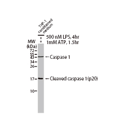
Caspase 1 antibody [N1N3] (GTX101322)

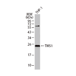
TMS1 antibody [N1C3] (GTX102474)

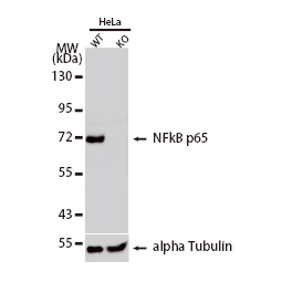
NFkB p65 antibody (GTX107678)

Gasdermin D antibody [N1N3] (GTX116840)

NLRP3 antibody (GTX133569)

IL1 beta antibody (GTX74034)

G protein-coupled receptors (GPCRs) constitute the largest superfamily of human cell surface transmembrane receptors with over 820 genes. They respond to a variety of ligands that include hormones, neurotransmitters, metabolites, ions, photons, and mechanical forces, with subsequent intracellular signaling relayed through G protein-dependent and -independent mechanisms. GPCR-triggered pathways are responsible for a plethora of physiological and pathophysiological effects, which is why approximately a third of all prescribed drugs target their activity. Of the more than 350 human GPCRs that are not sensory receptors, perhaps 140 are considered “orphan” receptors with no known ligand or function. Emphasizing their clinical value, it is thought that ~60-85% of potentially therapeutic GPCRs have no drugs directed at them.

GeneTex is committed to establishing the most extensive product line of antibodies for human GPCRs, with all new production being recombinant monoclonal antibodies. These antibodies will be thoroughly characterized through enhanced “Five Pillar” validation strategies that feature knockdown/knockout lysates, comparable antibodies, cell fractionation, detection of endogenous GPCR expression, and overexpression. In addition, specialized testing using human GPCR arrays will be utilized when feasible. The goal is to generate the most diverse, expansive, and meticulously verified antibody reagents for GPCR biology research.

Physiological Functions of GPCRs

GPCRs play a crucial role in the regulation of tissue/cell physiology and homeostasis in the immune, nervous, endocrine, and cardiovascular systems, among others. They also function in a multitude of pathological processes, including cancer.

GPCRs in cancers

Many GPCRs serve as potential biomarkers for early cancer diagnosis. Furthermore, GPCRs are active in various aspects of cancer progression, including proliferation, apoptosis, angiogenesis, migration, and invasion. Therefore, the pharmacological inhibition of GPCRs and their downstream targets presents a promising avenue for developing novel, mechanism-based strategies for cancer therapy.

GPCRs in the immune system

Inflammatory cells such as leukocytes, monocytes, macrophages, and dendritic cells express more than one GPCR and sense a wide range of chemoattractants and chemokines. These receptors are crucial for the migration and infiltration of immune cells. Abnormal GPCR expression can lead to immune system dysfunction manifesting as inflammatory and autoimmune disease.

GPCRs in the nervous system

The nervous system utilizes membrane receptors to detect extracellular stimuli. By expressing GPCRs with diverse ligand-recognition capabilities, the nervous system can selectively filter and respond to specific signals. GPCRs are involved in chronic neurodegenerative diseases including but not limited to Alzheimer’s disease, Huntington’s disease, and Parkinson’s disease.

GPCRs in homeostasis

GPCRs play a crucial role in maintaining metabolic balance by influencing key processes such as glucose homeostasis and insulin secretion, appetite, calcium sensing, heart rate, and blood pressure.


Table 1. GPCRs related to different disorders
Disorder GPCRs
Cancer Bradykinin receptor
Chemokine receptors
Endothelin receptors
Frizzled receptors
Protease-activated receptors Prostaglandin receptors
Immunological disorders Adenosine receptors
Anaphylatoxin receptors (C3aR, C5aR, and C5L2)
Cannabinoid receptors
Chemokine receptors
Histamine receptors
Neurokinin receptors
Prostaglandin receptors
Protease-activated receptors

   Comparable Abs  

Disorder GPCRs
Neurodegenerative diseases Alzheimer’s disease Adenosine A2A receptor
Adrenergic receptors
Corticotrophin-releasing hormone receptors
Metabotropic glutamate receptors
Serotonin receptors
delta-opioid receptor
Parkinson’s disease Adenosine A2A receptor
Adrenergic receptors
Metabotropic glutamate receptors serotonin receptor
Huntington’s disease Cannabinoid receptors
Metabotropic glutamate receptors

 Comparable Abs  

Disorder GPCRs
Metabolic disorders Obesity Bile acid receptor
Melanocortin receptors
Type 2 diabetes Free fatty acid-binding receptors
Glucagon receptors
Incretin receptors (GLP1R and GIPR)
Somatostatin receptors
Cardiovascular diseases Adenosine receptors
Angiotensin II receptors

 Comparable Abs 

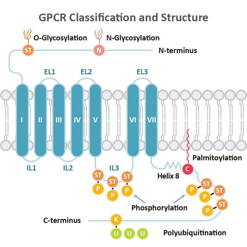
GPCR Classification and Structure

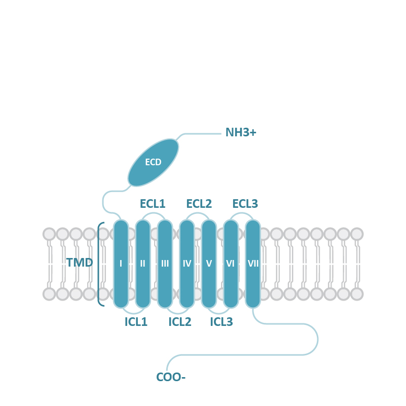
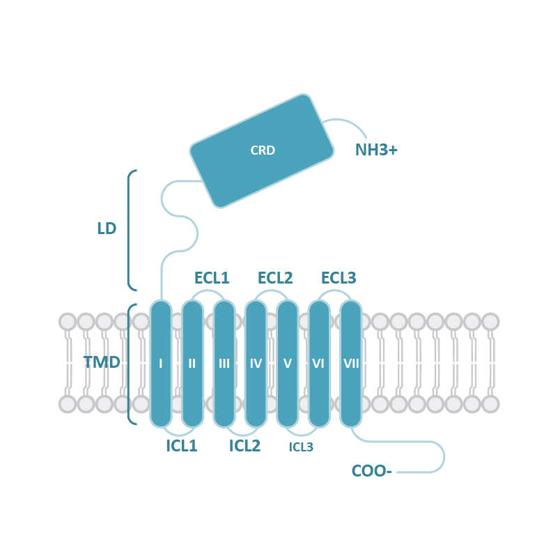
GPCRs share structural characteristics that include seven transmembrane (7TM) domains linked by both intra- and extracellular loops, an extracellular N-terminus, and an intracellular C-terminus. The loops, as well as the intra- and extracellular domains, are all subject to post-translational modifications. One widely used GPCR classification system is based on sequence homology and evolutionary relationships. This organizes GPCRs into six families designated A-F.

Table 2. GPCR families
Family Class General Structure
(Adapted from Qu et al., 2020)
Ligand Interaction
(Adapted from Xu et al., 2024)
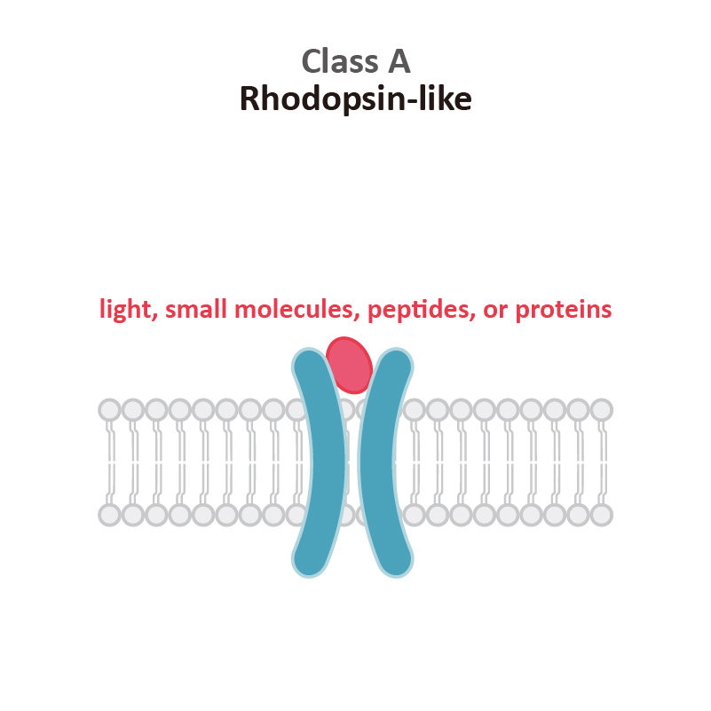
Class A

Rhodopsin-like (R)

Class A (Rhodopsin-like) GPCRs account for more than 85% of human GPCRs. This class is distinguished structurally by an additional palmitoylated 8th alpha helix.

 Class A

 Comparable Abs  

Family Class General Structure
(Adapted from Qu et al., 2020)
Ligand Interaction
(Adapted from Xu et al., 2024)
Class B1

Secretin (S)

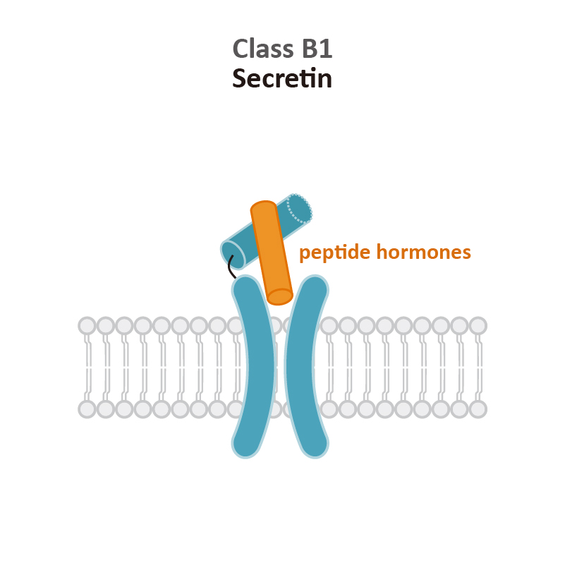
Class B1 (Secretin) GPCRs are characterized by their large extracellular domains (ECDs) that are able to bind large peptidic ligands such as hormones or neuropeptides.

Class B2

Adhesion (A)

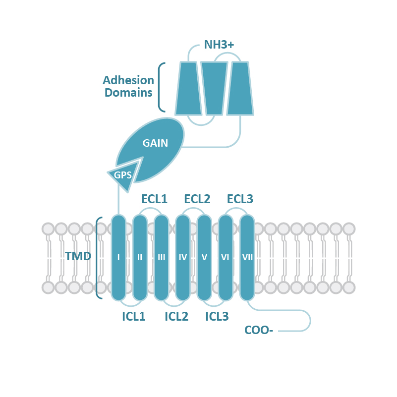
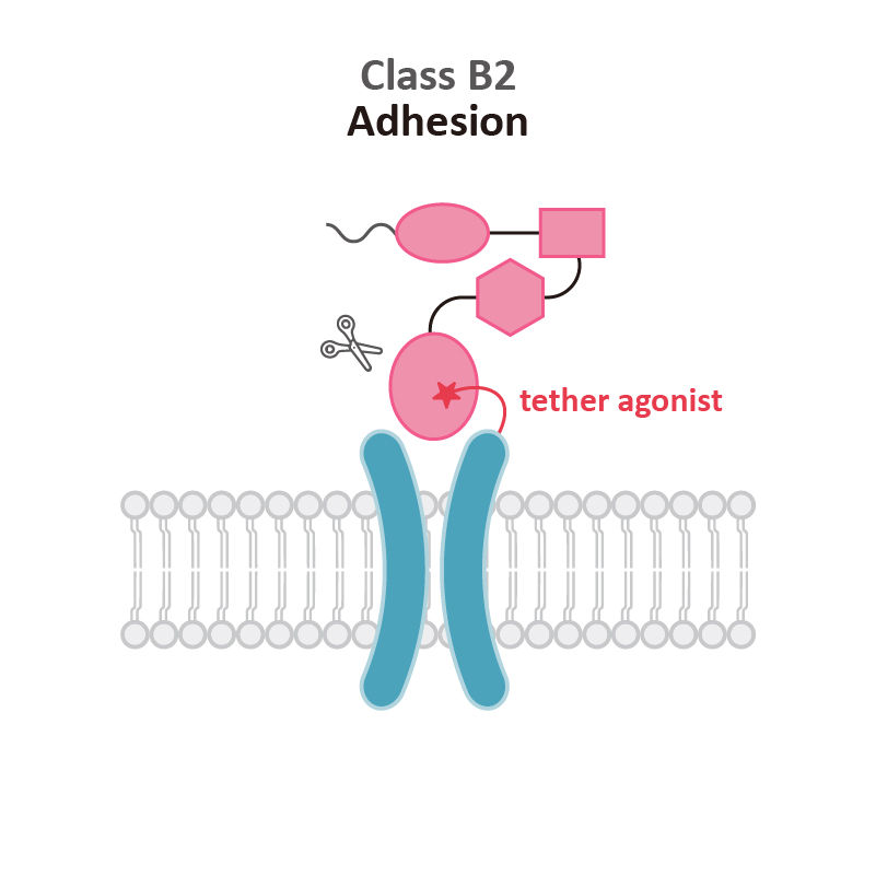
Similar to Class B1, Class B2 (Adhesion) GPCRs feature a large ECD. Signaling results from the pre-digestion of the GPCR autoproteolysis-inducing (GAIN) domain at the GPCR proteolytic site (GPS) motif. Mechanical force releases the Stachel peptide that then acts as a tethered agonist to bring about 7TM activation.

The individual N-terminal motifs of the subgroups reflect their unique roles in cell adhesion and migration.

Class B1

 Comparable Abs

Class B2

  Comparable Abs  
Family Class General Structure
(Adapted from Qu et al., 2020)
Ligand Interaction
(Adapted from Xu et al., 2024)
Class C

Glutamate (G)

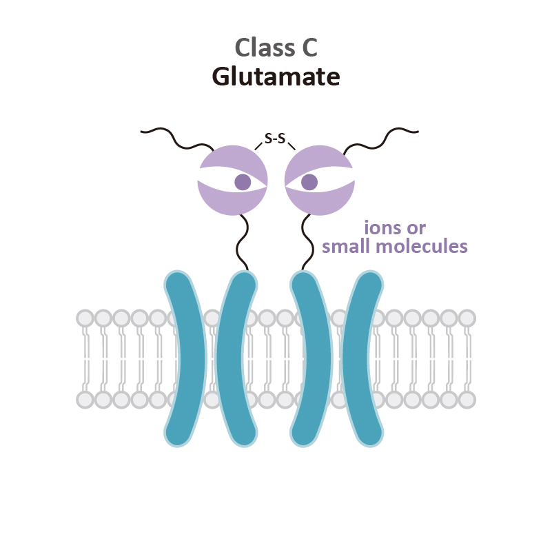
Class C (Glutamate) GPCRs are distinctive for large ECDs, which include a Venus Fly Trap (VFT) domain and a cysteine-rich domain (CRD), and their mandatory homo- or heterodimerization.

Class F

Frizzled (F)

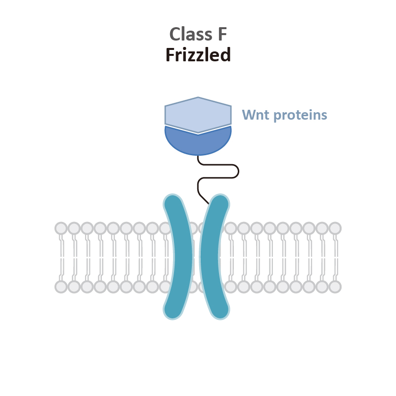
Class F (Frizzled) GPCRs feature a cysteine-rich domain (CRD) and a linker domain (LD) in the ECD. Members of this class play roles in development and regeneration through activation of the downstream Wnt or Hh signaling transduction pathways.

Class C

  Comparable Abs  

 Class F


Regulation of GPCR Signaling

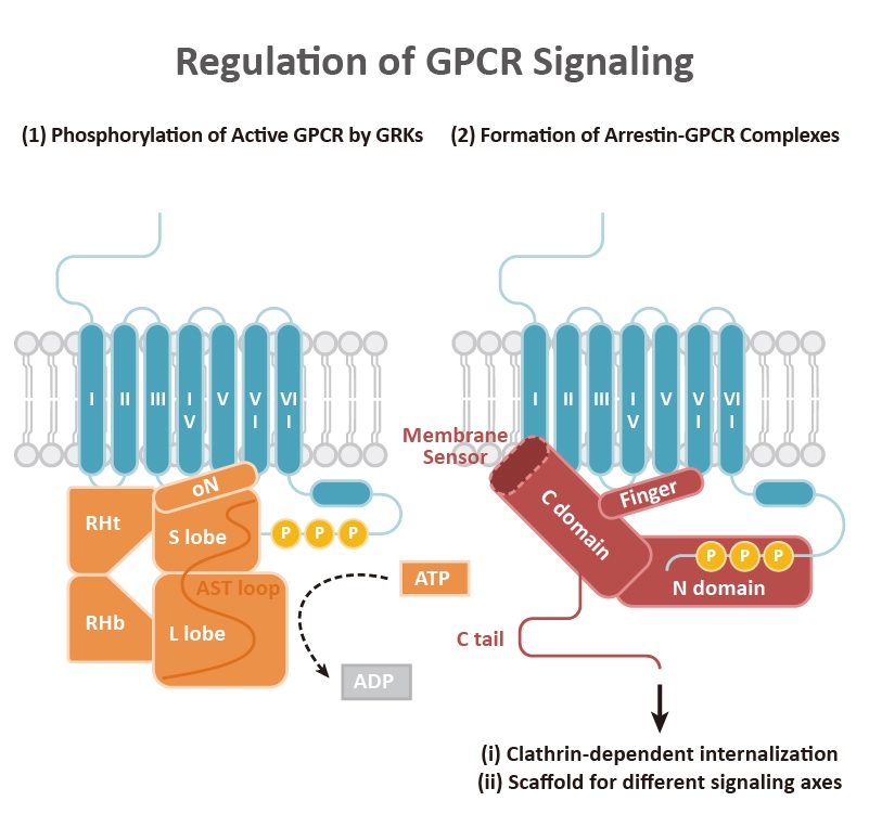
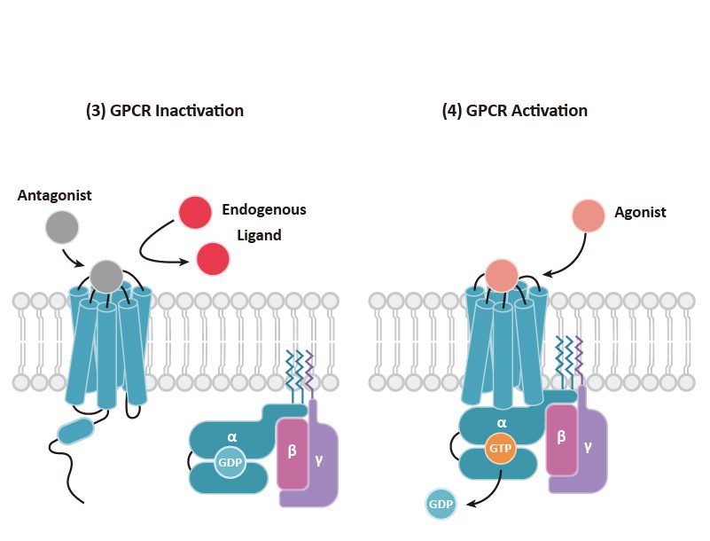
GPCR signaling begins with engagement of the receptor by a ligand, which leads to allosteric changes in the intracellular domains of the GPCR. In general, two types of effectors can then associate with the GPCR: (a) a Gα subunit that complexes with β and γ subunits to form the G protein trimer, and (b) β-arrestins that can direct their own signaling. It is becoming clear that GPCR signaling emanates not only from the cell surface but also from other intracellular compartments (e.g., endosomes, the Golgi apparatus, and the ER, among others). Signaling cascades involving kinases and transcription factor regulation orchestrate the subsequent cellular response.

Classically, GPCR signaling undergoes termination through GTP hydrolysis and dissociation of Gα-Gβγ. G protein-coupled receptor kinases (GRKs) phosphorylate the C-terminal tail of GPCRs, facilitating the binding of the β-arrestins that, as mentioned above, can trigger their own signaling as well as contribute to GPCR desensitization and internalization. The GRK family, comprising seven kinases (GRK1-7), is categorized into three subfamilies: (1) the GRK1 subfamily consists of rhodopsin kinase (GRK1) and GRK7, (2) the GRK2 subfamily consists of β-adrenergic receptor kinase-1 and -2 (GRK2 and GRK3), and (3) the GRK4 subfamily consists of GRK4-6. GRK2, GRK3, GRK5, and GRK6 are key regulators of GPCRs. Beyond GRKs and arrestins, GPCR functions and signal transduction are influenced by GPCR-interacting proteins (GIPs) such as receptor activity-modifying proteins (RAMPS), regulators of G protein signaling (RGS) proteins, GPCR-associated sorting proteins (GASPs), Homer proteins, and PDZ-scaffold proteins.


Table 3. mRNA Expression of GRKs and β-arrestins in tissues
Protein High-expressing tissues
GRK2 Bone marrow, spleen, lymph node, tonsil, and thymus
GRK3 Adipose tissue, spleen, cerebral cortex, tonsil, and hippocampus
GRK5 Heart muscle, lymph node, parathyroid gland, placenta, and gallbladder
GRK6 Bone marrow, lymph node, spleen, thymus, and granulocytes
β-arrestin1 Monocytes, cerebral cortex, pancreas, amygdala, and spleen
β-arrestin2 Bone marrow, spleen, granulocytes, liver, and monocytes

SUPPORT

outstanding technical support

PRODUCT

we offer a full product guarantee

DELIVERY

we offer free delivery to UK universities and non profit organisations