Lateral flow assays are one of the many types of immunoassays available for health monitoring. As point of care diagnostic tests, they provide simple “yes or no” answers for rapid and accurate disease surveillance.
Lateral flow tests for diagnostics
Lateral flow assays (LFAs) can be used to test for antibodies throughout the immune response. Monitoring specific antibody isotypes allows clinicians to see if the patient is in the initial infection stage or has developed antibodies, suggesting immunity against the disease. Combined with controls, LFAs have the potential to provide robust results, but with the added benefit of simple “yes or no” results in a rapid format.
Why use a Lateral flow test?
Like other serological tests, LFAs can identify antibodies or antigens in a sample of blood serum or another bodily fluid. They may also be used to detect allergy.
Why antibody test?
The antibody-based LFA test can provide rapid and unambiguous results at the point of care. Almost instant results can provide reassurance and allow immediate action. Rapid results are particularly important for healthcare and key workers needing to assess exposure for a safe working environment.
How does a Lateral flow assay work?
Like any serological immunoassay, the key principle is that the infectious agent/pathogenic antigen is attached to a surface, either directly or using a capture antibody. However, the process uses capillary action to “flow” the biological sample across a solid matrix allowing the reactant antibodies to bind to the antigen and be visualized with a secondary antibody conjugated to a reporter molecule such as a 40nm Gold molecule.
Rapid assays like lateral flow tests provide quick yes or no results in an easy to use format. They allow for point of care analysis as an alternative to more time-consuming immunoassays that need to be performed in laboratories. Rapid tests are used to qualitatively detect patient-generated antibodies to enable clinical diagnosis and indicate treatment.
How do they work?
Lateral flow or rapid tests use the principle of immunochromatography – whereby the components of the test flow laterally through a matrix by capillary action allowing the antigens to combine with antibodies to facilitate the detection and visualization of those complexes.
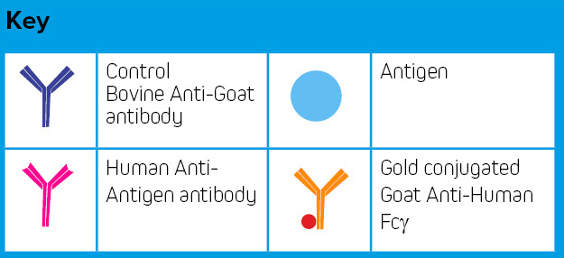
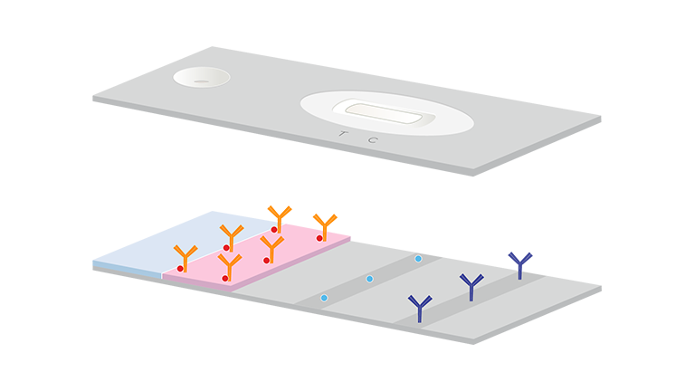
Step 1. Lateral flow test strips commonly consist of a sample pad, a conjugate pad that has been impregnated with conjugated antibodies, for example, gold conjugated Goat Anti-Human Fcγ specific antibodies and two “lines” which have been applied to nitrocellulose with antigen (T), and control antibodies, for example, Bovine Anti-Goat (C).
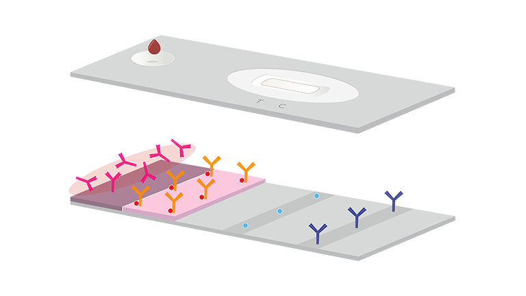
Step 2. The sample is introduced to the sample pad via a hole in the test strip cassette.
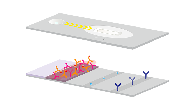
Step 3. Capillary action draws the sample into the conjugate pad and rehydrates the impregnated antibody conjugates. Any antibodies in the sample to which the conjugated antibody is specific will form complexes.
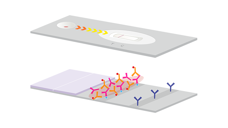
Step 4. The sample and complexed antibodies migrate onto the antigen-coated nitrocellulose (T) test “line” where they interact.
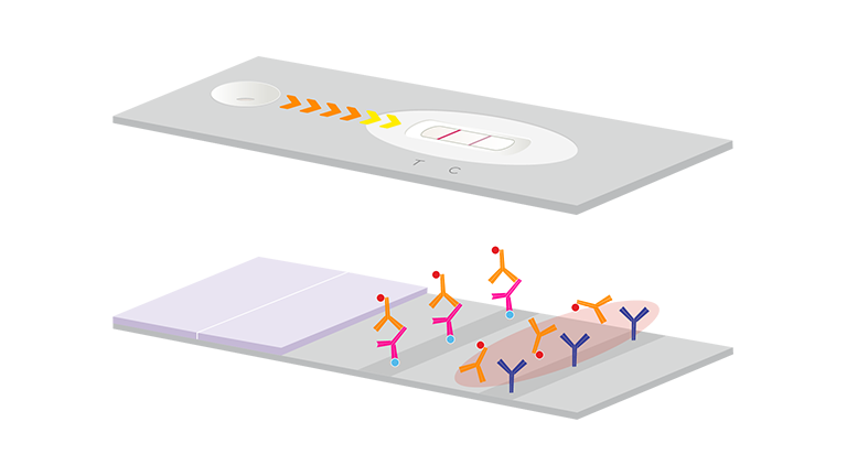
Step 5. If the antibodies in the sample are specific to the coated antigen the antibody complexes will be deposited and develop color over the line giving a visual indication of a “positive” test. If there are no antibodies specific to the antigen in the test sample, no colored line will develop. Un-complexed gold conjugated Goat Anti-Human antibodies continue to migrate through the test strip to the control line (C).
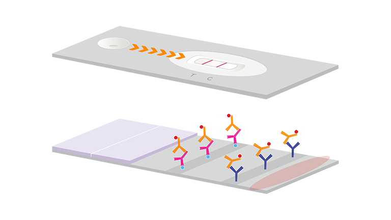
Step 6. The free conjugated Goat Anti-Human antibodies can be captured by the coated Bovine Anti-Goat IgG and develop a colored line to indicate the test has been performed correctly. No control line would indicate an invalid test.