Nucleotides for applications on Proteins/Enzymes

Probes for Protein Kinases and Protein Phosphatases

Protein phosphorylation is a regulatory mechanism that is extremely important in most cellular processes such as protein synthesis, cell division, signal transduction, cell growth, development and aging as many enzymes and receptors are activated and deactivated via phosphorylation/dephosphorylation due to specific protein kinases and protein phosphatases[1-4].

Phosphorylation and dephosphorylation can modify the function of a protein in almost every conceivable way; for example by increasing or decreasing its biological activity, by stabilizing it or marking it for destruction, by facilitating or inhibiting movement between subcellular compartments, or by initiating or disrupting protein– protein interactions. It is believed that about 30% of the proteins encoded by the human genome contain covalently bound phosphate and abnormal phosphorylation is recognized as a cause or consequence of many human diseases as well[5].

Jena Bioscience offers a selection of modified biomolecules and small-molecule reporters such as labeled ATPs as phosphoryl donor to analyze the complex processes of protein phosphorylation and dephosphorylation.


Name Cat. No. Size
DDAO Phosphate APC-001-1 1 mg
DDAO Phosphate APC-001-5 5 x 1 mg
γ-[(PEG3-Amino)-imido]-ATP-Biotin NU-970-BIO 50 μl (1 mM)
γ-(6-Aminohexyl)-ATP-ATTO-590 NU-833-590 80 μl (1 mM)
γ-[(Propargyl)-imido]-ATP CLK-T11-1 1 mg
γ-[(6-Azidohexyl)-imido]-ATP CLK-T12-1 1 mg
γ-(2-Azidoethyl)-ATP NU-1701S 100 μl (10 mM)
γ-(2-Azidoethyl)-ATP NU-1701L 5 x 100 μl (10 mM)
ATPγS NU-406-5 5 mg
ATPγS NU-406-25 25 mg
ATPγS NU-406-50 50 mg
ATP-acetyl-hex-Biotin NU-277 16 x 0.01 μmol (16 x approximately 11.5 μg)
ATP-acetyl-Desthiobiotin NU-276 16 x 0.01 μmol (16 x approximately 10.1 μg)

Selected References

[1] Li et al. (2013) Elucidating human phosphatase-substrate networks. Sci. Signal. 6 (275):rs10.
[2] Sacco et al. (2012) The human phosphatase interactome: an intricate family portrait. FEBS Lett. 586:2732.
[3] Xiao et al. (2016) Global discovery of protein kinases and other nucleotide-binding proteins by mass spectrometry. Mass Spectrometry Reviews 35:601.
[4] Casey et al. (2018) Interrogating protein phosphatases with chemical activity probes. Chem. Eur. J. 24:1.
[5] Cohen (2002) The origins of protein phosphorylation. Nature Cell Biology 4:E127.

Puromycin conjugates for specific C-terminal protein labeling in vitro and monitoring of global protein synthesis in vivo

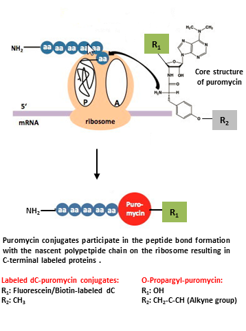
The antibiotic puromycin is a structural analog of aminoacyl-tRNA that has been traditionally used as an inhibitor of protein synthesis since it specifically incorporates at the C-terminus of nascent polypeptide chains thereby stopping translation. Based on this observation, a new and significantly faster approach, using labeled puromycin conjugates, can be applied to both, specific C-terminal labeling of full-length protein in vitro[1,2,3,4] and non-radioactive monitoring of global protein synthesis in vivo[5,6,7].

Labeled dC-puromycin conjugates: Specific C-terminal protein labeling of full-length protein in vitro
C-terminal labeled full-length protein can be produced by in vitro translation in the presence of low concentrations of labeled dC-puromycin conjugates[1].
These proteins have been successfully used to analyze protein-protein interactions (pull-down assay, FCCS, protein-protein microarrays) and protein-DNA interactions (DNA microarrays)[2,3,4].
In contrast to traditionally applied posttranslational labeling approaches, additional time consuming purification steps can be avoided due to the synchronization of protein expression and labeling.

O-Propargyl-puromycin: Monitoring of global protein synthesis in vivo
Current studies of the cellular protein level rely on indirect methods such as DNA and mRNA microarrays or classical radioactive metabolic labeling with 35S-methionine.
O-Propargyl-puromycin is cell-permeable and readily incorporated into newly translated proteins thus providing a sensitive, nonradioactive method for direct monitoring of global protein synthesis[5].
The resulting C-terminal alkyne labeled proteins can be detected via Cu(I)-catalyzed click chemistry that offers the choice to introduce a Biotin group (Azides of Biotin → Click Chemistry) for subsequent purification tasks or a fluorescent group (Azides of fluorescent dyes → Click Chemistry) for subsequent microscopic imaging.


Name Cat. No. Size
Biotin-dC-puromycin NU-925-BIO-S 100 μl (0,1 mM)
Biotin-dC-puromycin NU-925-BIO-L 5 x 100 μl (0,1 mM)
6-FAM-dC-puromycin NU-925-6FM-S 100 μl (0,1 mM)
6-FAM-dC-puromycin NU-925-6FM-L 5 x 100 μl (0,1 mM)
O-Propargyl-puromycin NU-931-05 0,5 mg (1 μmol)
O-Propargyl-puromycin NU-931-5 10 x 0,5 mg (10 μmol)

Selected References

[1] Miyamoto-Sato et al. (2000) Specific bonding of puromycin to full-length protein at the C-terminus. Nucleic Acids Res. 28 (5):1176.
[2] Nemoto et al. (1999) Fluorescence labeling of the C-terminus of proteins with a puromycin analogue in cell-free translation systems. FEBS Letters 462:43.
[3] Doi et al. (2002) Novel Fluorescence Labeling and High-Throughput Assay Technologies for In Vitro Analysis of Protein Interactions. Genome Research 12:487.
[4] Kawahashi et al. (2007) High-throughput fluorescence labeling of full-length cDNA products based on a reconstituted translation system. J. Biochem. 141 (1):19.
[5] Liu et al. (2012) Imaging protein synthesis in cells and tissues with an alkyne analog of puromycin. Proc Natl Acad Sci USA. 109 (2):413.
[6] Stark et al. (2004) A General Approach to Detect Protein Expression In Vivo Using Fluorescent Puromycin Conjugates. Chemistry & Biology 11:999.
[7] Godman et al. (2011) Novel insights into the regulation of skeletal muscle protein synthesis as revealed by a new nonradioactive in vivo technique. The FASEB Journal 25:1028.

Non-radioactive Protein Phosphorylation Analysis

In addition to the traditional methods for protein phosphorylation analysis (labeling with radioactive (“hot”) ATP[1], phospho-specific antibodies[2] or ATP depletion measurement[3]) we here summarize recent techniques using non-radioactively modified ATP analogs whose modified gamma-phosphate can be transferred by a kinase to its peptide/protein substrate in vitro.

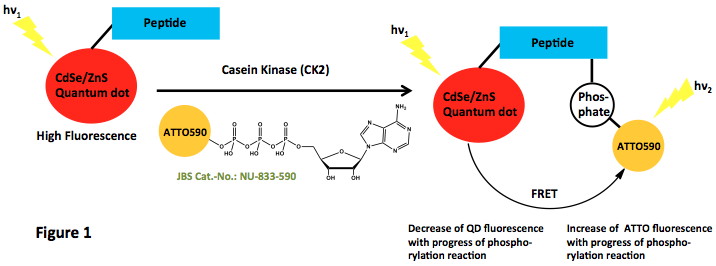
Direct labeling with fluorescently labeled ATP: Freeman et al.[4] implemented quantum dots (QDs) as an optical label for FRET-based analysis of the model system casein kinase (CK2) / alkaline phosphatase (ALP) (Figure 1). ATTO590-labeled ATP served as phosphate donor for the phosphorylation of the CK2 substrate peptide.

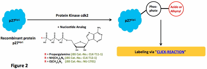
Indirect labeling with functionalized ATP analogs: Lee et al.[5] prepared azide and alkyne γ-modified ATP analogs and tested their ability to phosphorylate p27kip1 with wild type protein kinase cdk2. The phosphorylated protein can subsequently be CLICK-labeled, e.g. with fluorescent dyes (Figure 2):

Allen et al.[6] reported kinase-catalyzed thiophosphorylation with ATPγS. The resulting labeled kinase substrates were recognized by new thiophosphate ester-specific antibodies (not shown).


Name Cat. No. Size
γ-[(PEG3-Amino)-imido]-ATP-Biotin NU-970-BIO 50 μl (1 mM)
γ-(6-Aminohexyl)-ATP-ATTO-590 NU-833-590 80 μl (1 mM)
γ-[(Propargyl)-imido]-ATP CLK-T11-1 1 mg
γ-[(6-Azidohexyl)-imido]-ATP CLK-T12-1 1 mg
γ-(2-Azidoethyl)-ATP NU-1701S 100 μl (10 mM)
γ-(2-Azidoethyl)-ATP NU-1701L 5 x 100 μl (10 mM)
ATPγS NU-406-5 5 mg
ATPγS NU-406-25 25 mg
ATPγS NU-406-50 50 mg

Selected References

[1] Lehel et al. (1997) A chemiluminescent microtiter plate assay for sensitive detection of protein kinase activity. Anal. Biochem. 244:340.
[2] Till et al. (1994) Use of synthetic peptide libraries and phosphopeptide-selective mass spectrometry to probe protein kinase substrate specificity. J. Biol. Chem. 269:7423.
[3] Kupcho et al. (2003) A homogeneous, nonradioactive high-throughput fluorogenic protein kinase assay. Anal. Biochem. 317:210.
[4] Freeman et al. (2010) Probing protein kinase (CK2) and alkaline phosphatase with CdSe/ZnS quantum dots. Nano Lett. 10:2192.
[5] Lee et al. (2009) Synthesis and reactivity of novel γ-phosphate modified ATP analogues. Bioorg. Med. Chem. Lett. 19:3804.
[6] Allen et al. (2007) A semisynthetic epitope for kinase substrates. Nature Methods 4 (6):511.

Kinase Inhibition by Di-Nucleoside Phosphates

Protein kinases catalyze the phosphorylation of substrate proteins by transfer of the γ-phosphate of ATP to the acceptor amino acid. Therefore, they play a key role in cell signaling and regulate biological processes such as proliferation, differentiation, and apoptosis. The malfunctioning of these proteins is the root of many diseases.

Protein kinase inhibitors have been paid much attention in the recent years, especially, in drug discovery. There are basically five classes of kinase inhibitors:

  • ATP-site inhibitors
  • Peptide-site inhibitors
  • Bisubstrate inhibitors directed simultaneously at the ATP-site and the peptide-site
  • Regulatory domain targeting inhibitors
  • Docking site blocking inhibitors

The combination of structural elements of peptide-site and ATP-site inhibitors in one molecule renders bisubstate inhibitors particularly interesting for mechanistic studies on nucleotide kinases.

The general formula of Jena Bioscience’s bisubstrate inhibitors is N-Pn-N’ where P is a phosphate chain of the length n and N and N’, respectively, are ribo- or deoxy-nucleotides.

For a review please see:
Guranowski (2003) Analogs of diadenosine tetraphosphate. Acta Biochimica Polonia 50:947.


Name Cat. No. Size
AP2A NU-936-1 1 mg
AP2A NU-936-5 5 mg
AP3A – Solution NU-506S 50 μl (10 mM)
AP3A – Solution NU-506L 5 x 50 μl (10 mM)
AP3A – Solid NU-506-5 5 mg
AP3A – Solid NU-506-25 25 mg
AP4A – Solution NU-507S 100 μl (10 mM)
AP4A – Solution NU-507L 5 x 100 μl (10 mM)
AP4A – Solid NU-507-5 5 mg
AP4A – Solid NU-507-25 25 mg
AP5A NU-508S 50 μl (10 mM)
AP5A NU-508L 5 x 50 μl (10 mM)
AP6A NU-509S 20 μl (10 mM)
AP6A NU-509L 5 x 20 μl (10 mM)
AP3G (A cap) NU-941-1 1 mg
AP3G (A cap) NU-941-5 5 mg
AP4G NU-503S 50 μl (10 mM)
AP4G NU-503L 5 x 50 μl (10 mM)
AP5G NU-504S 50 μl (10 mM)
AP5G NU-504L 5 x 50 μl (10 mM)
AP4(8-Iodo-G) NU-510S 30 μl (10 mM)
AP4(8-Iodo-G) NU-510L 5 x 30 μl (10 mM)
AP5(8-Iodo-G) NU-511S 30 μl (10 mM)
AP5(8-Iodo-G) NU-511L 5 x 30 μl (10 mM)
AP4U NU-528S 150 μl (10 mM)
AP4U NU-528L 5 x 150 μl (10 mM)
AP5U NU-505S 50 μl (10 mM)
AP5U NU-505L 5 x 50 μl (10 mM)
AP4dT NU-501S 50 μl (10 mM)
AP4dT NU-501L 5 x 50 μl (10 mM)
AP5dT NU-502S 50 μl (10 mM)
AP5dT NU-502L 5 x 50 μl (10 mM)

Nucleotides for G-Protein Signaling

Many signal transduction processes are regulated by GTP binding proteins (G-proteins) like small GTPases → Proteins of the Ras superfamily or heterotrimeric G-proteins → Proteins. The G-proteins act as molecular switches cycling between an inactive GDP-bound and an active GTP-bound state[1].

GTP and GDP analogs with various modifications have become indispensable tools to study G-protein signaling. In general, GTP and GDP analogs containing a modified phosphate moiety are resistant to enzymatic hydrolysis or are hydrolyzed at much smaller rates compared to their natural counterparts (non-hydrolyzable nucleotides).

2’/3′-Mant-,TNT- and Ant-modified GTP and GDP analogs combine fluorescence with close mimicry of the properties of natural nucleotides in respect of protein binding and interaction. They display environmentally sensitive fluorescence and therefore give strong signals upon a binding event which makes them perfect probes for stopped-flow and equilibrium analysis.

For detailed application data please refer to the corresponding data sheets.

Table 1: GTP and GDP analogs for G-protein signaling analysis.

GTP Analog Modification
instrinsically fluorescent Mant-GTP ribose moiety (2’/3′-OH)
TNP-GTP
Ant-GTP
Non-hydrolyzable GTPαS α-phosphate moiety
GTPγS γ-phosphate moiety
Mant-GTPγS*
GpCpp α, β-phosphate moiety
GppCp β, γ-phosphate moiety
GppNHp
Mant-GppNHp*
TNP-GppNHp*


GTP Analog Modification
instrinsically
fluorescent
Mant-GTP ribose moiety (2’/3′-OH)
TNP-GTP
Non-hydrolyzable GpCp α, β-phosphate moiety
GppNH2 β-phosphate moiety
GDPβS


* These nucleotides possess both non-hydrolyzable and intrinsically fluorescent properties.


Name Cat. No. Size
GDP – Solid NU-1172-1G 1 g
GDP – Solid NU-1172-5G 5 g
GpCp NU-414-5 5 mg
GpCp NU-414-25 25 mg
GppNH2 NU-1136S 150 μl (10 mM)
GppNH2 NU-1136L 5 x 150 μl (10 mM)
GDPβS NU-427-5 5 mg
GDPβS NU-427-25 25 mg
Mant-GDP NU-204S 150 μl (10 mM)
Mant-GDP NU-204L 5 x 150 μl (10 mM)
TNP-GDP NU-217S 1 μmol
TNP-GDP NU-217L 5 x 1 μmol
GTP – Solid NU-1012-100 100 mg
GTP – Solid NU-1012-1G 1 g
GTP – Solid NU-1012-10G 10 g
GTP – Solid NU-1012-100G 100 g
GTP – Solid – Purity 85 % NU-1047-200 200 mg
GTP – Solid – Purity 85 % NU-1047-1G 1 g
GTP – Solid – Purity 85 % NU-1047-10G 10 g
GTP – Solid – Purity 85 % NU-1047-100G 100 g
GTPαS NU-409S 25 μl (100 mM)
GTPαS NU-409L 5 x 25 μl (100 mM)
GpCpp NU-405S 100 μl (10 mM)
GpCpp NU-405L 5 x 100 μl (10 mM)
GppCp NU-402-5 5 mg
GppCp NU-402-25 25 mg
GppNHp – Tetralithium salt NU-401-10 10 mg
GppNHp – Tetralithium salt NU-401-50 50 mg
GppNHp – Trisodium salt NU-899-50 50 mg
GppNHp – Trisodium salt NU-899-10 10 mg
GTPγS NU-412-2 2 mg
GTPγS NU-412-10 10 mg
GTPγS NU-412-20 20 mg
Mant-GTP NU-206S 150 μl (10 mM)
Mant-GTP NU-206L 5 x 150 μl (10 mM)
Ant-GTP NU-230S 150 μl (10 mM)
Ant-GTP NU-230L 5 x 150 μl (10 mM)
TNP-GTP NU-220S 1 μmol
TNP-GTP NU-220L 5 x 1 μmol
Mant-GppNHp NU-207S 10 μl (10 mM)
Mant-GppNHp NU-207L 5 x 10 μl (10 mM)
TNP-GppNHp NU-218S 0,1 μmol
TNP-GppNHp NU-218L 5 x 0,1 μmol
Mant-GTPγS NU-209S 10 μl (10 mM)
Mant-GTPγS NU-209L 5 x 10 μl (10 mM)
GppNHp – Trisodium salt NU-899-50 50 mg
GppNHp – Trisodium salt NU-899-10 10 mg

Selected References

[1] Vetter et al. (2001) The guanine nucleotide-binding switch in three dimensions. Science 294 (5545):1299.

Tubulin Assembling Nucleotides

Microtubules are a non-covalent helical polymer formed by the globular protein tubulin and act as “conveyer belts” inside the cells. They move vesicles, granules and organelles (mitochondria, chromosomes).

During in vitro microtubule assembly, tubulin heterodimers join end-to-end to form protofilaments (linear tubulin polymer rows), which associate laterally to form microtubules. GTP must be bound to both α and β subunits for a tubulin heterodimer to associate with other heterodimers.

When a tubulin molecule adds to the microtubule, the GTP is hydrolyzed to GDP. Microtubules will also form normally with nonhydrolyzable GTP analogs (like GpCpp [GMPCPP]) attached. However, in this case they will not be able to depolymerize. Thus, the normal role of GTP hydrolysis is to promote the constant growth of microtubules as they are needed by a cell.

In living cells, microtubules exist in an unusual dynamic equilibrium with tubulin subunits, which is called dynamic instability. Individual microtubules alternate between polymerization and de-polymerization periods, leading to rapid exchange between tubulin subunits and microtubule polymer. Dynamic instability responds to the needs of the cell in terms of microtubule (de)formation and distribution.

Properties of tubulin-assembling nucleotides

Nucleotide analogProperties
ITPhighly promotive
GTPstandard for reactivity of other analogs
GpCpp (GMPCPP)moderately promotive, completely suppresses dynamic instability - microtubules do not depolymerize
caged-GpCpp (caged-GMPCPP)caged" derivative of GpCpp (GMPCPP) - blocked by a photo-labile group 6-Methylthio-GTP
8Br-GTPmoderately promotive
dGTPmoderately promotive
mant-GTPslightly inhibitory
XTPslightly inhibitory
6-Thio-GTPhighly inhibitory
Name Cat. No. Size
ITP NU-1203S 15 μl (100 mM)
ITP NU-1203L 5 x 15 μl (100 mM)
GTP – Solution NU-1012 1 ml (100 mM)
GpCpp NU-405S 100 μl (10 mM)
GpCpp NU-405L 5 x 100 μl (10 mM)
NPE-caged-GpCpp NU-306S 10 μl (10 mM)
NPE-caged-GpCpp NU-306L 5 x 10 μl (10 mM)
6-Methylthio-GTP NU-1130S 100 μl (10 mM)
6-Methylthio-GTP NU-1130L 5 x 100 μl (10 mM)
8-Bromo-GTP NU-118S 50 μl (10 mM)
8-Bromo-GTP NU-118L 5 x 50 μl (10 mM)
dGTP – Solution NU-1003L 1 ml (100 mM)
dGTP – Solution NU-1003-10ML 10 ml (100 mM)
Mant-GTP NU-206S 150 μl (10 mM)
Mant-GTP NU-206L 5 x 150 μl (10 mM)
XTP – Sodium salt NU-935-2 2 mg
XTP – Sodium salt NU-935-10 10 mg
XTP – Triethylammonium salt NU-602S 15 μl (100 mM)
XTP – Triethylammonium salt NU-602L 5 x 15 μl (100 mM)
6-Thio-GTP NU-1106S 150 μl (10 mM)
6-Thio-GTP NU-1106L 5 x 150 μl (10 mM)
8-Morpholinyl-GTP NU-906S 50 μl (10 mM)
8-Morpholinyl-GTP NU-906L 5 x 50 μl (10 mM)

Selected References

Läppchen et al. (2005) GTP Analogue Inhibits Polymerization and GTPase Activity of the Bacterial Protein FtsZ without Affecting Its Eukaryotic Homologue Tubulin. Biochemistry 44 (21):7879.
Muraoka et al. (1999) Effects of Purinenucleotide Analogues on Microtubule Assembly. Cell Structure and Function 24:305.
Hyman et al. (1992) Role of GTP Hydrolysis in Microtubule Dynamics: Information from a Slowly Hydrolyzable Analogue, GMPCPP. Mol. Biol. of the Cell 3:1155, and references therein.

Nucleotides for Application in Structural Biology

There are currently two main methods to determine the three-dimensional structure of a protein: Nuclear Magnetic Resonance (NMR) Spectroscopy and X-ray crystallography. While structure determination with the NMR method is limited to proteins with an upper molecular weight of approximately 25 kDa, the X-ray method is suitable to resolve the structure of larger proteins or macromolecular complexes.

Co-crystals of human TMP-kinase and heavy-atom containing nucleotide analogs

The first step in the determination of an X-ray crystal structure, that is often also the most difficult one, is the growth of diffraction-quality protein crystals. Co-crystallization with protein specific ligands (e.g. substrates, cofactors, small-molecules) is already an integral part of most initial screening → Crystallography & Cryo-EM and optimization → Crystallography & Cryo-EM strategies[1].
For crystallization of enzyme-nucleotide complexes, non-hydrolyzable nucleotide analogs have become indispensable tools. This is due to their modified phosphate moiety that renders them resistant to hydrolysis and allows formation of stable nucleotide-protein complexes for structure determination[2,3].

Subsequent structure solution techniques (multiple isomorphous replacement (MIR) or multiple wavelength anomalous dispersion (MAD)) still involve the incorporation of heavy atoms into protein crystals. The search for suitable heavy-atom derivatives is the second major bottle neck in structure determination since binding of heavy atoms often results in disrupting the crystal lattice. Mercurated and Selenium-containing nucleotides however, may provide an alternative method that allows rational incorporation of heavy atoms into a large number of nucleotide- or DNA-binding proteins.

Selected References

[1] Hassell et al. (2007) Crystallization of protein-ligand complexes. Acta Crystallogr. D. Biol. Crystallogr. 63:72.
[2] Xia et al. (2011) Structural insights into complete metal ion coordination from ternary complexes of B family RB69 DNA polymerase. Biochemistry 50:9114.
[3] Jiang et al. (2011) Use of chromophoric ligands to visually screen co-crystals of putative protein-nucleic acid complexes. Current Protocols in Nucleic Acid Chemistry 7.15.1-7.15.8:S46.

Name Catalogue Number Size
ADP-ribose-pNP NU-955 1 mg

For research use only!

Shipping: shipped on gel packs

Storage Conditions: store at -20 °C
Short term exposure (up to 1 week cumulative) to ambient temperature possible.

Shelf Life: 12 months after date of delivery

Molecular Formula: C21H26N6O16P2 (free acid)

Molecular Weight: 680.41 g/mol (free acid)

Exact Mass: 680.09 g/mol (free acid)

CAS#: 939028-75-8

Purity: ≥ 95 % (HPLC)

Form: solid

Color: white to off-white

Selected References:
Nottbohm et al. (2007) A Colorimetric Substrate for Poly(ADP-Ribose) Polymerase-1, VPARP, and Tankyrase-1 Angew. Chem. Int. Ed 46:2066.

Structural formula of ADP-ribose-pNP
Name Catalogue Number
ATPys-BDP-FL, also known as BODIPY® FL ATPγS NU-978
8-Azido-ATP-y-Biotin NU-252-BIO
ATP-acetyl-hex-Biotin, Biotin-ATP probe, Biotin-hex-acyl-ATP (BHAcATP) NU-277
ATP-acetyl-Desthiobiotin, Desthiobiotin-ATP probe, Desthiobiotin-acyl-ATP (DBAcATP) NU-276

Acyl-ATP probes for identification of ATP-binding proteins in native proteomes

The comprehensive identification of ATP-binding proteins, such as the important class of kinases, and the dynamic analysis of nucleotide-protein interactions at the proteomic scale are fundamental for better understanding of the regulatory mechanisms of nucleotide-binding proteins[1].

ATP binding activities as well as specific binding sites of proteins can be detected globally in any biological sample or tissue from any species by using Biotin/Desthiobiotin-conjugated acyl-ATP probes (AcATP)[2-6].

The method is based on the following steps:

  • AcATP binds to the ATP pocket of ATP binding proteins and places the acyl group in close proximity to conserved lysine residues.
  • Nucleophilic attack by the lysine amino group, resulting in a covalent attachment of the acyl reporter and concomitant release of ATP.
  • Enrichment of Biotin/Desthiobiotin-tagged-proteins on streptavidin-coated solid support.
  • Digestion with proteases, isolation/purification of labeled peptides and identification / quantification using LC-MS/MS.

Selected references:

[1] Manning et al. (2002) The protein kinase complement of the human genome. Science 298:1912.
[2] Villamor et al. (2013) Profiling protein kinases and other ATP binding proteins in Arabidopsis using Acyl-ATP probes. Molecular & Cellular Proteomics 12 (9):2481.
[3] Xiao et al. (2013) Proteome-wide discovery and characterizations of nucleotide-binding proteins with affinity-labeled chemical probes. Anal. Chem. 85 (6):3198.
[4] Okerberg et al. (2019) Chemoproteomics using nucleotide acyl phosphates reveals an ATP binding site at the dimer interface of procaspase-6. Biochemistry 58 (52):5320.
[5] Nordin et al. (2015) ATP acyl phosphate reactivity reveals native conformations of Hsp90 paralogs and inhibitor target engagement. Biochemistry 54 (19):3024.
[6] Adachi et al. (2014) Proteome-wide discovery of unknown ATP-binding proteins and kinase inhibitor target proteins using an ATP probe. J. Proteome Res. 13 (12):5461.

Name Catalogue Number
GTPγS-BDP-FL, also known as BODIPY® FL GTPγS NU-973

SUPPORT

outstanding technical support

PRODUCT

we offer a full product guarantee

DELIVERY

we offer free delivery to UK universities and non profit organisations