Cancer Research

Breast Cancer Immunohistochemistry Markers

IHC markers are used in both clinical and research settings to provide insights into a variety of processes that are of interest to breast cancer and disease research at large. Breast cancer, the most common malignancy in women worldwide, is classified by using microscopic morphologic criteria along with standard clinical immunohistochemistry markers for HER2 and ER/PR in order to predict if a patient’s tumor will respond to Herceptin or hormone therapy. IHC is further useful for determining rates of cell differentiation, elucidating molecular pathways, and highlighting the proteomics of tumor growth and metastatic potential. For example, IHC markers help distinguish in-situ from invasive carcinomas (KRT14 and KRT5), subtype ductal from lobular carcinomas (E-cadherin, p120, FOXA1, GATA3), determine mammary origin of a metastatic carcinoma (CK7, CK20, Mammaglobin A), and highlight tumor proliferation and apoptosis (Ki-67, BCL2, TP53). Furthermore, the myoepithelial markers SMA (Smooth Muscle Actin), Calponin, p63 and SMMHC can be used to determine whether or not a cancer has invaded, since benign and early lesions have an intact myoepithelial layer surrounding breast glands.

Targeting pathways through IHC:
IHC markers can be used to identify the deregulation or pathogenic activation of various cell-cycle pathways in breast cancer. These include BCL2, an anti-apoptotic protein which promotes immortality in cells, and TP53, which normally promotes apoptosis but is defective and easier to detect with IHC when mutated. Markers can also be useful for studying angiogenesis within tumors, examining proteins with a potential impact on tumor growth and survival such as CD31, CD34 and VEGFA.

Advanced and metastatic tumors:
SIAH2 and EGFR overexpression are predictors of advanced stage and metastatic potential in breast cancers, and antibodies to these targets have been found to perform well in metastatic breast carcinomas (Van Reesema, 2016). Additional useful targets include GATA3, which sees very strong and diffuse staining in breast metastases (Sangoi, 2017), as well as GCDFP-15/PIP.

TNBC:
For triple negative (ER, PR, HER2-negative) breast cancers with elevated MYC expression, PIM1 inhibitors that target PIM1 kinase (a protein that regulates cell death and tumorigenesis) are being studied as potential therapeutic agents (Brasó-Maristany, 2016; Zhao, 2017). Another useful IHC marker for TNBC is the ΔNp63 isoform of transcription factor TP63, a protein involved in regulating cell adhesion that is regularly overexpressed in triple negative tumors (Nekulova, 2016).

The Genetics of Breast Cancer:
For the 10% of patients with hereditary breast cancer syndromes, mutations have been identified within predisposing genes including BRCA1, BRCA2, PTEN, TP53, CDH1, GATA3 and STK11 (Gaynor, 2013; Nik-Zainal, 2017; Balmaña, 2009), all highly penetrant genes. BRCA1 and BRCA2 are repair enzymes involved in double-stranded DNA break repair, and mutations in these genes lead to genomic instability and increased rates of variation and are associated with a high risk of developing breast, ovarian, and fallopian tube cancers (Genetics of Breast Cancer: A Topic in Evolution, Annals of Oncology 26:1291, 2015).
In 2-3% of cases, mutations are found in moderate penetrance genes such as CHEK2, BRIP1, ATM, and PALB2 (Ripperger, 2008). Although these variations are often not detectable by IHC and require sequencing, proteins to amplified or overexpressed oncogenes can be detected using antibodies. Oncogenes such as MYC and tumor suppressors that are knocked out during tumorigenesis can also be confirmed by IHC.

BRCAness:
A subset of patients with breast cancers that are considered to be sporadic have tumors with dysfunctional or absent BRCA1 or BRCA2 but lack inherited mutations in these genes. The progression of their disease is similar to those with germline variants in these proteins, but the mechanisms of selective inactivation and somatic mutation are less clear and have been described as the BRCAness phenotype. Recent evidence suggests that inherited polymorphisms and epigenetic silencing in genes that interact with the BRCA repair genes, specifically PALB2 and RAD51C, are correlated with the same mutational signature and may result in BRCAness phenotype tumors in some patients (Polak, 2017; Foo, 2017).

APOBEC Mutation Signatures in Breast Cancer:
In a large cohort of breast cancers, localized hyper-mutation signatures have been detected that are attributed to the potential dysfunction of repair genes including polymerases and also enzymes in the APOBEC family (Nik-Zainal, 2012). This process of mutation, named “kataegis” by Nik-Zainal et al, has been described as regional clusters of mutations that exist in great abundance and within very close proximity to each other. Importantly, the same type of mutation, C>T or G>A, occurs multiple times typically in specific trinucleotides in the same immediate region on the same chromosomal strand, and these repetitive, successive variations seem to indicate a directed and tracked process of mutagenesis (Morganella, 2016). Nik-Zainal et al found that among these signatures, some (labeled 2 and 13) were present in up to 50% of tumors examined across more than 500 breast samples (Nik-Zainal, 2016; Nik-Zainal, 2012). Incidentally, they have also been found in other cancers such as lung adenocarcinoma (Campbell, 2016). The APOBEC family of cytidine deaminases has been proposed as one potential source of such localized, pathogenic hypermutation due to the specific function of these proteins in somatic hypermutation for antibody production in concert with AICDA (AID) (McBride, 2008; Hwang, 2015). A study in yeast has found distinct similarities between APOBEC function and kataegis mutation clusters (Lada, 2012). Furthermore, Kanu et al found that APOBEC3B is activated in tumors with loss of the PTEN tumor suppressor and amplification of transcription factor ERBB2. These events lead to DNA replication stress and chromosomal instability and consequential upregulation of APOBEC3B. The high rate of APOBEC mutation signature in these tumors implies that its upregulation may act as an important potential source of driving mutagenesis (Kanu, 2016). This connection has also been seen in work examining APOBEC3A and APOBEC3B upregulation, inherited mutation, the frequent spontaneous deletion of the gene FHIT and risk for breast cancer (Middlebrooks, 2016; Periyasamy, 2015; Volinia, 2017; Burns, 2013).

MMR Defects and Familial Risk for Breast Cancer:
A study from 2018 showed an important link between known pathogenic mutations in well characterized Lynch Syndrome progenitor genes MSH6 and PMS2 and breast carcinogenesis. Roberts et al show that the same variants in these mismatch repair genes that are known to cause microsatellite instability and cancer in Lynch Syndrome patients are associated with a two to three fold increased risk for breast cancer and may constitute an inherited basis for developing the disease (Roberts, 2017).

ERBB2 / HER2


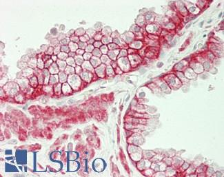
HER2 (ERBB2) is a tyrosine kinase/ epidermal growth factor receptor that is overexpressed and mutated in approximately 10-20% of breast cancers (Zaha, 2014). ERBB2 (HER2) exhibits positive membranous staining in malignant cells, where it promotes cancer cell growth. It is associated with more aggressive tumors. Immunohistochemistry staining of ERBB2 is used alongside the proteins ER (estrogen receptor), PR (progesterone receptor) and Ki-67 to classify different subtypes of breast cancer, and it is a widely used prognostic marker for breast cancer, as levels of ERBB2 expression predict a patient’s response to Herceptin therapy.
Staining: While this target can show either cytoplasmic or membranous staining, only membranous staining is relevant for cancer.


LSBio’s recommended antibody to HER2 for use in immunohistochemistry is HER2 Antibody LS‑B2133.

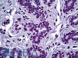
Anti-ERBB2 / HER2 antibody IHC of human breast, carcinoma. Immunohistochemistry of formalin-fixed, paraffin-embedded tissue after heat-induced antigen retrieval. Antibody LS-B2133 concentration 5 ug/ml.

Estrogen Receptor / ESR1


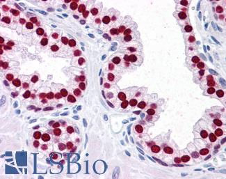
ER (Estrogen receptor / ESR1) is a biomarker that is prognostic for breast cancer and predicts response to hormonal therapy. Estrogen receptor is a nuclear hormone receptor with nuclear staining in malignant breast cancer cells, and usually has higher levels of expression in Luminal A subtypes compared to others (Jeselsohn, 2015). Levels of expression of estrogen receptor are used in conjunction with progesterone receptor (PR), HER2 and Ki-67 expression levels to subtype breast cancers for different downstream treatments. Most breast cancers have positive expression of ER protein, primarily in luminal tissues where the luminal A subtype sees high levels of expression (Jeselsohn, 2015; Yersal, 2014).
Staining: Staining for this target is expected to be nuclear.

LSBio’s recommended antibody to ER for use in immunohistochemistry is ER Antibody LS‑B10527.

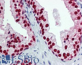
Anti-ER Alpha / Estrogen Receptor antibody IHC staining of human breast. Immunohistochemistry of formalin-fixed, paraffin-embedded tissue after heat-induced antigen retrieval. Antibody LS-B10527 dilution 1:100.

Progesterone Receptor / PGR


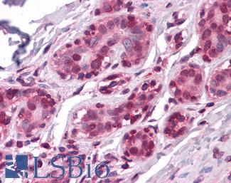
PR (progesterone receptor / PGR) is a biomarker that is prognostic for breast cancer and predicts response to hormonal therapy. Progesterone receptor is a nuclear hormone with nuclear staining in malignant breast cancer cells. Immunohistochemistry staining of PR is used alongside estrogen receptor (ER), HER2 and Ki-67 expression levels to subtype breast cancers for different downstream treatments. PR has been found to modulate the behavior of estrogen receptor protein and impede the growth of ER positive tumors, which coincides with the typically better prognosis of ER+, PR+ breast cancers when compared to other subtypes (Mohammed, 2015).
Staining: Staining for this target is expected to be nuclear.

LSBio’s recommended antibody to PR for use in immunohistochemistry is PR Antibody LS‑B2983.

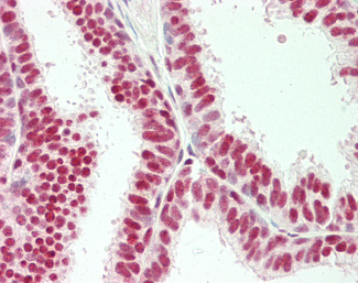
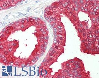
Anti-PGR / Progesterone Receptor antibody IHC of human uterus. Immunohistochemistry of formalin-fixed, paraffin-embedded tissue after heat-induced antigen retrieval. Antibody LS-B2983 concentration 20 ug/ml.

TP53


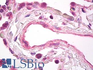
TP53 is a tumor suppressor that may be used as a marker for estrogen and progesterone receptor negative high grade breast cancers. It is associated with basal subtypes, where it may have aberrant or positive expression (Zaha, 2014). High levels of TP53 protein are often found in triple negative breast cancers (TNBC) alongside high levels of Ki-67. In TNBC, TP53 staining correlates with tumor grade and worse prognosis (Pan, 2017).
Staining: TP53 staining is typically nuclear, but antibodies to this target can also show weaker cytoplasmic staining.

LSBio’s recommended antibody to TP53 for use in immunohistochemistry is TP53 Antibody LS‑C172956.

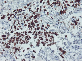
IHC of paraffin-embedded Carcinoma of Human lung tissue using anti-TP53 mouse monoclonal antibody.

EGFR


EGFR protein regulates a number of fundamental cell cycle pathways and is involved in migration and tumor invasion. Gene amplification of EGFR is a frequent occurrence in some types of breast cancer and also in numerous other tissues. Roughly half of triple negative and also IBC (inflammatory breast cancer) cases show overexpression of this protein (Masuda, 2012). Expression is generally associated with the basal subtype but is also seen in some luminal B tumors, and is correlated with lower ER / PR expression (Changavi, 2015).
Staining: This protein has membranous and cytoplasmic staining in IHC.

LSBio’s recommended antibody to EGFR for use in immunohistochemistry is EGFR Antibody LS‑B2914.

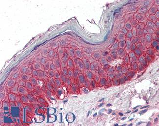
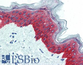
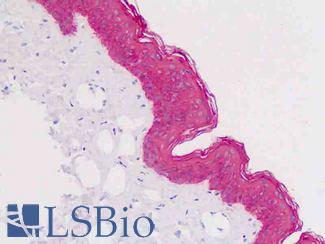
Anti-EGFR antibody IHC of human skin. Immunohistochemistry of formalin-fixed, paraffin-embedded tissue after heat-induced antigen retrieval. Antibody LS-B2914 concentration 5 ug/ml.

BRCA1


BRCA1 is a multifunctional protein involved in DNA repair, tumor suppression and cell replication, whose dysfunction through inherited or somatic mutation or through epigenetic silencing is the oncogenic source of a certain percentage of breast cancers (Romagnolo, 2015). Mutations in this gene confer high risk for both breast cancer and ovarian cancer, and thus immunohistochemistry staining and testing for wild type and mutated forms of this repair gene are necessary for diagnosis and downstream treatment (Anantha, 2017).
Staining: This protein has nuclear and cytoplasmic staining in IHC depending upon the isoform.

LSBio’s recommended antibody to BRCA1 for use in immunohistochemistry is BRCA1 Antibody LS‑B3772.

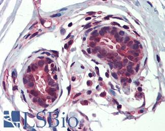
Anti-BRCA1 antibody IHC of human breast. Immunohistochemistry of formalin-fixed, paraffin-embedded tissue after heat-induced antigen retrieval. Antibody LS-B3772 concentration 5 ug/ml.

APOBEC3B


APOBEC3B is a repair gene and cytidine deaminase involved in somatic hypermutation. The deregulation or dysfunction of APOBEC3B and other members of the APOBEC family has been implicated as a direct source of mutagenesis in breast and other cancers (Nik-Zainal, 2012; McBride, 2008; Hwang, 2015). APOBEC3B is upregulated in a number of breast cancers, and this upregulation correlates with a mutation spectrum (kataegis) in these tumors consistent with APOBEC function and may be a simultaneous or downstream consequence of ERBB2 amplification (Nik-Zainal, 2012; Kanu, 2016).
Staining: Staining for APOBEC3B is expected to be nuclear.

LSBio’s recommended antibody to APOBEC3B for use in immunohistochemistry is APOBEC3B Antibody LS‑B12051.

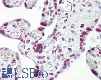
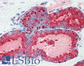
Human Uterus: Formalin-Fixed, Paraffin-Embedded (FFPE)

Ki-67 (MKI67)


Ki-67 (MKI67) is a marker for proliferation in breast cancer cells and many other tumor and tissue types. The degree of expression of Ki-67 is used in conjunction with ER, PR and HER2 expression levels to subtype breast cancers. Higher levels of this protein are correlated with increased rates of proliferation (Zaha, 2014), absence of ER and PR and poor prognosis, and staining for Ki-67 in immunohistochemistry is thus important for determining the necessary treatment (Zorka, 2014).
Staining: Staining for this target is expected to be nuclear.
LSBio’s recommended antibody to Ki-67 for use in immunohistochemistry is Ki-67 Antibody LS‑B3321.

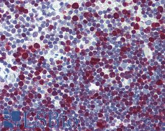
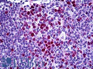
Anti-MKI67 / Ki-67 antibody IHC of human thymus. Immunohistochemistry of formalin-fixed, paraffin-embedded tissue after heat-induced antigen retrieval. Antibody LS-B3321 dilution 1:50.

CNN1 (Calponin)


CNN1 (calponin) is a myoepithelial marker in breast cancer with functional roles in proliferation, angiogenesis and migration (O’Kelly, 2008). In immunohistochemistry staining, calponin is found circulating the nucleus in the cytoplasm of myoepithelial cells (Zaha, 2014).

LSBio’s recommended antibody to Calponin for use in immunohistochemistry is Calponin Antibody LS‑B4304.

Anti-CNN1 / Calponin antibody IHC of human myometrium. Immunohistochemistry of formalin-fixed, paraffin-embedded tissue after heat-induced antigen retrieval. Antibody LS-B4304 concentration 5 ug/ml.

Smooth Muscle Actin (SMA / ACTA2)


Smooth Muscle Actin (SMA / ACTA2) is a myoepithelial marker for normal and cancerous breast tissue. Smooth muscle actin is often used alongside SMMHC, calponin and cytokines like CK5 and CK17 to distinguish myoephithelial cells from ductal carcinoma cells, which are negative for these proteins (Zaha, 2014). High levels of smooth muscle actin expression are correlated with poorer prognosis in breast cancers positive for EGFR and ERBB2; breast cancers with overexpression of these proteins see upregulation of ACTA2 likely due to its regulation by the upstream transcription activator STAT1. Overexpression of ACTA2 is correlated with an increase in invasive potential through its functions in the epithelial to mesenchymal transition (Jeon, 2016).
Staining: Staining for this target is cytoplasmic.

LSBio’s recommended antibody to SMA for use in immunohistochemistry is SMA Antibody LS‑B7351.

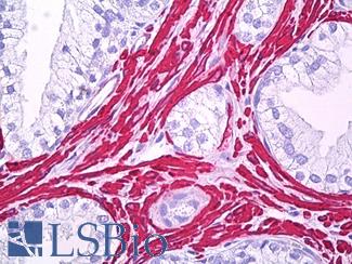
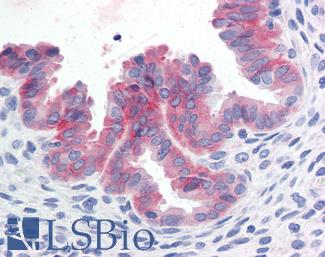
Anti-Smooth Muscle Actin antibody IHC of human prostate, smooth muscle. Immunohistochemistry of formalin-fixed, paraffin-embedded tissue after heat-induced antigen retrieval. Antibody LS-B7351 dilution 1:50.

SMMHC


SMMHC (smooth muscle myosin heavy chain / MYH11) is a myoepithelial breast cancer marker. It is overexpressed in HER2-positive, estrogen receptor- and progesterone receptor-negative breast tumors and under-expressed in triple negative breast cancers (Li, 2016). As a myoepithelial marker, MYH11 tends to provide more sensitive results with less myofibroblast cross-reactivity when compared to biomarkers like calponin and smooth muscle actin. It can be used to illustrate ductal wall stability, as ductal tumor cells usually lack expression of MYH11 (Zaha, 2014).
Staining: Staining for this target is expected to be cytoplasmic.

LSBio’s recommended antibody to SMMHC for use in immunohistochemistry is SMMHC Antibody LS‑B5148.

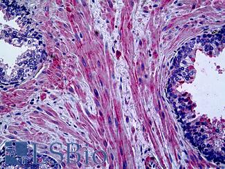
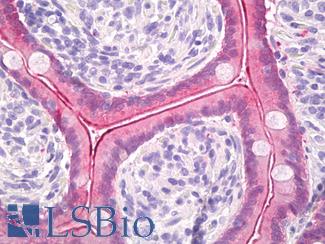
Anti-Myosin, Smooth Muscle Heavy Chain antibody IHC of human prostate. Immunohistochemistry of formalin-fixed, paraffin-embedded tissue after heat-induced antigen retrieval. Antibody LS-B5148 concentration 20 ug/ml.

CK5 (KRT5)


CK5 (KRT5) is a core basal-like and myoepithelial marker alongside EGFR in the immunohistochemistry of breast cancer (Yang, 2011). It has been found to have increased expression in luminal cells of adenoid cystic carcinoma (Nakai, 2016) and in KRT5 positive, estrogen receptor (ER) and progesterone receptor (PR) negative cells within luminal tumors have been correlated with treatment resistance and increased invasiveness (Axlund, 2013; Kabos, 2011).
Staining: Staining for this target is expected to be cytoplasmic.

LSBio’s recommended antibody to CK5 for use in immunohistochemistry is CK5 Antibody LS‑B3359.

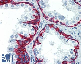
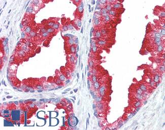
Anti-KRT5 / Cytokeratin 5 antibody IHC of human prostate. Immunohistochemistry of formalin-fixed, paraffin-embedded tissue after heat-induced antigen retrieval. Antibody LS-B3359 concentration 10 ug/ml.

CK7 (KRT7)


CK7 (KRT7) is a luminal cell biomarker for breast cancer (Abd El-Rehim, 2004). Cytokeratins are intermediate filaments that are involved in cytoskeletal integrity. This keratin is helpful for distinguishing Paget’s disease (breast cancer in the skin) from melanoma. In Paget’s disease, CK7 highlights malignant breast cancer cells in the epidermis (Karakas, 2011; Smith, 1997).
Staining: Staining for this target is expected to be cytoplasmic.

LSBio’s recommended antibody to CK7 for use in immunohistochemistry is CK7 Antibody LS‑B7164.

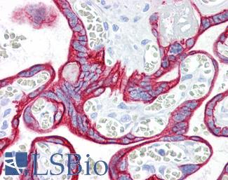
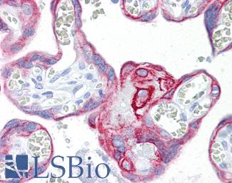
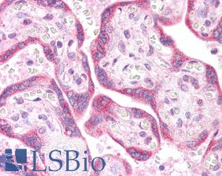
Anti-KRT7 / Cytokeratin 7 antibody IHC of human placenta. Immunohistochemistry of formalin-fixed, paraffin-embedded tissue after heat-induced antigen retrieval. Antibody LS-B7164 concentration 10 ug/ml.

CK14 (KRT14)


CK14 (KRT14) is a basal-like and myoepithelial marker for normal and cancer tissues of the breast. CK14 expression is linked with metastasis; a study from 2016 by Kevin J. Cheung et al found that metastatic polyclonal tumors shared high levels of KRT14 expression, indicating it as a biomarker for divergent invasive cancers in distant organs that followed an initial “collective invasion” into the stroma (Cheung, 2016). They found a drastic increase in CK14 expression in circulating tumor cells relative to primary tumors. CK14 may thus be an effective target not just as a myoepithelial marker but also for highly invasive metastatic polyclonal breast cancers.
Staining: Staining for this target is predominantly cytoplasmic.

LSBio’s recommended antibody to CK14 for use in immunohistochemistry is CK14 Antibody LS‑B3916.

Anti-KRT14 / Cytokeratin 14 antibody IHC of human skin. Immunohistochemistry of formalin-fixed, paraffin-embedded tissue after heat-induced antigen retrieval. Antibody LS-B3916 dilution 1:25.

CK20 (KRT20)


CK20 (KRT20) is an epithelial marker often used together with KRT7 (CK7) when trying to determine a metastatic carcinoma’s point of origin, and a CK7 positive, CK20 negative staining pattern is present in many breast cancers (Tsao, 2007). CK20 may be also be a useful marker for circulating tumor cells in breast cancer patients, and higher levels of expression in the peripheral blood have been found to be correlated with ER-negative high grade tumors and with lymph node metastases (Tunca, 2012; Lasa, 2013). Cytokeratins are intermediate filaments that contribute to cytoskeletal integrity in epithelial cell types, and cytokeratin 20 localizes to the cytoplasm.
Staining: Staining for this target is expected to be cytoplasmic.

LSBio’s recommended antibody to CK20 for use in immunohistochemistry is CK20 Antibody LS‑B10488.

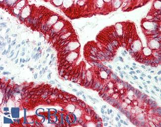
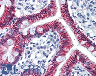
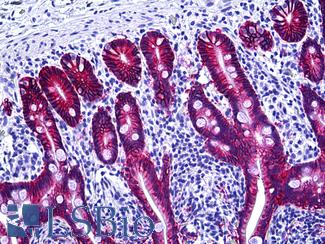
Anti-KRT20 / Cytokeratin 20 antibody IHC staining of human small intestine. Immunohistochemistry of formalin-fixed, paraffin-embedded tissue after heat-induced antigen retrieval. Antibody LS-B10488 dilution 1:200.

E-Cadherin (CDH1)


E-cadherin (CDH1), a cadherin involved in cell to cell adhesion, sees consistent loss of expression in invasive lobular carcinoma of the breast and this coincides with the induction of metastatic and invasive properties in lobular tumors (Ciriello, 2015). CDH1 is often knocked out in ERBB2/HER2- negative, estrogen receptor-positive invasive lobular cancers (Hugo, 2017) and this downregulation is a predictor of poor prognosis (Li, 2017). A study from 2017 looking at the MDA-MB-468 human breast cancer cell line found that e-cadherin knockdown is specifically correlated with invasion, while higher expression profiles are more closely linked with higher proliferation (Ping, 2016), highlighting its utility as a marker in immunohistochemistry staining of invasive lobular tumors.
Staining: Cadherins typically show membranous staining in immunohistochemistry.

LSBio’s recommended antibody to E‑cadherin for use in immunohistochemistry is E‑cadherin Antibody LS‑B4674.

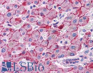
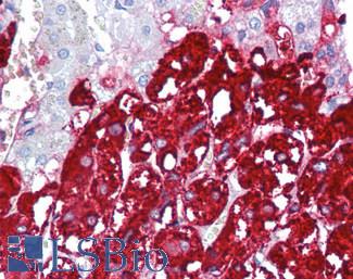
Anti-E Cadherin antibody IHC of human liver. Immunohistochemistry of formalin-fixed, paraffin-embedded tissue after heat-induced antigen retrieval. Antibody LS-B4674 concentration 5 ug/ml. This image was taken for the unconjugated form of this product. Other forms have not been tested.

p120 (CTNND1)


p120 (CTNND1) may be used alongside e-cadherin (CDH1) in immunohistochemistry as a marker for lobular breast carcinoma and has been found to be a useful biomarker for early lobular lesions (Dabbs, 2007). Alongside downregulation of e-cadherin, immunohistochemistry staining of p120 shows it to be strongly positive in lobular tumors, with redistribution from the membrane to the cytoplasm (Li, 2014; Li, 2010).
Staining: Staining for this target is expected to be nuclear.

LSBio’s recommended antibody to p120 for use in immunohistochemistry is p120 Antibody LS‑B14422.

Human Prostate: Formalin-Fixed, Paraffin-Embedded (FFPE)

FOXA1


FOXA1 is a transcription factor involved in differentiation that is necessary for estrogen receptor (ER/ESR1) chromatin interaction and regulates many of the same genes. It is a useful marker for luminal breast cancers, as it is often expressed in luminal subtypes independent of ER expression and it does not see expression in basal subtypes (Bernardo, 2013). In ER-negative tumors, a lack of FOXA1 expression is associated with worse prognosis, as loss of FOXA1 is associated with the consequential expression of metastases-prone, basal-specific proteins that it normally acts to directly repress (Bernardo, 2013; Albergaria, 2009; Gong, 2014).

LSBio’s recommended antibody to FOXA1 for use in immunohistochemistry is FOXA1 Antibody LS‑B4356.

Anti-FOXA1 antibody IHC of human prostate. Immunohistochemistry of formalin-fixed, paraffin-embedded tissue after heat-induced antigen retrieval. Antibody LS-B4356 concentration 5 ug/ml.

GATA3


The protein GATA3 is a transcription factor and is a useful biomarker for breast cancer. It has been found to be significantly mutated in luminal breast cancers (Goncalves, 2014), and it is also regularly highly expressed in breast carcinoma. It stains at high levels of expression across many subtypes including triple-negative breast cancers (Kandalaft, 2016) and for both primary and metastatic carcinomas (Sangoi, 2016), and it is considered a biomarker for the Luminal A subtype of epithelial cells (Yersal, 2014).
Staining: Staining for this target is expected to be nuclear.

LSBio’s recommended antibody to GATA3 for use in immunohistochemistry is GATA3 Antibody LS‑B4163.

Anti-GATA3 antibody IHC of human breast. Immunohistochemistry of formalin-fixed, paraffin-embedded tissue after heat-induced antigen retrieval. Antibody LS-B4163 dilution 1:200.

VEGFA


VEGFA protein is a marker for tumorigenesis that is involved in angiogenesis and proliferation and is regularly upregulated in breast and other cancers. VEGFA is consistently expressed throughout tumorigenesis and metastasis at higher levels than normal tissue, and seems to be particularly overexpressed in triple-negative breast cancers (Taneja, 2010). VEGFA is a growth factor in vascular endothelial cells, where it activates cellular proliferation, angiogenesis and migration (O’Leary, 2016). VEGFA is involved in activating PI3K-Akt, MEK-ERK and other downstream signaling pathways, and as an actor in angiogenesis it promotes an increase in microvessel density when overexpressed in cancer (Taneja, 2010).
Staining: Staining for this target is a secreted cytokine, and it can show both cytoplasmic and extracellular staining.

LSBio’s recommended antibody to VEGFA for use in immunohistochemistry is VEGFA Antibody LS‑B7747.

Anti-VEGF antibody IHC of human lung, vascular endothelium. Immunohistochemistry of formalin-fixed, paraffin-embedded tissue after heat-induced antigen retrieval. Antibody LS-B7747 dilution 5 ug/ml.

BCL2


BCL2 is a marker of apoptosis in breast cancer, where overexpression of this protein is associated with a more favorable prognostic outcome in estrogen receptor- (ER) positive patients. Despite this, the function of BCL2 is anti-apoptotic and it is considered an oncogene (Hwang, 2017), and ER-negative, BCL2-positive patients have been shown to have worse outcomes (Zaha, 2014). It is a useful marker for specifically the luminal A subtype of breast cancer (Yersal, 2014; Eom, 2016). BCL2 staining is typically cytoplasmic, but can also localize to membranes.

LSBio’s recommended antibody to BCL2 for use in immunohistochemistry is BCL2 Antibody LS‑B1173.

Anti-BCL2 / Bcl-2 antibody IHC of human spleen. Immunohistochemistry of formalin-fixed, paraffin-embedded tissue after heat-induced antigen retrieval. Antibody LS-B1173 concentration 20 ug/ml.

CD31


CD31 is an angiogenesis marker in breast cancer, and increased staining of vessels within lymph nodes is an indicator of potential lymph node metastases of invasive ductal carcinoma (Popiela, 2008; Zaha, 2014). This protein is expressed in endothelial cells within capillaries and vessels. CD31 is a cellular adhesion molecule, and staining is typically membranous.

LSBio’s recommended antibody to CD31 for use in immunohistochemistry is CD31 Antibody LS‑B3446.

Anti-CD31 antibody IHC of human placenta. Immunohistochemistry of formalin-fixed, paraffin-embedded tissue after heat-induced antigen retrieval. Antibody LS-B3446 dilution 1:200.

CD34


CD34 is a hematopoietic stem cell marker/adhesion protein that is also expressed in endothelium, where staining is typically membranous. This protein is also occasionally expressed in fibroblasts, and is present in the cytoplasm of mesenchymal cells surrounding benign glandular tissue in the breast. Consistent loss of expression in breast mesenchymal fibroblasts is associated with high grade, malignant breast cancers (Hua, 2011). Loss of expression of this protein in the stroma coincides with expression of smooth muscle actin and the adoption of invasive properties in ductal tumor cells (Catteau, 2013).

LSBio’s recommended antibody to CD34 for use in immunohistochemistry is CD34 Antibody LS‑B2652.

Anti-CD34 antibody IHC of human placenta. Immunohistochemistry of formalin-fixed, paraffin-embedded tissue after heat-induced antigen retrieval. Antibody LS-B2652 concentration 10 ug/ml.

SIAH2


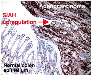
SIAH2 is a novel target in breast and other cancers; in a study on advanced and metastatic breast cancer, Siewertz van Reesama et al found that both SIAH and EGFR outperform standard breast cancer biomarkers in predicting advanced stages of metastasis. SIAH2 is a downstream member of the RAS pathway, where it is required for signal transduction (Van Reesema, 2016). SIAH2 expression acts as a sign of activation of the RAS pathway (Van Reesema, 2016), and its deficiency has been found to reduce MAPK signaling (Ahmed, 2008). SIAH2 has been found to be overexpressed in invasive breast cancers versus ductal carcinomas and normal tissue, where it may cause aberrant activation of the ERK pathway, encouraging proliferation and simultaneous dysregulation of apoptotic signaling (Sun, 2016).
Staining: Staining for this target can be both cytoplasmic and nuclear.

LSBio’s recommended antibody to SIAH2 for use in immunohistochemistry is SIAH2 Antibody LS‑C141889.

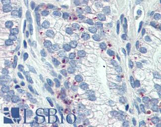
Staining of SIAH2 in human colorectal adenocarcinoma.

Prolactin-induced protein (PIP / GCDFP-15)


Prolactin-induced protein (PIP / GCDFP-15) is a secreted protein produced by benign and malignant salivary gland and breast tissue. PIP is overexpressed in many breast carcinomas, where increased expression has been found to increase the cell adhesive and proliferative properties of malignant cells (Vanneste 2015; Naderi 2015).
Staining: This target typically stains secretions (may appear cytoplasmic, membranous, and/or extracellular).

LSBio’s recommended antibody to PIP / GCDFP‑15 for use in immunohistochemistry is PIP / GCDFP‑15 Antibody LS‑B2498.

Anti-PIP / GCDFP15 antibody IHC of human breast. Immunohistochemistry of formalin-fixed, paraffin-embedded tissue after heat-induced antigen retrieval. Antibody LS-B2498 concentration 5 ug/ml.

Mammaglobin A (SCGB2A2)


Mammaglobin A (SCGB2A2) is a breast cancer biomarker that is tissue specific to breast and highly expressed in breast cancers. Overexpression of this protein has been correlated with proliferative and invasive properties in tumor cells (Picot, 2016), and coincides with simultaneous upregulation of Ki-67 (MKI67) protein – these two markers combined can be a useful pair in tissue specific immunohistochemistry staining of breast tumors (Gargano, 2006).
Staining: Staining for this target is has been demonstrated to be cytoplasmic and in secretions.

LSBio’s recommended antibody to SCGB2A2 for use in immunohistochemistry is SCGB2A2 Antibody LS‑B3007.

Anti-SCGB2A2 / Mammaglobin A antibody IHC of human breast. Immunohistochemistry of formalin-fixed, paraffin-embedded tissue after heat-induced antigen retrieval. Antibody LS-B3007 concentration 4 ug/ml.

PIM1


PIM1 is a marker in breast tumors with particular relevance as a critical proliferative regulator in triple-negative breast cancers (TNBC). PIM1 was found to regulate proliferation and apoptosis in TNBC, with overexpression that correlates with MYC-target transcription and tumor proliferation. It is a potential novel drug target in TNBC treatment (Brasó-Maristany, 2016).
Staining: Staining for PIM1 is expected to be cytoplasmic.

LSBio’s recommended antibody to PIM1 for use in immunohistochemistry is PIM1 Antibody LS‑B5493.

Anti-PIM1 antibody IHC of human adrenal. Immunohistochemistry of formalin-fixed, paraffin-embedded tissue after heat-induced antigen retrieval. Antibody LS-B5493 concentration 5 ug/ml.

Colorectal Cancer Immunohistochemistry Markers

Colorectal carcinoma (CRC) affects 4% of the population, and up to 30% of patients have a family history of the disease. Identified hereditary syndromes such as Lynch syndrome (Hereditary nonpolyposis colorectal cancer or HNPCC) and Familial adenomatous polyposis (FAP), however, only account for 10% of patients (Liccardo, 2017) and risk factors such as age, diet and smoking, or diseases such as diabetes and chronic inflammatory bowel diseases have also been implicated as increasing risk for the development of CRC (Siegel, 2017).


Early detection and screening methods such as sigmoidoscopy, colonoscopy or stool-based tests for the detection of fecal blood (guaiac) or mutations (FIT-DNA) can reduce CRC deaths by detecting cancers and removing polyps at early stages, but once the cancer develops, treatment is often surgical with chemotherapy. Newer drugs for targeted therapy include EGFR inhibitors to slow the growth of the cancer, or VEGF inhibitors to prevent the formation of blood vessels necessary for tumor growth.


IHC is used to identify CRC in tumors of unknown primary. CRCs express nuclear transcription factor CDX2, which is highly specific for intestinal epithelial cells, and Villin, which is specific to adenocarcinomas of the GI tract. GPA33 codes for a membranous protein that is expressed in a majority of colorectal tumors, and it is an effective marker particularly for well-differentiated cancers, where it is also a recent target for antibody radioimmunotherapy (Baptistella, 2016; Cheal, 2017). SATB2 may be a useful marker for primary and metastatic colorectal carcinomas due to its high specificity and function in chromatin remodeling. Other markers useful for identifying a colonic origin to adenocarcinomas include: mCEA, CA19-9, Calretinin, CDH17, COX-2, MOC-31 (EPCAM), CK20 and CK7, as well as MUC2. Additionally, a large number of colorectal cancers see deregulation and hotspot mutations in the oncogenes KRAS, BRAF, and PIK3CA.

The genetics of colorectal cancer:
CRC can arise from different but often overlapping genetic pathways. At a glance, colorectal cancer is divided into distal colorectal cancers with chromosomal instability and proximal cancers with high levels of mutation characterized by microsatellite instability (MSI) (Mouradov, 2014), with some crossover in the mutational spectrum across different subtypes of colorectal tumors. The most commonly mutated genes involve members of the WNT signaling pathway, the MAPK pathway, the PI3K pathway, the P53 pathway and the TGFB pathway (Mouradov, 2014; Kandalaft, 2016).
Genes in these pathways are associated with multiple inherited syndromes and also colorectal cancers of unknown origin, and include both inherited and somatic mutations in the adenomatous polyposis coli gene (APC, mutated in Familial Adenomatous Polyposis disorder or FAP) or in MUTYH (Win, 2016). The APC protein is an inhibitor of beta-catenin (CTNNB1), involved in cell-adhesion and nuclear transcriptional regulation, and loss of APC results in increased transcription of proto-oncogenes. APC loss of function has also been implicated in the inception of chromosomal instability alongside SMAD4, TP53 and somatic KRAS mutation (Mouradov, 2014; Pino, 2010). Other genes with functions similar to APC that can also be mutated in CRC include AXIN1, AXIN2, TCF7L2 or NKD1 (Novellasdemunt, 2015).
Lynch syndrome (LS) is another inherited colorectal cancer syndrome and has identified progenitor genes in the mismatch repair (MMR) protein complexes, including MLH1, MSH2, MSH6, MSH3, PMS1, PMS2 and also EPCAM. Characteristic of LS is microsatellite instability (MSI), which is caused by loss of function or silencing in the aforementioned MMR genes. MSI is a hyper-mutation phenotype that frequently occurs in colorectal cancers, and it is described by the broad appearance of insertion and deletion mutations as well as single nucleotide polymorphisms across the genome (Boland, 2010). These mutations occur due to polymerase slippage and misrepair in mono-, di-, tri- and tetranucleotide repeat tracts when MMR protein function is impaired (Shah, 2010). The resulting translated frameshifts and amino acid substitutions affect hundreds to thousands of proteins (Boland, 2018). It is a phenomenon common to tumor subtypes in many tissues, and is particularly prevalent in lymphoma, glioblastoma, colorectal, ovarian, and endometrial cancers (Cortes-Ciriano, 2017; Hause, 2016). Patients with LS who have an inherited predisposition for MSI can likewise develop numerous simultaneous tumors throughout their body with an age of onset in the mid 40’s. In the colon, patients have a tendency to develop poorly differentiated, right-sided tumors.
Patients with microsatellite instability but no known inherited MMR protein defect may be considered Lynch-like or have a mutation phenotype subset caused by inherited dysfunction in the DNA polymerase epsilon catalytic subunit (POLE) (Mouradov, 2014; Jansen, 2016), or alternatively these tumors may belong to the CIMP subtype. Roughly 20% of colorectal cancers may fall into the CIMP (CpG Island Methylator Phenotype) category, described by systematic epigenetic dysregulation and frequent BRAF V600E mutation, leading to MLH1 hyper-methylation rather than or alongside somatic mutation and consequential microsatellite instability. CIMP is typically a mechanism for the formation of sessile serrated adenomas. Targets that are often downregulated in CIMP cases include MLH1, KRT20, and CDX2 (Rhee, 2016), and genes that see frequent upregulation include the mucins MUC6, MUC5AC, and MUC5B (Walsh, 2013) as well as histone methylation and epigenetic regulation genes DNMT1 and CBX2 (Zhu, 2017; Li, 2013; Chen, 2017; Choi, 2014).

GPA33 (A33)


GPA33 (A33) gene codes for a membranous protein that is widely expressed in colorectal cancers, appearing in up to 95% of cases but particularly in well differentiated tumors. (Baptistella, 2016). Upregulation of GPA33 may be a downstream effect of peroxisome proliferator-activated receptor (PPARG) activation, which induces expression of KLF4, a transcription factor that targets GPA33 (Rageul, 2009). Antibodies to GPA33 used in the radiotherapy of colorectal cancers have been proposed as an effective means of treatment for tumors that express the protein (Cheal, 2017).
Staining: Staining for this protein is typically membranous in well differentiated tumors and in normal tissue, but may be primarily cytoplasmic or nuclear in poorly differentiated and mucinous tumors (Baptistella, 2016).

LSBio’s recommended antibody to GPA33 for use in immunohistochemistry is GPA33 Antibody LS‑B15164.

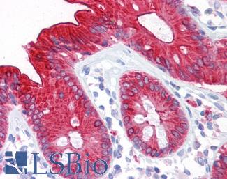
Human Colon: Formalin-Fixed, Paraffin-Embedded (FFPE)

CK20 (KRT20)


CK20 (KRT20) is a keratin that is expressed in epithelial cells in colorectal crypts, where levels of this protein have been found to increase gradually from the crypt bottom (where it is absent) to the top (Chan, 2009; Moll, 1990). It is frequently used as a differentiation marker in the colon. Chan et al found that CK20 is positively regulated by CDX1, a homeobox gene involved in promoting intestinal differentiation (Chan, 2009). In colorectal cancer, CK20 typically has strong expression paired with an absence of expressed CK7 (KRT7) (Chan, 2009; Harbaum, 2012) and its expression is often inversely correlated with levels of LGR5 (Shimokawa, 2017). In a study on several hundred primary colorectal tumors, Harbaum et al found that while the majority had high levels of CK20, microsatellite unstable (MSI) tumors tended to have reduced or depleted expression of the protein (Harbaum, 2012). Aggressive, poorly differentiated colorectal tumors and those with high rates of MSI may stain negatively for this marker in immunohistochemistry (Harbaum, 2012; Merlos-Suárez, 2011).
Staining: Staining is characteristically cytoplasmic.

LSBio’s recommended antibody to CK20 for use in immunohistochemistry is CK20 Antibody LS‑B5959.

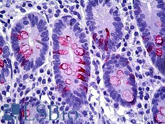
Anti-KRT20 / Cytokeratin 20 antibody IHC of human small intestine. Immunohistochemistry of formalin-fixed, paraffin-embedded tissue after heat-induced antigen retrieval. Antibody LS-B5959 dilution 1:50. This image was taken for the unconjugated form of this product. Other forms have not been tested.

CK7 (KRT7)


CK7 (KRT7) is a keratin expressed in a number of tissues including in the colon, where its expression is limited to glandular cells (proteinatlas.org). CK7 typically has negative expression in colorectal cancer, and as a marker in IHC antibody panels it is often paired with positive expression of CK20 in support of adenocarcinoma (Harbaum, 2012; Diagnostic Immunohistochemistry p. 521). However, KRT7 expression in colon cancer may vary with the subtype and aggressiveness of the tumor; while high levels of CK20 expression are linked with increased differentiation, conversely in some colorectal tumors higher levels of CK7 and low levels of CK20 have been found and are attributed to poor differentiation (Harbaum, 2012; Harbaum, 2011). Furthermore, microsatellite-stable colorectal cancers with BRAF mutation, a subtype known to be aggressive and have poor prognosis, have been found to express higher levels of CK7 than other typically negative subtypes (Landau, 2014).
Staining: Staining is characteristically cytoplasmic.

LSBio’s recommended antibody to CK7 for use in immunohistochemistry is CK7 Antibody LS‑B7163.

Anti-KRT7 / Cytokeratin 7 antibody IHC of human placenta. Immunohistochemistry of formalin-fixed, paraffin-embedded tissue after heat-induced antigen retrieval. Antibody LS-B7163 concentration 10 ug/ml.

Calretinin (CALB2)


Calretinin (CALB2) is a calcium-binding protein involved in calcium signaling. High levels of expression of calretinin in medullary carcinoma of the colon with strong focal staining in IHC have been documented, emphasizing its utility as an immunohistochemistry biomarker for detection of these cancers (Winn, 2008; Lin, 2014).
Staining: Staining is can be cytoplasmic, but also potentially membranous and nuclear.

LSBio’s recommended antibody to Calretinin for use in immunohistochemistry is Calretinin Antibody LS‑B4220.

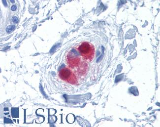
Anti-CALB2 / Calretinin antibody IHC of human colon, submucosal plexus. Immunohistochemistry of formalin-fixed, paraffin-embedded tissue after heat-induced antigen retrieval. Antibody LS-B4220 concentration 10 ug/ml.

CDH17


CDH17 is a cadherin involved in cell adhesion and proliferation and expressed in the glands of the colon, duodenum and small intestine as well as the appendix and gallbladder (Fagerberg, 2014; proteinatlas.org). In a study examining over 700 tumors across multiple tissues including the colon, Panarelli et al found that CDH17 has immunohistochemistry staining patterns in gastrointestinal cancers similar to CDX2 and is a highly specific marker for the intestinal epithelium (Panarelli, 2012). Furthermore, overexpression of this protein has been implicated in metastasis and invasive potential in colorectal cancer (Bartolome, 2014).
Staining: Staining is expected to be primarily membranous.

LSBio’s recommended antibody to CDH17 for use in immunohistochemistry is CDH17 Antibody LS‑B6022.

Anti-CDH17 / LI Cadherin antibody IHC of human small intestine. Immunohistochemistry of formalin-fixed, paraffin-embedded tissue after heat-induced antigen retrieval. Antibody LS-B6022 concentration 5 ug/ml.

Cyclooxygenase 2 (COX-2 / PTGS2)


Cyclooxygenase 2 (COX-2 / PTGS2) is a protein involved in inflammation, proliferation and cell renewal that is regularly upregulated in colorectal cancer and may play a role in its progression (Liu, 2017; Wang, 2009). Colorectal cancer patients with tumors expressing high levels of COX-2 who are treated with NSAIDs and COXIBs (COX-2 inhibitors) see up to a 50% decrease in their risk of developing colorectal cancer (Wang, 2009). Mutant APC mouse models that undergo inhibition of this protein have also seen a decrease in tumorigenesis (Brown, 2005).
Staining: Staining is expected to be cytoplasmic and potentially membranous.

LSBio’s recommended antibody to COX-2 for use in immunohistochemistry is COX-2 Antibody LS‑B1608.

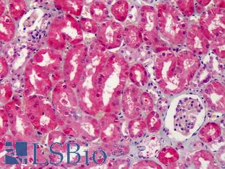
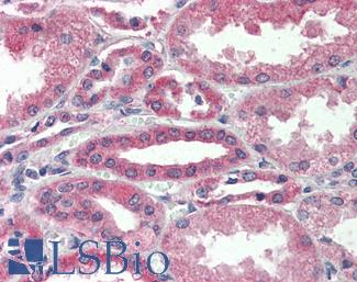
Anti-COX-2 antibody IHC of human kidney. Immunohistochemistry of formalin-fixed, paraffin-embedded tissue after heat-induced antigen retrieval. Antibody LS-B1608 concentration 5 ug/ml.

MUC2


MUC2 is a member of the mucin family of proteins and is a secreted protein that produces protective mucous in the gut (Allen, 1998). MUC2 is implicated in the progression of ulcerative colitis, where it is downregulated (Moehle, 2006), and also in the formation colorectal cancers (Betge, 2016). A MUC2 knockout model in mice demonstrated a propensity for colorectal cancer in its absence, as these mice frequently developed invasive colorectal adenocarcinomas (Velcich, 2002).
Staining: Staining is expected to be cytoplasmic and potentially in secretions.

LSBio’s recommended antibody to MUC2 for use in immunohistochemistry is MUC2 Antibody LS‑B5562.

Anti-MUC2 antibody IHC of human intestine. Immunohistochemistry of formalin-fixed, paraffin-embedded tissue after heat-induced antigen retrieval. Antibody LS-B5562 dilution 1:100.

CDX2


CDX2 codes for a homeobox protein involved in intestinal cell differentiation and tumor suppression in the colon (Platet, 2017), and loss of expression of this protein is correlated with colorectal tumorigenesis and poor survival (Platet, 2017; Dalerba, 2016). Platet et al found that higher levels of expression in the colon are correlated with a lack of cellular deformability and an increase in anti-metastatic structural properties (Platet, 2017). Furthermore, loss of expression of this protein may lead to tumor cells attaining stem cell attributes, as described by Lundberg IV et al in their findings that CDX2 downregulation is linked with upregulation of Yamanaka factor SOX2, involved in embryonic development and implicated in the induction of pluripotency (Lundberg, 2016; Takahashi, 2006). They found this to be a frequent phenomenon in MSI-positive (microsatellite unstable) and CIMP (CpG Island Methylator Phenotype) tumors. Jiang et al also found a link between methylation silencing of CDX2 (hypermethylation is a driving factor of CIMP) and colorectal cancer (Jiang, 2016). Downregulation of CDX2 may also be caused by FBXW7, a tumor suppressor with frequent mutations in cancer that manages the degradation of a large number of proteins including the Yamanaka factor Myc (Kumar, 2016; Takeishi, 2014). FBXW7 has been found to negatively regulate CDX2 expression by promoting its degradation together with the serine-threonine kinase GSK3B (Kumar, 2016).
Staining: Staining is expected to be nuclear.

LSBio’s recommended antibody to CDX2 for use in immunohistochemistry is CDX2 Antibody LS‑B1514.

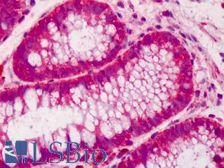
Anti-CDX2 antibody IHC of human colon. Immunohistochemistry of formalin-fixed, paraffin-embedded tissue after heat-induced antigen retrieval. Antibody LS-B1514 concentration 5 ug/ml. This image was taken for the unconjugated form of this product. Other forms have not been tested.

Villin


Villin (VIL1) is an actin-binding protein involved in the maintenance of microvilli in epithelial cells and is also involved in the epithelial-mesenchymal transition, and deregulation of this gene may contribute to tumorigenesis (Patnaik, 2016). Loss of expression of villin is seen in poorly differentiated colorectal carcinomas, particularly in microsatellite-unstable (MSI) tumors (Arango, 2012), and is linked to poor survival (Jaudah, 2013).
Staining: Staining is expected to localize to luminal membranes.

LSBio’s recommended antibody to Villin for use in immunohistochemistry is Villin Antibody LS‑B8547.

Anti-Villin antibody IHC of human small intestine, epithelial membrane. Immunohistochemistry of formalin-fixed, paraffin-embedded tissue after heat-induced antigen retrieval. Antibody LS-B8547 dilution 10 ug/ml.

MOC-31 (EPCAM)


MOC-31 is a clone of the protein EPCAM (TACSTD1) and is a useful biotarget for metastatic adenocarcinomas, including those originating in the colon. As an immunomarker, MOC-31 antibodies are used to distinguish adenocarcinoma from mesothelial cells with high sensitivity (Morgan, 1999; Kundu, 2011; Hecht, 2006). EPCAM itself is a protein involved in cell adhesion and has been implicated in the progression of a number of cancers (Dai, 2017; Baeuerle, 2007), included a proportion of individuals with Lynch Syndrome who hold inherited deletions in the gene. These deletions lead to silencing of the adjacent repair gene MSH2 via transcriptional read-through, which then causes microsatellite instability and colorectal cancer (as well as other malignancies that also frequently arise from Lynch Syndrome) (Kempers, 2010).
Staining: Staining is expected to be primarily membranous.

LSBio’s recommended antibody to EPCAM / MOC‑31 for use in immunohistochemistry is EPCAM / MOC‑31 Antibody LS‑B5565.

Anti-EPCAM antibody IHC of human intestine. Immunohistochemistry of formalin-fixed, paraffin-embedded tissue after heat-induced antigen retrieval. Antibody LS-B5565 dilution 1:100.

CEA / Carcinoembryonic Antigen


Carcinoembryonic (CEA / mCEA / pCEA) antigen includes a large class of proteins such as CEACAM5 that are biomarkers for colorectal cancer. These cell adhesion proteins have high expression in specifically metastatic cancer, making them suitable targets for immunohistochemistry of late tumors (Bajenova, 2014; Polat, 2014). The degree of expression of CEA is correlated with tumor stage, with particularly pronounced levels in well-differentiated adenocarcinomas relative to early tumors (which are expected to express the same levels as normal tissues) (Vukobrat-Bijedic, 2013).
Staining: Staining is expected to be membranous and cytoplasmic.

LSBio’s recommended antibody to CEA for use in immunohistochemistry is CEA Antibody LS‑B7173.

Anti-CEA antibody IHC of human colon. Immunohistochemistry of formalin-fixed, paraffin-embedded tissue after heat-induced antigen retrieval. Antibody LS-B7173 concentration 10 ug/ml.

SATB2


SATB2 is a transcription factor involved in chromatin remodeling that negatively regulates a number of stem cell markers such as AXIN2, CD44 and NANOG (Li, 2016; Dragomir, 2014). Loss of expression of SATB2 in colorectal cancer has been correlated with an increase in stemness and invasive potential (1). SATB2 may be a useful immunohistochemistry (IHC) marker for primary and metastatic colorectal cancers due to its high specificity (Dragomir, 2014).
Staining: Staining is expected to be primarily nuclear.

LSBio’s recommended antibody to SATB2 for use in immunohistochemistry is SATB2 Antibody LS‑B4981.

Anti-SATB2 antibody IHC of human brain, cortex. Immunohistochemistry of formalin-fixed, paraffin-embedded tissue after heat-induced antigen retrieval. Antibody LS-B4981 concentration 5 ug/ml.

Carbohydrate antigen (CA19-9/FUT)


Carbohydrate antigen (CA19-9/FUT) is a common metastatic colorectal cancer marker to tetrasaccharide carbohydrate (Sialyl-Lewis A), which has high levels of occurrence alongside CEACAM5 (CEA) expression levels in late tumors (Vukobrat-Bijedic, 2013; Polat, 2014). CA19-9 is an effective marker for circulating tumor cells, as increased levels of CA19-9 in serum coincide with the quantity of circulating tumor cells from colorectal metastases (Zhao, 2017).
Staining: Staining is primarily cytoplasmic.

LSBio’s recommended antibody to CA19-9 for use in immunohistochemistry is CA19-9 Antibody LS‑B5366.

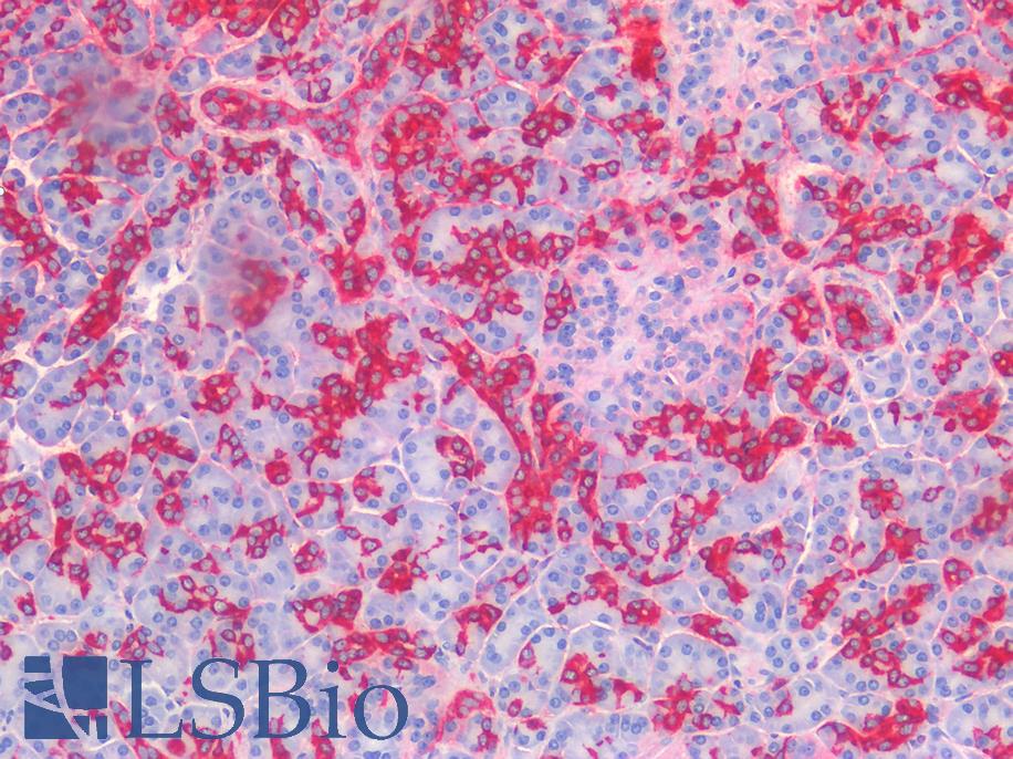
Human Pancreas: Formalin-Fixed, Paraffin-Embedded (FFPE)

TP53


TP53 is a tumor suppressor, repair gene and transcriptional regulator, and it is considered the most frequently mutated gene in cancer (Andrysik, 2017). Germline mutations in TP53 lead to Li-Fraumeni syndrome and cancer, including early-onset colorectal cancer (Yurgelun, 2015). In colorectal cancer, the expression of TP53 with gain of function mutations is a regular occurrence and is correlated with an increase in proliferative and invasive attributes in tumors of advanced stage (Andrysik, 2017; Sun, 2016). Furthermore, TP53 is often overexpressed in colorectal dysplasia resulting from ulcerative colitis (Kobayashi, 2017).
Staining: Staining is expected to be nuclear.

LSBio’s recommended antibody to TP53 for use in immunohistochemistry is TP53 Antibody LS‑B7722.

Anti-p53 antibody IHC of human placenta. Immunohistochemistry of formalin-fixed, paraffin-embedded tissue after heat-induced antigen retrieval. Antibody LS-B7722 dilution 10 ug/ml.

BRAF


BRAF, a MAPK/ERK signaling pathway kinase, is one of the most frequently somatically mutated genes in some cancers, particularly melanoma, thyroid carcinomas, and colorectal cancer (Chang, 2016; Zhang, 2014; Cantwell-Dorris, 2011). BRAF mutation occurs in roughly 10% of colorectal cancers, where detection of the V600E (rs113488022) mutation is useful for subtyping CIMP (CpG Island Methylator Phenotype), particularly when it accompanies the disappearance of the repair protein MLH1 (Cantwell-Dorris, 2011). These microsatellite-unstable (MSI) tumors typically propagate along the sessile serrated adenoma development pathway (Rhee, 2016; Cantwell-Dorris, 2011) and experience epigenetic silencing and loss of function frameshift in a large number of tumor suppressor and cell cycle regulation genes including TGFBR2, BAX, RNF43, CASP5 and KMT2C (MLL3) (Cortes-Ciriano, 2017). This is caused by the hypermethylation-induced silencing of mismatch repair protein MLH1, which occurs subsequent to and may even be caused by BRAF mutation (Fang M, 2014; Cantwell-Dorris, 2011). BRAF V600E is a pro-proliferative mutation, and while the adoption of V600E-mutant BRAF protein alone is not a pathogenic event (this mutation is the source of overproliferation in many benign nevi) (Wu, 2007), when combined with mutations in tumor suppressor genes it leads to carcinogenesis.
Staining: Staining may be nuclear, cytoplasmic or membranous.
LSBio’s recommended antibody to BRAF for use in immunohistochemistry is BRAF Antibody LS‑B1627.

Anti-BRAF antibody IHC of human brain, cortex. Immunohistochemistry of formalin-fixed, paraffin-embedded tissue after heat-induced antigen retrieval. Antibody LS-B3862 dilution 1:200.

PIK3CA


PIK3CA is a kinase frequently mutated in cancer, including up to 15% of colorectal cancers. This gene is regularly mutated in proximal cancers alongside KRAS mutation and MGMT silencing (Rosty, 2013), and frequently experiences the same somatic mutations characterized by H1047R, E545K, and E542K (Chang, 2016). The acquisition of somatic mutations in this gene may lower rates of survival in drug-resistant colorectal tumors that also harbor KRAS-mutations (Xu, 2017).
Staining: Staining is expected to be primarily cytoplasmic but ay also localize to membranes.

LSBio’s recommended antibody to PIK3CA for use in immunohistochemistry is PIK3CA Antibody LS‑B5363.

Anti-PIK3CA antibody IHC of human liver. Immunohistochemistry of formalin-fixed, paraffin-embedded tissue after heat-induced antigen retrieval. Antibody LS-B5363 concentration 5 ug/ml.

RECQL4


RECQL4 is a RECQ-like helicase involved in aging and DNA repair that forms a complex with TP53 (Qiao, 2016; Croteau, 2012). This protein has mutations, amplified copy numbers and upregulation in a number of cancers including colorectal, breast and gastric cancer (Qiao, 2016; Cancer Genome Atlas Network, 2012; Mo, 2016; Arora, 2016; Lao, 2013; Buffart, 2005; Giannakis, 2016). Amplification of this protein in metastatic colorectal cancer seems to be correlated with other oncogenes on chromosome 8, including Yamanaka factor MYC, SLA, PTK2, PTP4A3, TPD52, MOS (Buffart, 2005), as well as PLEC, EPPK1, RAD21, and PDS5B (Cancer Genome Atlas Network, 2012; Buffart, 2005; Giannakis, 2016). In gastric cancer cells, high levels of expression of RECQL4 have been linked to drug resistance and activation of the multi-drug resistant protein 1 (MDR1 / P-glycoprotein 1 / ABCB1 / CD243) (Mo, 2016). IHC (immunohistochemistry) staining of RECQL4 in colorectal cancer showed this protein localizing to the nucleus (Lao, 2013).
Staining: Staining is expected to be predominantly nuclear.

LSBio’s recommended antibody to RECQL4 for use in immunohistochemistry is RECQL4 Antibody LS‑B13621.

Human Colon: Formalin-Fixed, Paraffin-Embedded (FFPE)

Adenomatous Polyposis Coli (APC)


Adenomatous Polyposis Coli (APC) is a gene frequently mutated in colorectal cancer and inherited mutations in this gene are causative for classical familial adenomatous polyposis. This gene is involved in the regulation of cell adhesion through its interaction with CDH1 (e-cadherin) and CTNNB1 (beta-catenin) via the WNT signaling pathway (Markowitz, 2009), and it is considered a tumor suppressor. The loss of functional APC protein (often alongside mutations in TP53 or KRAS) is a frequent event in colorectal cancers and is associated with cellular over-proliferation, chromosomal instability and carcinogenesis (Pino, 2010).
Staining: Staining is expected to be primarily cytoplasmic.

LSBio’s recommended antibody to APC for use in immunohistochemistry is APC Antibody LS‑B2788.

Anti-APC antibody IHC of human colon. Immunohistochemistry of formalin-fixed, paraffin-embedded tissue after heat-induced antigen retrieval. Antibody LS-B2788 concentration 10 ug/ml.

MLH1


MLH1 is a primary MMR (mismatch repair) DNA repair gene, and loss of function of MLH1 protein leads to microsatellite instability and cancer in the colon and other organs (Papadopoulos, 1994). This can be caused by a “second hit” somatic mutation in MLH1 in patients with inherited heterozygous pathogenic mutations (Ollikainen, 2007), or alternatively through epigenetic silencing via hypermethylation of the MLH1 promoter in CIMP (CpG Island Methylator Phenotype) colorectal tumors (Boland, 2010). Inherited MLH1 mutation is one of the most frequent causes of Lynch Syndrome, and it is a pivotal gene in the mismatch repair functionality of cells (Cohen, 2014). IHC staining for MLH1 is therefore useful for determining the tumor subtype of colorectal cancer patients, as in Lynch Syndrome, Lynch-like cancers and in CIMP cases expression of this protein regularly disappears.
Staining: Staining is expected to be nuclear.

LSBio’s recommended antibody to MLH1 for use in immunohistochemistry is MLH1 Antibody LS‑B3475.

Anti-MLH1 antibody IHC of human small intestine. Immunohistochemistry of formalin-fixed, paraffin-embedded tissue after heat-induced antigen retrieval. Antibody LS-B3475 dilution 1:200.

MSH2


MSH2 is a DNA repair protein involved in mismatch repair and inherited or somatic mutations in this gene are a source of microsatellite instability (MSI) and cancer in the colon. Heterozygous germline mutations in MSH2 are the cause of disease in a large subset of Lynch Syndrome (LS) patients (Cohen, 2014); in LS, somatic mutation of the remaining functional copy of this protein leads to MSI, widespread mutation and LOF in a number of important tumor suppressor and apoptotic genes, and subsequently tumorigenesis throughout the colon (Lynch, 2009).
Staining: Staining is expected to be nuclear.

LSBio’s recommended antibody to MSH2 for use in immunohistochemistry is MSH2 Antibody LS‑B1973.

Anti-MSH2 antibody IHC of human breast. Immunohistochemistry of formalin-fixed, paraffin-embedded tissue after heat-induced antigen retrieval. Antibody LS-B1973 concentration 5 ug/ml.

MSH6


MSH6 is a DNA repair protein involved specifically in the mismatch repair of mutations that arise from replication and recombination, and somatic and inherited mutations in this gene lead to microsatellite instability and colorectal cancer. MSH6, along with MSH2, MSH3, MLH1, PMS1 and PMS2, is dysfunctional in the germline of many Lynch Syndrome patients (Houlleberghs, 2017; Giglia, 2016), and genetic testing along with immunohistochemistry staining of this protein is useful for diagnosis and subtyping of colorectal tumors (Giglia, 2016).
Staining: Staining is expected to be nuclear.

LSBio’s recommended antibody to MSH6 for use in immunohistochemistry is MSH6 Antibody LS‑B10739.

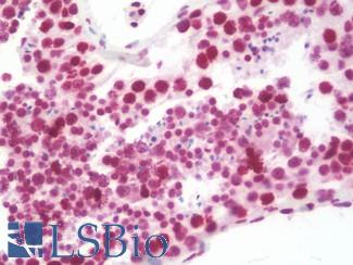
Anti-MSH6 antibody IHC staining of human testis. Immunohistochemistry of formalin-fixed, paraffin-embedded tissue after heat-induced antigen retrieval. Antibody LS-B10739 dilution 1:50.

PMS2


PMS2 is a DNA mismatch repair protein that is often mutated or downregulated in colorectal cancer. Inherited mutations in PMS2 are the cause of Lynch Syndrome and colorectal cancer in a subset of patients (Rosty, 2016; Cohen, 2014). Knockout of PMS2 results in microsatellite unstable tumors with malignant potential, and IHC staining for this protein can thus be used to subtype colorectal tumors (Worthley, 2005).
Staining: Staining is expected to be nuclear.

LSBio’s recommended antibody to PMS2 for use in immunohistochemistry is PMS2 Antibody LS‑B1980.

Anti-PMS2 antibody IHC of human breast. Immunohistochemistry of formalin-fixed, paraffin-embedded tissue after heat-induced antigen retrieval. Antibody LS-B1980 concentration 5 ug/ml.

AXIN1


AXIN1 is a protein involved in the WNT signaling pathway and acts alongside APC, CK1, AXIN2 and GSK3B in the regulation of B-Catenin (CTNNB1) phosphorylation and degradation (Mazzoni, 2014; Novellasdemunt, 2015). Mutations in this gene are prevalent in microsatellite-unstable (MSI) colorectal cancers, where loss of function of this protein may have a negative activating impact on WNT signaling and B-Catenin management and contribute to tumorigenesis (Novellasdemunt, 2015; Segditsas, 2006).
Staining: Staining may be cytoplasmic, nuclear and/or membranous.

LSBio’s recommended antibody to AXIN1 for use in immunohistochemistry is AXIN1 Antibody LS‑B2741.

Anti-AXIN1 / AXIN antibody IHC of human adrenal. Immunohistochemistry of formalin-fixed, paraffin-embedded tissue after heat-induced antigen retrieval. Antibody LS-B2741 concentration 75 ug/ml.

AXIN2


AXIN2, also known as conductin, is a member of the WNT signaling pathway and maintains the degradation and phosphorylation of B-Catenin (CTNNB1) alongside APC, AXIN1, CK1 and CSK3B in basal colorectal cells (Mazzoni, 2014; Novellasdemunt, 2015; Segditsas, 2006). AXIN2 is somatically mutated in microsatellite-unstable (MSI) colorectal cancers, where loss of function contributes to aberrant activation of WNT signaling and tumorigenesis. Germline mutations in this gene are also associated with a predisposition for colorectal carcinoma (Segditsas, 2006).
Staining: Staining may be cytoplasmic, nuclear and/or membranous.

LSBio’s recommended antibody to AXIN2 for use in immunohistochemistry is AXIN2 Antibody LS‑B7029.

Anti-AXIN2 / AXIL antibody IHC of human uterus. Immunohistochemistry of formalin-fixed, paraffin-embedded tissue after heat-induced antigen retrieval. Antibody LS-B7029 concentration 5 ug/ml.

TCF7L2


TCF7L2 is a member of the WNT signaling pathway and interacts with B-catenin. It is frequently mutated in MSI- (microsatellite instability) positive colorectal tumors, and also forms fusion transcripts with the neighboring VTI1A gene in some colorectal cancers (Nome, 2014). Homozygous germline mutations in this gene (mutations rs12255372 and rs7903146) have been associated with a risk for colorectal cancer as well (Folsom, 2008; Sainz, 2012; Hazra, 2008).
Staining: Staining may be nuclear and cytoplasmic.

LSBio’s recommended antibody to TCF7L2 for use in immunohistochemistry is TCF7L2 Antibody LS‑B7129.

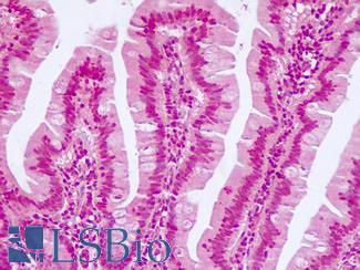
Anti-TCF7L2 antibody IHC of human small intestine.

Lung Cancer Immunohistochemistry Markers

Lung cancer is the second most common cause of cancer deaths in the U.S. Lung cancers exist as three subtypes: non-small cell lung cancers (NSCLC), which are comprised of the adenocarcinoma and squamous cell carcinoma subtypes that together account for 75% of cases, and small cell lung cancers (neuroendocrine origin) which account for the remainder.


The availability of targeted therapies makes it essential to correctly subtype NSCLCs: IHC markers such as NKX2-1 / TTF-1, Napsin A, and surfactant A (adenocarcinoma) or p63, CK5 / CK6, SOX2 and Desmoglein-3 (squamous cell carcinoma) can be used to identify the subtype in cases where the morphology is unclear, or with biopsies where there is insufficient material to make the diagnosis (Noh, 2012; Gurda, 2015). Small cell lung cancers can be identified with markers such as LMWK, CAM5.2, chromogranin, synaptophysin, CD56, and NKX2-1 / TTF-1 (Travis, 2012).


Although platinum-based chemotherapy has been the standard treatment for metastatic NSCLC, lung cancers with mutations associated with epidermal growth factor receptor (EGFR) and anaplastic lymphoma kinase (ALK) may respond to specific kinase inhibitors, so there is a need to identify the subset that show these mutations (Cooper, 2011; Malhotra, 2017; Kumar, Curr Probl Cancer 2017 Mar – Apr;41(2):111-124). Furthermore, among smoking-induced tumors, some with EGFR mutations form resistance to these kinase inhibitors and show overexpression of the AHR transcription factor. Upregulation of AHR may be providing resistance by activating downstream oncogenes such as Src protein. High AHR expression in these tumors is associated with worse prognosis and it may therefore be relevant to test for AHR levels through immunohistochemistry (Ye, 2017). Separately, recent immunotherapies with PD1/PDL1 checkpoint inhibitors of the interaction between the ligand PD-L1 with its receptor PD-1 have provided a further basis for patient selection and treatment (Cagle, Arch Pathol Lab Med 2016 Apr; 140: 322-325; Borghaei, NEJM 2015; 373:1627-1639).

The genetics of lung cancer:
NSCLCs experience a number of frequent mutations and pathogenic copy number alterations in RAS/RAF and tyorisine kinases and other common oncogenes and usually show high levels of chromosomal instability. KRAS undergoes activating mutations in up to 40% of lung adenocarcinomas (El-Telbany, 2018; Kanwal, 2017). Mutations in BRAF kinase (most commonly the V600E variant) occur at a less frequent but still significant rate. EGFR is another frequent target of lung cancer, and is regularly overexpressed and mutated (Kanwal, 2017). Data from recent sequencing initiatives by the Cancer Genome Atlas showed mutations in TP53 in 50-80% of samples across two pools of patients (504 and 1144 samples respectively), with alterations most frequent in squamous cell carcinomas. Copy number amplification of the Yamanaka transcription factor SOX2 was supported by CNV data in 20-50% of cases, while tumor suppressor proteins CDKN2A and CDKN2B saw recurrent deletions (20-30% of cases) (TCGA Lung Squamous Cell Carcinoma, Provisional; Campbell, 2016; Gao, 2013; Cerami, 2012; data is available at cBioPortal).
Other studies have shown rearrangements in ALK, ROS1 and RET kinases and amplifications of MET, FGFR1, and PDGFRA kinases (Zhu, 2017). Further frequently mutated pan-cancer oncogenes that often undergo driving mutations in lung adenocarcinoma include H3F3A and H3F3B, HER2 (ERBB2), PIK3CA, PTEN, and DDR2 (Rothschild, 2015). Finally, in the realm of histone methylation and proteins associated with epigenetic regulation, DOT1L, KMT2C (also implicated in tumor suppression), and SETD2 are among the most frequently mutated genes (Huang, 2017; Campbell, 2016).
In trying to uncover the origins of genomic damage and instability in lung cancer, recent research stemming from advances in sequencing shows that lung carcinomas can be grouped into the distinct repeat mutational signatures that they exhibit. Campbell et al discussed these signatures in a study on squamous cell and adenocarcinomas of the lung. In addition to known mutation patterns resulting from UV damage and smoking, they found evidence of APOBEC3B-specific repair gene dysfunction that is also seen in a large percent of breast cancers (Nik-Zainal, 2012; Morganella, 2015; Nik-Zainal, 2017; Nik-Zainal, 2016; Campbell, 2016). Additionally, they uncovered a cluster of lung cancers with microsatellite instability (MSI) and low levels of MLH1 similar to the CIMP and Lynch phenotypes of MSI-positive colorectal cancer.

PDL1 (CD274)


PDL1 (CD274), or programmed cell death-ligand 1, is an immune checkpoint receptor involved in immune escape in cancer, and it is upregulated in many cancers including NSCLC (Zhang, 2016). High levels of expression of PDL1 are associated with poor prognosis in NSCLC and pulmonary lymphoepithelioma-like carcinoma (Zhang 2016). Expression is also associated with EGFR mutations, the onset of which may directly lead to upregulation of PDL1 (Chen, 2015; Tang, 2015). Levels of expression of PDL1 detected through immunohistochemistry can predict response to PD1 therapy (Sorensen, 2016). Current clinical trials involving PDL1 inhibitors have shown impressive results in treating advanced stage cancers, particularly melanoma. Placental trophoblasts and macrophages are the best positive controls for this target.
Staining: This protein may have cytoplasmic or membranous staining in immune cells and tumor cells.

LSBio’s recommended antibody to PDL1 / CD274 for use in immunohistochemistry is PDL1 / CD274 Antibody LS‑B10562.

Anti-B7-H1 / PD-L1 / CD274 antibody IHC staining of human testis. Immunohistochemistry of formalin-fixed, paraffin-embedded tissue after heat-induced antigen retrieval. Antibody LS-B10562 dilution 1:100.

PD1 (PDCD1 / CD279)


PD1 (Programmed Death Receptor 1) is an immune checkpoint protein active in T cells that is a target alongside its ligand PDL1 and also CTLA-4 for immunotherapy in lung and other cancers (Alsaab, 2017). PD1 is involved in negatively regulating T cell inflammatory activity and, when bound to receptors on tumor cells, can work to subdue tumor suppression by inhibiting the immune response. Targeted inhibition of PD1 itself can therefore function as an anti-cancer therapy by reactivating this response (Jin, 2011; Francisco, 2010; Fife, 2011).
Staining: PD1 is expected to have membranous staining in immune cells and tumor cells.

LSBio’s recommended antibody to PD1 (PDCD1) for use in immunohistochemistry is PD1 (PDCD1) Antibody LS‑B7883.

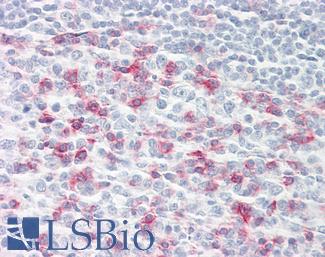
Anti-CD279 / PD-1 antibody IHC of human tonsil. Immunohistochemistry of formalin-fixed, paraffin-embedded tissue after heat-induced antigen retrieval. Antibody LS-B7883 dilution 20 ug/ml.

AHR


Aryl hydrocarbon receptor (AHR) is a transcription factor involved in xenobiotic metabolism and autophagy and it is expressed in many tissues including lung. In cancer, AHR is involved in migration and proliferation, and is upregulated in pancreatic, liver, head and neck, lung, thyroid and gastrointestinal tumors. AHR appears to function in both tumor suppression and tumorigenesis depending on the cell type and tissue (Tsai, 2017; Safe, 2013). In the lung, AHR expression is correlated with smoking-induced tumors, where carcinogens such as BaP directly bind to AHR and induce xenobiotic metabolism. Activation of AHR leads to upregulation of CYP1A1 and other cytochrome P450 enzymes, which are responsible for metabolizing carcinogens but can sometimes create a more damaging substance in error (Tsay, 2013). Higher expression of AHR in lung carcinomas is associated with a worse prognosis, particularly in tumors with mutant EGFR. In these tumors, the activation of AHR induces oncogenic Src expression and functional proliferative signaling (PI3K/Akt), resulting in resistance to tyrosine kinase inhibitors that target EGFR (Ye, 2017).
Staining: AHR is expected to have nuclear and cytoplasmic staining.

LSBio’s recommended antibody to AHR for use in immunohistochemistry is AHR Antibody LS‑A3018.

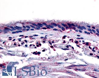
Anti-Aryl Hydrocarbon Receptor antibody LS-A3018 IHC of human respiratory epithelium and bronchial smooth muscle. Immunohistochemistry of formalin-fixed, paraffin-embedded tissue after heat-induced antigen retrieval.

Surfactant Protein A (SFTPA1)


Surfactant protein A (SFTPA1) is involved in host defense in the lung and its expression is necessary for tubular myelin formation and in reducing surface tension within alveoli (Lopez-Rodriguez, 2016). Germline mutations in SFTPA1 that result in impaired surfactant secretion and respiratory insufficiency have been implicated in a hereditary predisposition for lung fibrosis and cancer (Nathan, 2016). The specific nature of surfactant A to lung tissue has made it a useful marker in lung diseases including adenocarcinoma (Takahashi, 2006). It has comparable sensitivity to marker NKX2-1 /TTF-1 and is particularly useful in distinguishing NKX2-1-positive lung from thyroid tumors (Tan, 2008).
Staining: Staining of SFTPA1 is expected to be cytoplasmic in lung tissue pneumocytes.

LSBio’s recommended antibody to Surfactant Protein A for use in immunohistochemistry is Surfactant Protein A Antibody LS‑B2543.

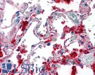
Anti-SFTPA1 / Surfactant Protein A antibody IHC of human lung. Immunohistochemistry of formalin-fixed, paraffin-embedded tissue after heat-induced antigen retrieval. Antibody LS-B2543 concentration 10 ug/ml.

SOX2


SOX2 is a Yamanaka factor and master regulator involved in embryogenesis and transdifferentiation (Takahashi, 2006; Whyte, 2013). In squamous cell carcinoma of the lung, SOX2 frequently has amplified copy number (~20% of cases) and overexpression of the gene is found in roughly 90% of cases (Fukazawa, 2015; Rudin, 2012). Upregulation of SOX2 is likely a primary determinant of the transformation of basal cells into carcinomas of various subtypes, and the variable upstream inactivation of different tumor suppressors seems to decide which subtype. Specifically, in the lung SOX2 overexpression results in squamous cell carcinoma following loss of PTEN, CDKN2A/CDKN2B and/or STK11, while upregulation subsequent to TP53 inactivation results in adenocarcinoma (Murray, 2016; Ferone, 2016).
Staining: Staining for this protein is expected to be nuclear.

LSBio’s recommended antibody to SOX2 for use in immunohistochemistry is SOX2 Antibody LS‑B4562.

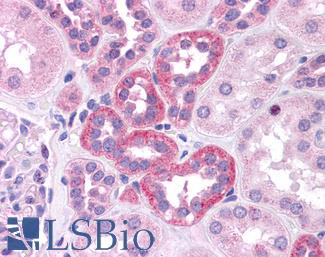
Anti-SOX2 antibody IHC of human kidney. Immunohistochemistry of formalin-fixed, paraffin-embedded tissue after heat-induced antigen retrieval. Antibody LS-B4562 concentration 5 ug/ml.

APOBEC3B


APOBEC3B is a cytidine deaminase involved in DNA repair and also somatic hypermutation. The deregulation of APOBEC family members including APOBEC3B has been implicated as a frequent driving source of mutagenesis in breast cancer and in other tissues (Nik-Zainal, 2012; McBride, 2008; Hwang, 2015). Campbell et al have also identified APOBEC mutation signatures in lung cancer, and further studies have found evidence of APOBEC3B upregulation in lung cancer and a correlation between increased expression and worse prognosis in NSCLC , making it an interesting and potentially important target for immunohistochemistry (Campbell 2016; Sasaki, 2014; Shumei, 2016).
Staining: Staining of APOBEC3B is expected to be nuclear.

LSBio’s recommended antibody to APOBEC3B for use in immunohistochemistry is APOBEC3B Antibody LS‑B12051.

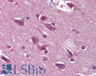
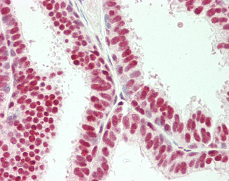
Human Uterus: Formalin-Fixed, Paraffin-Embedded (FFPE)

MET


MET (hepatocyte growth factor receptor / HGFR) is a receptor tyrosine kinase that is normally active in embryonic stem cells and progenitor cells. During embryogenesis, MET expression allows cells to burrow into adjacent areas in order to generate new embryonic structures, and in adult tissues, this invasive potential is a normal and essential component of wound repair. MET also promotes cell growth and inhibits apoptosis. In cancers, Met activation is associated with an aggressive, invasive phenotype and confers a poor prognosis, triggering tumor growth and ingrowth of new blood vessels. MET deregulation is common in human cancers, and contributes to therapeutic resistance, as it can be induced by chemotherapeutic agents and radiation. Hence Met inhibition, either targeting the receptor or HGFR ligand, is an area of interest, both as a primary and adjuvant means of treating cancer.
Staining: MET is expected to have membranous and cytoplasmic staining.

LSBio’s recommended antibody to c-MET for use in immunohistochemistry is c-MET Antibody LS‑B2812.

Anti-c-Met antibody IHC of human uterus. Immunohistochemistry of formalin-fixed, paraffin-embedded tissue after heat-induced antigen retrieval. Antibody LS-B2812 concentration 10 ug/ml.

CK5 (KRT5)


CK5 (KRT5) is positive in mesothelioma and in squamous carcinoma and rare in adenocarcinoma. It is a useful marker when used alongside CK6 and TP63 and in comparison with NKX2-1 staining for subtyping squamous cell versus adenocarcinomas of the lung (Carney, 2015; Rekhtman, 2011)
Staining: Staining may be membranous and cytoplasmic.

LSBio’s recommended antibody to KRT5 / CK5 for use in immunohistochemistry is KRT5 / CK5 Antibody LS‑B9009.

Anti-KRT5 / CK5 / Cytokeratin 5 antibody IHC staining of human skin, epidermis. Immunohistochemistry of formalin-fixed, paraffin-embedded tissue after heat-induced antigen retrieval.

CK6 (KRT6)


CK6 (KRT6) stains positively in mesothelioma and squamous carcinoma and is rare in adenocarcinoma. KRT6 is useful as a marker when used together with TP63 and KRT5 for subtyping squamous cell versus adenocarcinomas of the lung (Carney, 2015; Rekhtman, 2011).
Staining: Staining is expected to be cytoplasmic.

LSBio’s recommended antibody to KRT6 / CK6 for use in immunohistochemistry is KRT6 / CK6 Antibody LS‑C392262.

Formalin-fixed, paraffin-embedded human bladder carcinoma stained with Cytokeratin 6 antibody (SPM269). This image was taken for the unmodified form of this product. Other forms have not been tested.

Chromogranin A (CHGA)


CHGA (Chromogranin A) encodes a protein involved in the production of secretory vesicles in endocrine and neuroendocrine cells. Levels of this protein correlate with the number of secretory vesicles present, and staining of this protein is useful for subtyping neuroendocrine tumors in multiple tissues including the lung (Gut, 2016; Syversen, 2004). Serum of patients with small cell lung cancer can be checked for circulating CHGA, and in this context it has been shown to have more sensitivity than neuroendocrine marker NSE (Taneja, 2004). Sensitivity of CHGA is expected to increase with clinical stage (Seregni, 2001).
Staining: Staining for Chromogranin A is expected to be cytoplasmic.

LSBio’s recommended antibody to Chromogranin A for use in immunohistochemistry is Chromogranin A Antibody LS‑B6265.

Anti-CHGA / Chromogranin A antibody IHC of human adrenal. Immunohistochemistry of formalin-fixed, paraffin-embedded tissue after heat-induced antigen retrieval. Antibody LS-B6265 dilution 1:200.

NKX2-1 (TTF-1)


NKX2-1 (also known as TTF-1) is a transcription factor with a necessary function in the differentiation of lung, thyroid and forebrain cells, and it regulates the expression of surfactant proteins in the lung that are required for host defense and stability (Boggaram, 2009) . NKX2-1 is a useful cancer marker for distinguishing a primary adenocarcinoma from other metastases, particularly when combined with Napsin A in a marker panel (Anagnostou, 2008; Ye, 2011). It is often expressed in adenocarcinoma, where expression is correlated with EGFR mutation and also higher rates of survival in some patients (Wan, 2014; Anagnostou, 2008).
Staining: NKX2-1 is expected to have nuclear staining.

LSBio’s recommended antibody to NKX2-1 / TTF-1 for use in immunohistochemistry is NKX2-1 / TTF-1 Antibody LS‑B10666.

Anti-TTF1 / TTF-1 / NKX2-1 antibody IHC staining of human lung. Immunohistochemistry of formalin-fixed, paraffin-embedded tissue after heat-induced antigen retrieval. Antibody LS-B10666 dilution 1:100.

CD56 (NCAM1)


CD56 (NCAM1 / neural cell adhesion molecule 1) is a cell adhesion protein expressed in natural killer cells where its expression acts as a reliable marker of activation (Pruitt, 2011; Van Acker, 2017). It is often considered the most specific and sensitive of small cell neuroendocrine markers in the lung (Rekhtman, 2010), and is particularly useful in diagnosing small cell cancers with intense crush artefact (Kontogianni, 2005).
Staining: Staining is expected to be primarily cytoplasmic.

LSBio’s recommended antibody to NCAM1 / CD56 for use in immunohistochemistry is NCAM1 / CD56 Antibody LS‑B5569.

Anti-CD56 antibody IHC of human intestine, ganglion cells. Immunohistochemistry of formalin-fixed, paraffin-embedded tissue after heat-induced antigen retrieval. Antibody LS-B5569 dilution 1:100.

DSG3


DSG3 (Desmoglein-3) identifies squamous differentiaton and is a highly specific and sensitive marker for pulmonary squamous cell carcinomas, particularly in subtyping from other classes of lung cancer (Savci-Heijink, 2009). Desmosomal protein is expressed in squamous epithelium where it forms part of the desmosomal structures that comprise cell junctions. Upregulation of DSG3 has been correlated with metastasis in a number of cancers including lung, potentially due to DSG3’s role in cell-adhesion and migration mechanics via regulation of the WNT/B-Catenin/TCF signaling pathway (Weed, 2015).
Staining: Staining for DSG3 is expected to be membranous.

LSBio’s recommended antibody to DSG3 for use in immunohistochemistry is DSG3 Antibody LS‑B14031.

Human Tonsil: Formalin-Fixed, Paraffin-Embedded (FFPE)

Synaptophysin (SYP)


SYP (Synaptophysin) is a protein associated with neuronal synaptic vesical membranes that regulates endocytosis. In immunohistochemistry, Synaptophysin acts as a small cell neuroendocrine marker, and it is a very sensitive marker of neuroendocrine differentiation (Wiedenmann, 1986; Fisseler-Eckhoff, 2012).
Staining: Staining for SYP is expected to be cytoplasmic in neuroendocrine cells.

LSBio’s recommended antibody to Synaptophysin for use in immunohistochemistry is Synaptophysin Antibody LS‑B3393.

Anti-SYP / Synaptophysin antibody IHC of human adrenal. Immunohistochemistry of formalin-fixed, paraffin-embedded tissue after heat-induced antigen retrieval. Antibody LS-B3393 dilution 1:50.

Napsin A


Napsin A (NAPSA) is a secreted pepsidase produced in type II pneumocytes and renal tubular epithelium. It is involved in processing surfactant B in the lung and acts as a useful cytoplasmic marker for primary lung adenocarcinomas (Brasch, 2003; Ueno, 2004; Siddiqui, 2012). When paired with transcription factor TTF-1 and squamous markers TP63 and KRT5 in a marker panel, Napsin A is useful for distinguishing primary lung adenocarcinomas from other metastases (Ye, 2011; Turner, 2012; Siddiqui, 2012 ).
Staining: Staining for NAPSA is expected to be cytoplasmic.

LSBio’s recommended antibody to NAPSA / Napsin A for use in immunohistochemistry is NAPSA / Napsin A Antibody LS‑C142870.

NAPSIN1 Antibody (TMU-Ad02) – IHC of Napsin1 on human lung adenocarcinoma. This image was taken for the unconjugated form of this product. Other forms have not been tested.

EGFR


EGFR (Epidermal growth factor receptor) is an ERBB family receptor tyrosine kinase involved in regulating proliferation, apoptosis and other aspects of the cell cycle through the MAPK and PI3K/AKT signaling pathways. Mutations or overexpression of EGFR, often coinciding with increased copy number of the gene, occur in 40-90% of NSCLC tumors and mutations are recurrent in adenocarcinomas and in never-smokers (Bethune, 2010; Oronsky, 2017). As a biomarker, EGFR expression predicts response to tyrosine kinase inhibitor chemotherapy (TKI’s) alongside testing for common activating mutations in EGFR (such as the L858R substitution) (Ladanyi, 2008). Individuals with inactivating mutations or with wild-type EGFR expression do not tend to see benefits from TKI treatment, and it is necessary to test for levels of wild type EGFR and mutation status to determine the appropriate therapy (Oronsky, 2017).
Staining: EGFR is expected to have membranous and cytoplasmic staining in IHC.

LSBio’s recommended antibody to EGFR for use in immunohistochemistry is EGFR Antibody LS‑B2199.

Anti-EGFR antibody IHC of human prostate. Immunohistochemistry of formalin-fixed, paraffin-embedded tissue after heat-induced antigen retrieval. Antibody LS-B2199 concentration 5 ug/ml.

KRAS


KRAS is a RAS/MAPK signaling pathway kinase with driving hotspot mutations in many tissues. KRAS is mutated in roughly 30% of lung adenocarcinomas and is less frequent in in squamous NSCLC (~5%), and it does not have detectable mutations in small cell lung cancer (Bhattacharya, 2015; Westcott, 2013). The most common KRAS mutation in lung cancer is the substitution G12C, but G12V and G12D also frequently occur (Bhattacharya, 2015).
Staining: Staining is expected to be primarily cytoplasmic.

LSBio’s recommended antibody to KRAS for use in immunohistochemistry is KRAS Antibody LS‑B4683.

Anti-KRAS antibody IHC of human placenta. Immunohistochemistry of formalin-fixed, paraffin-embedded tissue after heat-induced antigen retrieval. Antibody LS-B4683 concentration 5 ug/ml. This image was taken for the unconjugated form of this product. Other forms have not been tested.

Prostate Cancer Immunohistochemistry Markers

Prostate cancer is the most common non-skin cancer in men, and the third leading cause of cancer death after lung and colorectal cancer. Immunohistochemistry (IHC) is used to facilitate the diagnosis of prostate carcinoma, to determine whether or not foci are invasive, and to determine if a patient’s cancer will respond to androgen therapy (Pentyala, 2016).


Early detection of prostate carcinoma relies on both clinical detection (rectal exam or transrectal ultrasound) and performing serum measurements of proteins such as prostate-specific antigen (PSA / KLK3), a glycoprotein secreted by epithelial cells of the prostate gland. Prostein is often used alongside PSA to increase sensitivity in identifying prostate metastases. IHC with antibodies to NKX3-1 are also useful in identifying prostate as a site of potential origin in a metastasis of unknown primary (Kandalaft, 2015). When prostate biopsies are taken, IHC with markers such as high molecular weight cytokeratins, p63, EPCAM, TGFbeta, and AMACR can determine whether the basal cell myoepithelial layer is intact or has been infiltrated by the tumor. Furthermore, markers such as GalNac-T3 (GALNT3), PSMA (FOLH1), hepsin (TMPRSS1), and PCA3 have been useful to distinguish between prostate cancer and benign prostatic hyperplasia (BPH).


The genetics of prostate cancer:
The progression of genomic alterations that drives prostate cancer involves a number of well described pathways, including the AR (Androgen Receptor) signaling pathway with mutations in up to 70% of prostate cancers, the WNT signaling pathway, and the PI3K pathway. Specifically, mutations in AR-pathway genes AR, FOXA1, NCOR1 and NCOR2 are frequent, as are mutations in the transcription factor ERG including its frequent fusion to TMPRSS2 (Boysen, 2015; Adamo, 2015; Alumkal, 2010; Tomlins, 2005). 40-50% of tumors harbor variations in TP53 or PTEN (Robinson, 2015), and in addition roughly 10% of prostate tumors show somatic alterations in SPOP, involved in protein ubiquitination and DNA double strand break repair. The dysfunction via mutation of this protein, which also has a role in the transcriptional regulation of AR, has been found to cause genomic rearrangements and subsequently carcinogenesis. These tumors may represent a distinct subclass of prostate cancer characterized by SPOP-induced genomic instability (Boysen, 2015; Robinson, 2015).
Other important novel drivers have been uncovered through recent gene profiling of prostatic adenocarcinomas, illustrating genomic changes in genes involved in prostate development, cell cycle, chromatin modification, androgen signaling, and other metabolic processes. (Dhingra, 2017). These include mutations in the transcription factors ERF, CREB3L1 and POU2F2 (Oct-2, a regulator of neuronal differentiation; Theodorou, 2009) as well as genomic alterations in PCA, chromosomal deletions in NKX3-1 and PTEN, TP53, CHD1, or CDKN1B, genomic amplifications of MYC, PIK3CA, FGFR1, and NSD3 (WHSC1L1) , and recurrent somatic and germline SNPs (single nucleotide polymorphisms) in FOXA1, MSMB, ELAC2, MED12, and IDH1.

ACPP (PSAP)


ACPP (PSAP) is a dephosphorylating enzyme produced in prostatic glandular epithelium. Increased serum levels of ACPP were shown to be associated with prostate cancer in the 1930’s, particularly in patients with bone metastases. This enzyme was one of the first tumor serum markers, and it was the main prostate cancer biomarker until the discovery of PSA (Gutman, 1938; Muniyan, 2013; Rao, 2008). Higher ACPP expression in tumors has been correlated with increasing tumor stage (Gunia, 2009), although ACPP is thought to be a tumor suppressant by means of vitamin D-associated slowing of prostate growth (Lin, 1992). Lower expression in normal tissue is therefore thought to be a risk factor for the development of cancer (Stewart, 2005; Kong, 2013). Recently, this target has been used successfully to sensitize patient dendritic cells in order to stimulate an immune response to cancer (Fong, 2001). Although it is highly expressed in prostatic glandular epithelium, ACPP is not as specific as PSA, and has been occasionally associated with other tumors such as lung carcinoid tumors (Azumi, 1991).
Staining: ACPP is expected to have cytoplasmic staining in prostate tissue.

LSBio’s recommended antibody to ACPP / PAP for use in immunohistochemistry is ACPP / PAP Antibody LS‑B3108.

Anti-Prostatic Acid Phosphatase antibody IHC of human prostate. Immunohistochemistry of formalin-fixed, paraffin-embedded tissue after heat-induced antigen retrieval. Antibody LS-B3108 concentration 5 ug/ml.

AMACR


AMACR/P504S is a mitochondrial and peroxisomal enzyme that is overexpressed in prostate intraepithelial neoplasia and prostate cancer (Luo, 2002; Evans, 2003; Lloyd, 2013). AMACR functions to oxidize fatty acids, drugs such as ibuprofen, and bile acid intermediates. AMACR stimulates prostate cancer growth, but unlike other prostate cancer markers, the effects of this target have been demonstrated to be independent of androgen signaling (Zha, 2003). Because of the enzymatic activity of this target, it is a potential mechanistic contributor to the relationship between high consumption of dietary branched chain fatty acids (present in meats and dairy products) and increased risk of prostate cancer (Lloyd, 2008; Zhu, 2005; Stacewicz-Sapuntzakis, 2008; Capurso, 2017).
Staining: Staining for AMACR is expected to be cytoplasmic.

LSBio’s recommended antibody to AMACR / P504S for use in immunohistochemistry is AMACR / P504S Antibody LS‑B3468.

Anti-AMACR antibody IHC of human prostate. Immunohistochemistry of formalin-fixed, paraffin-embedded tissue after heat-induced antigen retrieval. Antibody LS-B3468 dilution 1:200.

CDKN1B


CDKN1B, commonly known as p27 (or KIP1), is an important nuclear regulator of cell cycle progression. p27 protein is specifically involved in g1 arrest, where it functions to stop or slow down the cell division cycle. Loss of p27 nuclear expression and/or translocation from the nucleus to cytoplasm has been associated with poor prognosis and potential disease progression in a variety of cancers, such as squamous carcinomas of head and neck, melanomas, and lung carcinomas (Hnit, 2015; Vallonthaiel, 2016; Dobashi, 2017; Chu, 2008; Denicourt, 2007; Tsihlias, 1998). This target may also be associated with hereditary cancers: mutations in CDKN1B have been linked to an increased risk of prostate cancer (Chang, 2004). Decreased expression of p27 has been associated with growth in pituitary adenomas, and circulating p27 autoantibodies (hence decreased p27 expression) have been correlated with poor prognosis in osteosarcoma (Li, 2016; Martins, 2016; Bamberger, 1999; Teixeira, 2000). Conversely, the indolent course of thyroid papillary carcinomas has been attributed to the presence of p27 (Garcia-Rendueles, 2017). Tumor cells that are quiescent (non-dividing) are protected from the cytotoxicity of many chemotherapeutic agents. Therefore, disrupting the normal nuclear expression of p27, which prevents cells from entering the mitotic pathway, may enhance response rates to chemotherapy, making this target important in the search for new cancer treatments (Becker, 2017; Barzegar, 2017).
Staining: CDKN1B is expected to have predominantly nuclear staining in normal prostate tissue and may have loss of expression or show cytoplasmic translocation in prostate tumors.

LSBio’s recommended antibody to CDKN1B for use in immunohistochemistry is CDKN1B Antibody LS‑B5740.

Anti-CDKN1B / p27 Kip1 antibody IHC of human tonsil. Immunohistochemistry of formalin-fixed, paraffin-embedded tissue after heat-induced antigen retrieval. Antibody LS-B5740 concentration 5 ug/ml.

ELAC2


ELAC2 (HPC2 / Prostate Cancer, Hereditary, 2) is a nuclear and mitochondrial endonuclease enzyme that functions in tRNA processing (Rossmanith, 2011). Missense variants in ELAC2 are associated with a small percentage of familial prostate cancers (Rebbeck, 2000; Tavtigian, 2000; Xu, 2000; Xu, 2001).
Staining: ELAC2 may have nuclear or cytoplasmic staining.

LSBio’s recommended antibody to ELAC2 for use in immunohistochemistry is ELAC2 Antibody LS‑B5670.

Anti-ELAC2 antibody IHC of human breast, epithelium. Immunohistochemistry of formalin-fixed, paraffin-embedded tissue after heat-induced antigen retrieval. Antibody LS-B5670 dilution 1:100.

FOLH1 (PSMA)


Folate Hydrolase 1 (FOLH1, also known as Prostate-Specific Membrane Antigen or PSMA) is a membrane-associated protein that is highly expressed in prostatic epithelium. It increases in expression progressively with increasing grade in prostatic intraepithelial neoplasia and prostatic carcinoma. Decreased expression is associated with poor survival in prostate cancer (Bostwick, 1998; Murphy, 1998). The function of FOLH1 is not well understood (Kaittanis, 2018). Recently, this target has been shown to be expressed in neovascular endothelium in a number of non-prostatic carcinomas, including lung, pancreatic, and renal cell carcinomas, and glioblastomas. Positive expression in endothelium may predict a positive response to chemotherapy (Baccala, 2007; Nguyen, 2016; Stock, 2017; Wang, 2015; Wernicke, 2011).
Staining: FOLH1 is expected to have membranous and cytoplasmic staining in prostate tissue.

LSBio’s recommended antibody to FOLH1 / PSMA for use in immunohistochemistry is FOLH1 / PSMA Antibody LS‑B2542.

Anti-FOLH1 / PSMA antibody IHC of human prostate. Immunohistochemistry of formalin-fixed, paraffin-embedded tissue after heat-induced antigen retrieval. Antibody LS-B2542 concentration 10 ug/ml.

FOXA1


Fork-head box protein A1 is a nuclear transcription factor that is activated in embryogenesis of the liver. It is associated with the function of nuclear hormone receptors including estrogen, and is integral to tamoxifen effectiveness in breast cancer. FOXA1 is also associated with the function and distribution of androgen receptors in prostate tissue, and mutations in FOXA1 have been demonstrated in prostate cancer (Sahu, 2011; Hurtado, 2011; Barbieri, 2012). This target has also been associated with aggressive prostate cancers, and may be an independent predictor of recurrence (Tsourlakis, 2017).
Staining: FOXA1 is expected to have nuclear expression in lung tissue.

LSBio’s recommended antibody to FOXA1 for use in immunohistochemistry is FOXA1 Antibody LS‑B6101.

Anti-FOXA1 antibody IHC of human prostate. Immunohistochemistry of formalin-fixed, paraffin-embedded tissue after heat-induced antigen retrieval. Antibody LS-B6101 concentration 5 ug/ml.

MSMB


Beta-microseminoprotein (MSMB), also known as prostate secretory protein 94 (PSP94), is a secreted immunoglobulin binding protein present in seminal fluid that is synthesized in prostate epithelium (Hara, 1989). The single nucleotide polymorphism rs10993994 in MSMB has been associated with an increased risk for prostate carcinoma (Whitaker, 2010), leading to the theory that MSMB plays a protective role in prevention of prostate carcinoma (Anklesaria, 2018).
Staining: This protein is expected to have cytoplasmic staining in prostate tissue.

LSBio’s recommended antibody to MSMB / MSP for use in immunohistochemistry is MSMB / MSP Antibody LS‑B12982.

Human Prostate: Formalin-Fixed, Paraffin-Embedded (FFPE)

Prostein (SLC45A3 / p501S)


Prostein (also known as prostate cancer-associated protein 6 / P501S / SLC45A3) is a protein present in the golgi apparatus of benign and malignant prostatic glandular epithelium, and shows perinuclear cytoplasmic localization in immunohistochemical experiments (Xu, 2001; Sheridan, 2007). Because it is highly specific for prostate glandular cells, this target is useful for differentiating extra-prostatic metastases from other carcinomas such as urothelial carcinomas or colorectal carcinomas (Xu, 2001; Lane, 2008; Chuang, 2007; Sheridan, 2007). Although it may show diminished expression in some aggressive prostate cancers, this target is sometimes expressed in PSA-negative prostate carcinomas, and these two targets used in combination can lead to increased sensitivity in the identification of prostate cancer metastases (Perner, 2013; Sheridan, 2007).
Staining: Prostein is expected to have cytoplasmic staining in prostate tissue.

LSBio’s recommended antibody to Prostein / SLC45A3 for use in immunohistochemistry is Prostein / SLC45A3 Antibody LS‑B1623.

Anti-SLC45A3 / Prostein antibody IHC of human prostate. Immunohistochemistry of formalin-fixed, paraffin-embedded tissue after heat-induced antigen retrieval. Antibody LS-B1623 concentration 5 ug/m

Prostate Specific Antigen (KLK3)


Most commonly known as prostate specific antigen (PSA), Kallikrein-Related Peptidase 3 (KLK3) is a secreted protease produced in prostate glandular epithelium that is thought to function in the liquefaction of seminal fluid. PSA is a highly sensitive and specific clinical marker for recurrent prostate carcinoma after prostatectomy (Oesterling, 1991). It is also useful for confirming suspected prostatic origin in the setting of metastatic carcinoma (Stamey, 1987; Oesterling, 1988).
Staining: PSA is expected to have cytoplasmic staining in prostate tissue.

LSBio’s recommended antibody to PSA / KLK3 for use in immunohistochemistry is PSA / KLK3 Antibody LS‑B3470.

Anti-KLK3 / PSA antibody IHC of human prostate. Immunohistochemistry of formalin-fixed, paraffin-embedded tissue after heat-induced antigen retrieval. Antibody LS-B3470 dilution 1:200.
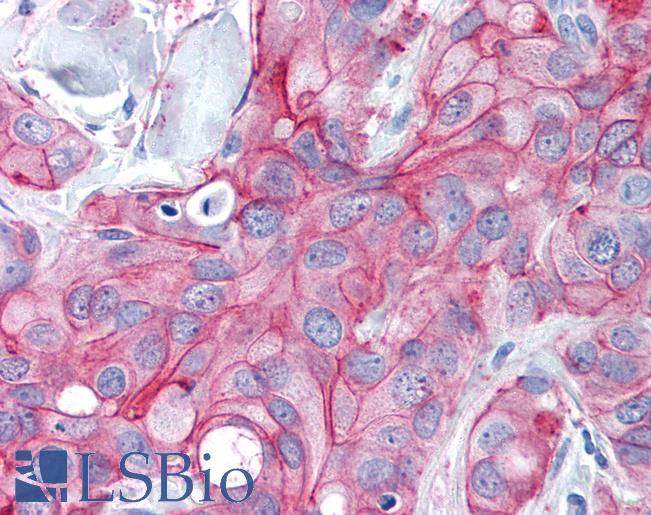
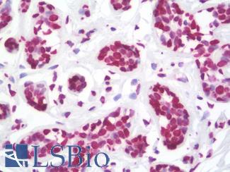
VEGFA


VEGFA (vascular endothelial growth factor A) is a secreted mitogenic cytokine that stimulates endothelial cells, promoting angiogenesis and increased vascular permeability through interactions with VEGF receptors. VEGFA is the target of antiangiogenic chemotherapueutic agents such as bevacizumab (Grothey, 2008), and measurement of serum VEGFA has been explored as a potential noninvasive means of monitoring efficacy of antiangiogenic therapy in cancers (Caporarello, 2017; Secord, 2014). VEGFA is expressed in osteoblasts and is important in normal bone development. It is thought to play a significant role in the unique propensity for bone involvement of metastatic-prostate cancer, which produces characteristic osteoblastic bone lesions with increased bone density rather than lytic bone lesions associated with other types of cancer (Lee, 2017; Roberts, 2013; Hu, 2016).
Staining: VEGFA is a secreted cytokine and can show both cytoplasmic and extracellular staining.

LSBio’s recommended antibody to VEGFA for use in immunohistochemistry is VEGFA Antibody LS‑B3579.

Anti-VEGFA antibody IHC of human kidney. Immunohistochemistry of formalin-fixed, paraffin-embedded tissue after heat-induced antigen retrieval. Antibody LS-B3579 dilution 1:50.
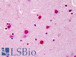
SUPPORT

outstanding technical support

PRODUCT

we offer a full product guarantee

DELIVERY

we offer free delivery to UK universities and non profit organisations