Resources

FAQs

How can I release the N-linked glycans from my glycoprotein?

There are two main techniques commonly used to liberate glycoprotein N-linked glycans for glycosylation analysis – hydrazinolysis and endoglycosidase treatment. Our Guide to Glycosylation Analysis covers some of the considerations for choosing between these release methods.

How can I stop the sialic acids falling off my glycans?

Sialic acids are tricky things. In our experience, the purer the glycan sample the more likely the sialic acid residues are to fall off. There are some sensible precautions though:

  1. Avoid the combination of acidic pH and elevated temperature. Wherever possible, keep the pH in the range 6-9 and the temperature below 30 C.
  2. Be careful when drying glycan samples by centrifugal evaporation or rotary evaporation. When you use these procedures try to keep the temperature below 25 C. Lyophilization (freeze-drying) can be safer with respect to sialic acid degradation but with this technique there is a greater danger of losing the entire sample when the vacuum is released.

Which are the best fluorescent tags for glycan analysis?

The following table shows the most widely used fluorescent glycan tags for glycan analysis by HPLC, CE and MS:


Fluorescent Glycan Label Main Applications Fluorescence Wavelengths and Common Analysis Methods
2-AB (2-amino-benzamide) Oligosaccharide Profiling by HPLC-FD and MS λex = 250 or 330 nm, λem = 428 nm HPLC (amide, C18, AEX, HPAE-FD, PGC), ESI-MS, MALDI-MS
Proc (procainamide) Oligosaccharide Profiling by HPLC-FD and sensitive MS λex = 310 nm, λem = 370 nm HPLC (amide, C18, AEX, PGC), ESI-MS
2-AA (2-amino-benzoic acid) Oligosaccharide Profiling by HPLC-FD and MS Monosaccharide Profiling by HPLC-FD λex = 250nm or 360nm, λem = 425 nm Oligosaccharides: HPLC (amide, C18, AEX, PGC), ESI-MS, MALDI-MS Monosaccharides: HPLC (C18)
APTS (8-aminopyrene-1,3,6-trisulfonate) Oligosaccharide Profiling by CE λex = 455 – 488 nm, λem = 510 – 520 nm CE
VTag IgG Glycopeptide Profiling by HPLC-FD λex = 250 nm, λem = 360 nm HPLC (amide), MALDI-MS
DMB (1,2-diamino-4,5-methylene-dioxybenzene) Profiling of Human vs Non-Human Sialylation by HPLC λex = 373 nm, λem = 448 nm HPLC (C18)

Key: AEX = anion exchange, HPAE-FD = high pH anion exchange with fluorescence detection, PGC = porous graphitic carbon

FAQs - Quantitative Sialic Acid Release and DMB Labelling Kit

Can I store the reagents if I don’t use them all?

In general we recommend against storing the left over solution because we cannot guarantee the chemical quality after storage. The reason that it is generally too risky to store this solution is that it contains a mixture of two reducing agents and a light sensitive dye – if this solution is not protected from the air (oxygen in the air with react with the reducing agents) and the light then the efficiency of the labelling reaction will be impacted and the quality of the resulting data will suffer. As this mixture is in an ampule – which is required for shipment and storage under inert atmosphere – and is now broken open there is no way to reseal unless you transfer the solution – we consider this too big a risk and potentially too variable of a process from lab to lab to suggest this to our clients as best practice.

Will X Buffer interfere with my analysis?

Some salts/buffers commonly used with proteins may interfere with the sialic acid analysis process. This is dependent on the amount of sample taken compared to the volume of buffer (as a large amount of buffer can affect the acidity of the solution during acid hydrolysis). In our experience buffers such as PBS are not a problem where the sample concentrations are above 1mg/mL and between 50 and 200 µg of sample is taken for analysis. This will need to be tested for each sample type and we recommend a buffer blank also be taken though to the process to establish if there are any components in the buffer that interfere with the quantitation of sialic acids.

After adding acid to my protein I have a gelatinous solution, can you help?

We have seen this happen before under 2 circumstances: 1) When the amount of protein in the sample is too high you can end up with a thick solution after acid treatment. We were able to solve this problem by using less starting material (you can run the assay with 50-200µg starting material). Consequently, if you find that your signal is too low when running the samples on LC, do not dilute them prior to injection. 2) Certain buffers can also lead to a thick solution. Our solution to this was to perform a buffer exchange into water prior to acid release. We use vivaspin cartridges with a molecular weight cut off of 10 kDa for buffer exchange.

Do you observe any degradation of the sialic acids during the hydrolysis of samples?

The conditions that we have suggested for the DMB kit were developed using the existing scientific literature as a starting point. The sialic acids are released from glycoproteins by mild acid hydrolysis (2M acetic acid, 80°C for 2h) using conditions that preserve the N-acetyl, N-glycolyl and O-acetyl groups. We looked for the best balance between sialic acid release and minimal degradation (both of the sialic acids and of the of the acetyl groups). It was important for us to maintain the acetyl groups and limit their migration as our clients are often interested in the relative abundance of the Neu5,9Ac2 and other acetylated species.

My sample is an IgG type sample, and it has low levels of sialylation. Therefore, what is the highest concentration of sample I can use without altering volumes of reagents in the kit?

We have performed sialic acid analysis with IgG-type samples where we used 50µg to 1 mg of starting material. For this type of sample the optimal amount of starting material was 200-600µg (linear proportion of sample amount range in validation study). We found that there is no need to increase the volume of any of the reagents. Furthermore, as indicated in the guide if you have low sialylation in your sample, you can run your samples without dilution on the LC.

The protocol suggests solubilizing the protein in 25µL of the 2M acetic acid solution and to transfer only 5 µL, why is this so?

The experiment was set up this way to ensure maximum solubility and efficiency in the first step (trying to dissolve something in 5µl while heating is not feasible). After this, you only require a portion of this sample for an adequate response on FLR-LC after DMB labelling hence only a 5µL portion is taken through.

We used one pot of fetuin and aliquoted the sample so that we have a triplicate – however, our results are below the acceptance criteria, can you help?

We have tried to aliquot fetuin standards from one vial before and our data was also poor. It is best practice not to aliquot the standards but rather use them as provided. This is the only way to ensure that no sample is lost.

Have you applied this method with complicated matrix, like formulation buffer?

We have used this method with many formulation buffers and we have yet to encounter a problem. Regardless, we always run a buffer blank in order to establish if there are any potential interfering species.

What is the difference between the GPEP and Fetuin standards? How do I choose one for my experiment?

Fetuin is a glycoprotein which has both NANA and NAGA. This standard has been studied well in our labs using the DMB assay and we have set acceptance criterion which is based on several years of historical data – this criterion is constantly being monitored and been updated as we collect more and more data about this standard.

GPEP is a glycopeptide with a biantennary disialylated glycan. This is a purified homogeneous glycopeptide that has been quantitated using qNMR. The acceptance criteria is set based on this qNMR value.

There are pros and cons to both standards. Fetuin is a glycoprotein which may match your own sample of interest better than a glycopeptide, however, the glycopeptide is quantified in a more robust way (since it is a smaller molecule that can be subject to NMR). Also, fetuin contains both sialic acids – which may be an important aspect for you, while, GPEP only has NANA. You will need to assess what is important for your assay – we always use both but this is a choice you will need to make for your specific situation.

Which steps in the DMB process are sensitive to error?

In terms of sensitive steps to take into account – when we use this kit in-house or for client services the hitch points are often the following:

  1. Accurate transfer of 5ul of the sample from the acid release step for the labelling experiment. Considerable care should be taken because of the small volume transferred
  2. After adding the labelling reagents that the samples are mixed well
  3. Perhaps the step with the most opportunity to impact quantitation is when preparing the LC samples for analysis. These must be prepared with care and mixed very well to ensure a uniform solution is injected into the LC
  4. Also in general – protecting the labelled samples from degradation by exposure to light is important – provided that they weren’t left sitting out for an extended period of time they should be fine – this is easily identified as the samples tend to go pink in colour

Why do you run triplicates?

We always run triplicates in order to ensure that the data we have is trustworthy. Especially with the quantitative assays it is easy to have some degree of operator error – for instance in this assay transferring 5ul for labelling is a pinch point. It allows the analyst to quickly see if there is an outlier

Guide to Glycosylation Analysis

Chemical Release

Hydrazinolysis is suitable for releasing both N- and O-linked glycans from glycoproteins. It involves the incubation of the dried glycoprotein with anhydrous hydrazine followed by a straightforward workup to purify the released glycans. For success, hydrazinolysis must be carried out with absolutely pure hydrazine under strictly controlled conditions. Advantages of hydrazinolysis include its versatility with the ability to release both N- and O-linked glycans in a virtually non-selective way.

Ludger products for chemical release of glycans:

Hydrazinolysis kit (LL-HYDRAZ-A2): For release of N + O-linked glycans. Released glycans have free reducing terminii to allow fluorescent tagging by reductive amination. The release conditions can be optimized for release of N-glycans, O-glycans or both N- and O-glycans.

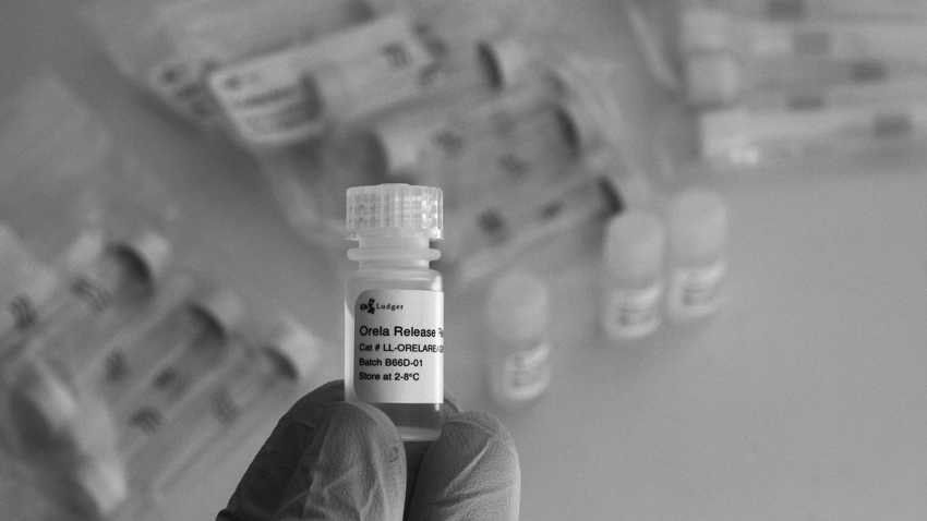
Orela kit (LL-ORELA-A2): For release of O-linked glycans. Released glycans have free reducing terminii to allow fluorescent tagging by reductive amination.

Enzyme (endoglycosidase) Release

Endoglycosidase release is generally a simpler method to implement than hydrazinolysis and suitable for many applications. There are a number of useful endoglycosidases, including Peptide N Glycosidase F (PNGase F), which releases most N-glycans from glycoproteins. Take care when using endoglycosidases as there are a number of conditions and substances that can lead to selective non-release of glycans. In particular, PNGaseF does not release N-glycans which have core fucose attached in the alpha1-3 position which can be found on N-glycans from plants and insects.

Ludger products for chemical release of glycans:

PNGase F (E-PNG-01 or E-PNG01-200): removes N glycans.

O-Glycosidase (E-G001): removes core 1 O glycans (Gal-β(1-3)GalNAc- alpha attached to the serine or threonine)

LudgerZyme Ceramide Glycanase kit (LZ-CER-HM-KIT): Ceramide glycanase deglycosylates a variety of glycosphingolipids by cleaving the β-glycosyl linkage.

Liberation of glycans from glycoproteins

The first step in glycan analysis of glyconjugates such as glycoproteins and glycosphingolipids is the release of the sugars from the molecules to which they are attached.

Great care should be taken in choosing the best glycan release method from the chemical and enzymatic techniques available.

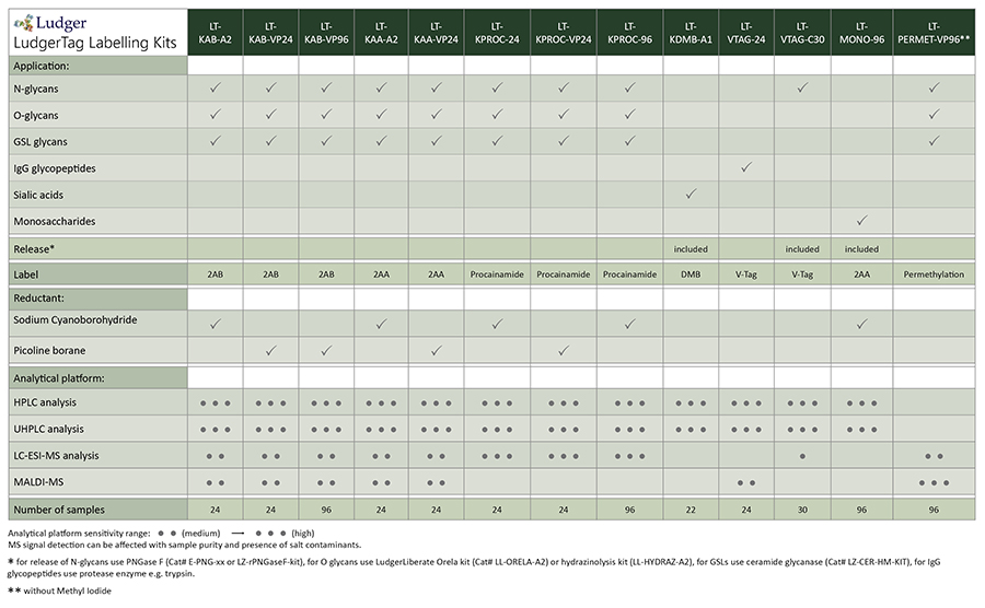
Ludger offers a range of kits for labeling glycans and glycopeptides. We have summarised the recommended applications for the LudgerTag kits in the following table:

Tagging of glycans with fluorescent or UV-active dye labels

Release of glycans by hydrazinolysis or endoglycosidases produces glycans with a reducing terminus that can be tagged with a fluorophore or chromophore label. Most glycan labels are aromatic amines which are conjugated to the glycans by reductive amination. The procedure for this is outlined in our overview of the glycan labeling protocol.

Glycan cleanup methods that are particularly useful during glycosylation analysis include the following:

  • Hydrophilic interaction
    This involves application of the glycan to a hydrophilic matrix in the presence of high levels of particular organic solvents. The glycans bind to the matrix and hydrophobic non-glycan contaminants are washed off with the solvent. The purified glycans are subsequently recovered by elution with an aqueous solvent.
    Examples: LudgerClean S or T1 cartridges, permethylation clean up plate, procainamide clean up plate
  • Electronic interaction
    This uses a special resin with a very flat (on a molecular) surface of sp2 hybridized electrons. The glycans are applied to the resin in an aqueous solution and are captured. Non-organic substances such as salts are washed away and the glycans are then recovered by elution using a mixed organic-aqueous solvent. The method works very well for a wide range of glycans and allows rapid purification of the carbohydrate.
    Examples: LudgerClean EB10 cartridges, EC50 cartridges
  • Ultrafiltration
    Specialized modified polyethersulfone membrane with a molecular weight cut-off of approximately 30 kDa designed for a removal of enzymes and other protein material from glycan solution.
    Examples: Post exoglycosidase clean-up spin columns and 96-well plate optimised for removal of exoglycosidase enzymes from fluorescently labelled and unlabelled glycan mixtures
  • Cation exchange Example: LudgerClean CEX cartridges
  • Hydrophobic interaction Example: LudgerClean PBM plate

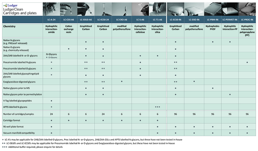
We have summarised the recommended applications for our LudgerClean products in the following table:

Purification of glycans after procedures such as labeling and enzyme treatments

Removal of contaminating substances such as salts, proteins, peptides, or excess derivatization reagents from your glycan samples is essential for the success of many treatments and analyses. Clean up can be performed after glycan labelling or after treatment with enzymes (e.g. after glycan release or treatment with exoglycosidases)

SUPPORT

outstanding technical support

PRODUCT

we offer a full product guarantee

DELIVERY

we offer free delivery to UK universities and non profit organisations