SNARE-Mediated Membrane Fusion and dPEG®, Part 3

Part 3: Modeling Membrane Fusion with SNARE Protein Analogs


Click the links for Part 1 and Part 2 of this three-part series.

Membrane fusion is a universal process in living organisms and is critical to proper cellular function. Examples of membrane fusion events include exocytosis, fertilization, envelopment of infecting viruses, and transport of proteins through the Golgi stack. Though essential, membrane fusion is not spontaneous because free energy is required to overcome steric hindrances, electrostatic repulsions, and hydration shells between two membranes as they move toward each other prior to fusion.

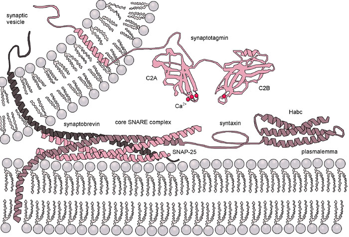
In cells membrane fusion is facilitated by a protein superfamily known as SNARE (for Soluble N-ethylmaleimide-sensitive-factor Attachment Receptor), of which there are 36 members in humans. The best studied SNARE-mediated membrane fusion process is exocytosis in neural cells. Indeed, the first proteins of the human SNARE family to be identified were synaptobrevin, syntaxin, and synaptosome-associated protein 25 (SNAP-25). Figure 1 shows a SNARE-mediated exocytosis.

Figure 1: Membrane Fusion via SNARE Protein Complex Mediation, By Danko Dimchev Georgiev, M.D. [GFDL or CC-BY-SA-3.0], via Wikimedia Commons

Because the native SNARE-mediated process is so complex, numerous research groups have worked to create simplified artificial systems that mimic SNARE-mediated membrane fusion. Numerous systems have been developed using both targeted and non-targeted membrane fusion. Pawan Kumar, Samit Guha, and Ulf Diederichsen have written an excellent review of these SNARE mimetic systems, focusing on targeted membrane fusion. The review is listed in the References section below with a link to the paper, which is behind a paywall.


Most of the targeted SNARE mimetic systems discussed in the review consist of a lipophilic anchor (cholesterol, POPE, and DOPE are discussed), a flexible linker, and a recognition sequence. The flexible linker most often used is a PEG linker, typically a PEG12, though PEG3 has also been used. The purpose of the PEG linker is to site the recognition sequence away from the lipid membrane but to maintain flexibility so that the recognition sequence can move freely in space until it pairs with its target sequence. Example systems using Quanta BioDesign’s dPEG®12 linkers (most notably PN10283, Fmoc-N-amido-dPEG®12-acid) have been discussed in Part 1 and Part 2 of this series.

How dPEG® Can Help You

Quanta BioDesign’s dPEG® linkers provide the traditional advantages of PEG (water solubility, low immunogenicity, reduced aggregation and precipitation of proteins) but with a critical difference. Traditional PEGs are dispersed, but Quanta BioDesign’s patented dPEG® constructs are discrete molecules. Constructs made from dispersed PEGs confound analysis because of the intrinsic heterogeneity of the PEG. Each of Quanta BioDesign’s dPEG® constructs is a single molecular entity. There is no range of linker sizes or molecular weights. Thus, products made from dPEG® are well-defined, easily characterized, and amenable to standard analytical techniques.

Whether you are studying SNARE protein-mediated membrane fusion, building a new drug delivery platform, or creating a new peptide or protein drug, if you need PEG, then you need dPEG®. Quanta BioDesign invented and patented the dPEG® construct, and we manufacture our products in the United States.

References and Additional Reading

(1) Pawan Kumar, Samit Guha, and Ulf Diederichsen. SNARE protein analog-mediated membrane fusion. J Peptide Sci (2015), 21(8), 621-629. DOI: 10.1002/psc.2773

(2) Lando L. G. Schwenen, et al. Resolving single membrane fusion events on planar pore-spanning membranes. Scientific Reports (2015), 5:12006. DOI: 10.1038/srep12006

(3) Jörg Malsam and Thomas H. Söllner. Organization of SNAREs within the Golgi Stack. Cold Spring Harb Perspect Biol (2011), 3:a005249. DOI: 10.1101/cshperspect.a005249

(4) Yu A. Chen and Richard H. Scheller. SNARE-mediated membrane fusion. Nature Reviews Mol. Cell Biol (February 2001), 2, 98-106.

(5) Reinhard Jahn, Thorsten Lang, and Thomas C. Südhof. Membrane Fusion. Cell (February 21, 2003), 112, 519-533. DOI:10.1016/S0092-8674(03)00112-0

(6) Joseph G. Duman and John G. Forte. What is the role of SNARE proteins in membrane fusion? Am J Physiol Cell Physiol 285: C237–C249, 2003; DOI: 10.1152/ajpcell.00091.2003.

(7) Reinhard Jahn and Richard H. Scheller. SNARES — Engines for Membrane Fusion. Nature Reviews Mol Cell Biol (September 2006), 7, 631-643. DOI:10.1038/nrm2002

SUPPORT

outstanding technical support

PRODUCT

we offer a full product guarantee

DELIVERY

we offer free delivery to UK universities and non profit organisations