Two-Photon Microscopy
Two-Photon Microscopy (TPM), a microscopic system utilising Two-Photon absorption (TPA), has become an indispensible tool in the study of biology and medicine thanks to the comparative advantages to conventional confocal microscopy and is expected to shift the paradigm of the researches.
These are a few of the advantages
- The near-infrared radiation used for excitation can penetrate more easily into biological samples without damage. The ability to penetrate a material and to be focused accurately in three dimensions makes TPM ideal for imaging thick samples, even in vivo.
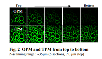
- The rate of Two-Photon excitation is squarely proportional to the illuminating intensity, resulting in localised excitation, which makes it possible to obtain hundreds of sectional images, from which 3-dimensional images can be obtained.
- The large separation between excitation and emission spectra excludes the possible interference from the excitation light and scattering, which allows selective detection of the fluorescence photons.
- Photobleaching and photodamaging are limited to a volume of sub-femtoliter, which allows visualisation over a long period of time.

TPM imaging is a different technique from standard confocal imaging. It requires very sophisticated setup and operation knowhow. Currently, most fluorescence probes presently available for TPM have small TP action cross sections (ϕ δ ), insufficient to obtain clear fluorescence images, demanding impractically high concentrations of probe and / or intense laser irradiation to observe the effects of Two-Photon absorption. This lack of efficient Two-Photon Probes has been one of the major bottlenecks to the advance of TPM in spite of their superior characterisation and performance.
A Two-Photon Probe (TPP) is a specially designed TPA molecule dedicated to a specific purpose and target. In the case of biological imaging, dealing with live tissues, it should meet some basic conditions set out as below:
• Appreciable water solubility
• Significant TP action cross section
• High photo-stability
• Reasonable cell permeability
• Ability to be conjugated to the target
• Low toxicity
One of the biggest challenges in meeting the conditions above is the incorporation of all the required functionalities within a single small molecule. Even though there are general design rules within the theoretical basis, it is not a simple job when you consider the complexities closely linked to the biological test environments. Therefore, it is crucial to find the right combination of fluorophore, receptor, linking group, and other functional domains when designing TPP molecules appropriate for a specific purpose.
To view the complete range of two photon probes click here